Homocysteine, Folate and Vitamin B12 in Colombian Patients with
Coronary Disease
Gilberto Garcia, Juanita Trejos, Beatriz Restrepo, Patricia Landázuri Facultad de Ciencias de La Salud - Universidad Del Quindio - Colômbia - South America
Summary
Objective: To determine the occurrence of association between homocysteine, folate, or vitamin B12 plasma levels and acute coronary syndrome in Colombian patients.
Methods: Case control study: cases were 50 patients with acute coronary syndrome and controls were 50
outpatients without coronary syndrome. Homocysteine, folate and vitamin B12 levels were determined by means of chemiluminescence immunoassay. Cholesterol and lipoproteins, triglycerides, BUN, creatinine, hemoglobin and hematocrit were also measured.
Results: Mean homocysteine plasma concentrations were significantly different between cases (12.4 µmol/l ± 6.0) and controls (9.7 µmol/l ± 2.4), p=0.01. The folic acid levels of the cases were lower than those of the control patients (10.5 ng/ml ± 3.5 vs 12.6 ng/ml ± 3.6, respectively, p=0.01). An inverse relationship was found between folate and homocysteine levels. No relationship was observed between vitamin B12 levels and homocysteine levels. There was a significant difference in triglyceride levels between case and control groups (136.91 ± 67.27 vs 174.3 ± 77.6, respectively, p=0.01). The odds ratio for hyperhomocysteinemia in acute coronary syndrome was 4.45 (95% confidence interval: 1.5 - 13.3).
Conclusion: The present study found a significant association between homocysteine levels and acute coronary syndrome in Colombian patients, similarly to the European and North American populations. There was a negative correlation between homocysteine plasma levels and folate levels. No association between plasmatic levels of homocysteine and those of vitamin B12 was observed. (Arq Bras Cardiol 2007;89(2):71-76)
Key words: Homocysteine; folate; vitamin B12; coronary disease; lipids; pteroypolyglutamic acids.
Mailing address: Patricia Landázuri •
Av. Bolivar 12 N - Armenia (Quindio) - Colombia - South America E-mail: [email protected]
Manuscript received September 19, 2006; revised manuscript received January 01, 2007; accepted March 03, 2007.
Introduction
Cardiovascular diseases are the major cause of morbidity and mortality worldwide and are the second cause of death in Colombia1. Obesity, hypercholesterolemia, smoking and
hypertension have been recognized as major risk factors for cardiovascular diseases; however, they do not fully explain the pathogenesis and causality of these diseases. A new class of emerging risk factor for cardiovascular diseases is homocysteine plasma levels2. Homocysteine is a sulphur-containing amino
acid that results as an intermediary product of the methionine degradation pathway. Life style and genetic defects such as folic acid and vitamin B12 or B6 deficiencies can play a role as cofactors that increase homocysteine levels. Some studies have shown that up to 10% of coronary events can be attributed to the increase in homocysteine levels3. Additional studies
have shown a negative correlation between homocysteine levels and folic acid and vitamin B12 and B6 plasma levels 4-5. Thus, hyperhomocysteinemia is a new cardiovascular risk
factor that could be modified by reducing homocysteine
intake or by supplementing the diet with vitamin B12 or B66.
Hyperhomocysteinemia can be present as a cardiovascular risk factor or not, depending on the geographic population studied; thus, the increased risk due to hyperhomocysteinemia seems to be different in Indians as compared to europeans7.
There are few studies about this relationship in latin-american populations; therefore, this study aims to determine the occurrence of association between homocystinemia and acute coronary syndrome in colombian patients.
Methods
myocardial infarction with or without electrocardiographic signs of elevated ST segment. Patients out of the age range or with history of coronary heart disease resulting from the use of drugs such as carbamazepine, amphetamines, estrogens, or from cocaine consumption, or who were under current or chronic use of certain drugs that influence homocysteine levels, such as methotrexate, trimethoprim, cholestyramine and cyclosporine were excluded. Patients with chronic or acute renal insufficiency, diabetes mellitus, hypothyroidism, and other forms of atherosclerosis or chronic heart failure were also excluded. Controls were subjects treated in the outpatient clinic of the Hospital, who had no cardiovascular disease or other chronic diseases such as renal failure. The same exclusion criteria for the cases were also applied to the control subjects. All subjects were from Colombia; the colombian population stems mostly from a mixture of europeans (caucasoids), africans (negroids) and amerindians (mongoloids)8. The city
of Armenia is geographically located between the central and eastern branches of the Andean mountains. The population of Armenia (600,000 inhabitants) belongs to the self-called “paisa community”8. In this population the mixture of
negroid and amerindian populations has been documented to be low8-11. Jimenez et al12have estimated ancestral racial
components in the “paisa community” at 85% caucasian and 15% amerindian.
Laboratory assays - Venous blood samples from the cases were obtained at baseline from eight to twelve hours after symptom presentation and before the beginning of thrombolysis or anticoagulant treatment, and transferred into EDTA-coated tubes (Becton Dickinson). Plasma was immediately obtained by centrifugation at 4°C, for 15 minutes at 3000 rpm. Aliquots of plasma were stored for batch analysis at 20°C within 1 hour of sampling. In the controls, samples were obtained after an overnight 12-hour fast.
Plasma levels of homocysteine, vitamin B12 and folic acid were assayed by the Immulite analyzer (Diagnostic Products Corporation, Los Angeles, California) which is a bench top immunoassay analyzer, with continuous random-access capabilities, that uses enzyme-amplified chemiluminescence chemistry for antibody or antigen detection. Analysis was performed according to the manufacturers’ protocols. Normal range of homocysteine levels was 5-15 µmol/l, hyperhomocysteinemia was considered mild if values were between 15.1 to 30 µmol/l, moderate between 30.1 to 100 µmol/L and severe if > 100 µmol/l. Folic acid levels were considered normal between 6 to17 ngr/ml, and those of vitamin B12 were considered normal between 187 to 900 pgr/ ml13. Additional laboratory assays were glycemia and lipids.
Definition of variables - Hypertension was defined as a systolic blood pressure above 140 mm Hg and diastolic blood pressure above 90 mm Hg; sedentary life style was defined as less than three days of exercise per week, and smoking status denoted at least one cigarette per month. Hypercholesterolemia was defined as cholesterol > 200 mg/dl.
Statistical analysis - For the examination of risk factors for acute coronary syndrome (cases) the data were examined with univariate analysis. The mean and standard deviation (SD) and median of homocysteine, lipid profile, folic acid, and vitamin B12 levels of the different groups were measured.
When appropriate, multivariate analysis adjusted for potential confounders (current smoking daily or most days; age; and coffee, tea and alcohol intake) was performed. For variables with normal distribution the Student’s t test for unpaired values was performed to compare the continuous variables between the groups. For variables with non-normal distribution the Mann-Whitney test was performed. The Chi-square test was performed to calculate the differences in discrete variables between the groups, and the odds ratio with a 95% confidence interval was also calculated. Correlation between continuous variables was analyzed by regression analysis using the SPSS software version 12.
Ethical considerations - The study was revised and approved by the Institutional Review Board (Ethics Committee) of the University of Quindio and an informed consent was signed by the subjects who agreed to participate in the study.
Results
Baseline characteristics of the study participants - The general characteristics of the study population are shown in Table 1. Age, gender and BMI were not significantly different between the groups (all p>0.05). The etiologic diagnosis of the acute coronary syndrome for the cases shows that most of the cases had unstable angina. Major cardiovascular risk factors such as hypertension, smoking status, sedentary life stile, and a positive family history for hypertension or cardiovascular disease were more prevalent in the cases than in the controls, p<0.05 for all risk factors.
Biochemical markers - Results of glycemia, lipid and creatinine levels in both groups are shown in Figure 1. Mean levels of total cholesterol and low-density lipoproteins (LDL) were higher in the cases and lower in the controls but without a statistically significant difference. High-density lipoprotein (HDL) levels were lower in cases compared to controls, but this difference was not statistically significant either. It is noteworthy that triglyceride levels were significantly higher in controls (p = 0.02). The levels of other markers such as very-low-density lipoproteins (VLDL), glycemia and creatinine were not significantly different between cases and control patients. No positive correlation was observed between cholesterol and homocysteine levels, either in the case group or in the control group. Similar findings were obtained when LDL, HDL, VLDL and triglyceride levels were correlated.
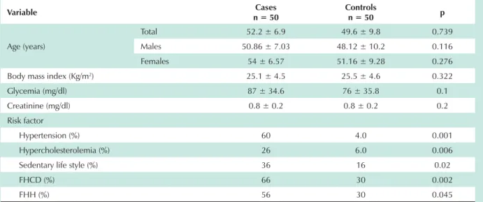
Homocysteine, folic acid and vitamin B12 levels in cases and controls - The mean levels of homocysteine, folic acid and vitamin B12 in cases and controls are shown in Table 2. Homocysteine levels in controls were within normal limits (9.7 µmol/l ± 2.4) but were higher in cases (12.4 µmol/l ± 6.0). The differences in mean homocysteine levels between cases and controls were statistically significant (p=0.004). For distribution of homocysteine levels, the normal range (1-15 µmol/l) was found in 80% of cases and 98% of controls. The upper range of homocysteine levels (>15 µmol/l) was found in 20% of cases and in 2% of controls. Levels higher than 20 µmol/l were found in cases but not in controls (Figure 2).
Table 1- Baseline characteristics of the study population and prevalence of cardiovascular risk factors
Variable Cases
n = 50
Controls
n = 50 p
Age (years)
Total 52.2 ± 6.9 49.6 ± 9.8 0.739 Males 50.86 ± 7.03 48.12 ± 10.2 0.116 Females 54 ± 6.57 51.16 ± 9.28 0.276 Body mass index (Kg/m2) 25.1 ± 4.5 25.5 ± 4.6 0.322
Glycemia (mg/dl) 87 ± 34.6 76 ± 35.8 0.1
Creatinine (mg/dl) 0.8 ± 0.2 0.8 ± 0.2 0.2
Risk factor
Hypertension (%) 60 4.0 0.001
Hypercholesterolemia (%) 26 6.0 0.006
Sedentary life style (%) 36 16 0.02
FHCD (%) 66 30 0.002
FHH (%) 56 30 0.045
p - level of statistical significance; FHCD - family history for coronary disease; FHH - family history for hypertension.
Table 2 - Levels of homocysteine, folic acid and vitamin B12
Variable Cases
n = 50
Controls
n = 50 p
Homocysteine
(µmol/l) 12.4 ± 6.0 9.7 ± 2.4 0.01 Folic acid
(ng/ml) 10.7 ± 3.9 13.1 ± 4.2 0.005 Vitamin B12
(pg/ml) 391.9 ± 229.3 368.3 ± 178.3 0.56
p - level of statistical significance.
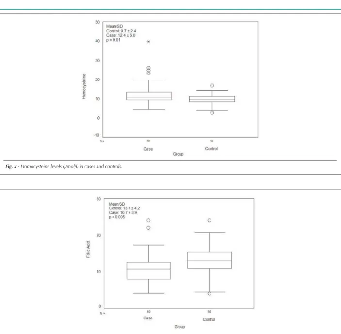
20% had intermediary levels, and 72% had folic acid levels in the upper range. In relation to cases, 16% had low levels, 40% had intermediary levels, and only 44% had levels in the upper range (Figure 3). A statistically significant negative relation was observed between homocysteine levels and folic acid levels. There were no significant differences between cases (381±197 pg/ml) and controls (368±178 pg/ml) in relation to mean levels of vitamin B12. Twelve percent of the controls but 80% of the cases were in the lower to normal range of vitamin B12 levels. Upper levels were most frequent in cases than in controls. There was no significant correlation between levels of vitamin B12 and homocysteine, either in cases or in controls.
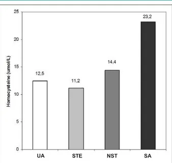
Odd ratio for hyperhomocysteinemia in acute coronary syndrome - The odds ratio for hyperhomocysteinemia in acute coronary syndrome was 4.45 (95% confidence interval: 1.5-13.3). Abnormal higher levels of homocysteine were found in 56% of the patients with unstable angina, in 34% of the patients with elevated ST, in 5% of the patients with stable angina and in 5% of the patients with non-elevated ST (Figure 4). After adjustment for confounding variables (gender, age), only homocysteine levels had a strong positive correlation with coronary disease.
Discussion
Several studies have been performed to demonstrate association between homocysteine levels and cardiovascular diseases2,3. Many of them have found a significant association5,14,15
but others have failed to show this association4,16,17. Differences
in the characteristics of the study population such as nutritional habits, the use of vitamin supplements or ethnic differences could explain the contradictory results18,19.
There is some evidence that the mean homocysteine concentration varies between countries. Genuine systematic
Fig. 2 -Homocysteine levels (µmol/l) in cases and controls.
Fig. 3 -Folic acid levels in cases and controls.
differences in the characteristics of the populations studied may account for the different estimates of effect in various studies. We tested the hypothesis that the mean homocysteine level was higher in the colombian group of patients with acute coronary syndrome than in controls, as occurs in caucasian populations. In the group of patients with acute coronary syndrome, we found a significant association between this disease and hyperhomocysteinemia; most of our patients with acute coronary syndrome had abnormally elevated levels of homocysteine. Studies in north american and european population have estimated that a 10% risk of coronary artery disease may be attributable to elevated homocysteine3,17. The
mechanisms of how hyperhomocysteinemia induces coronary disease are poorly understood but there are evidences of an endothelial oxidative damage20, endothelial smooth muscle
proliferation21, oxidation of low-density lipoproteins22 and
reduction of the nitric oxide production through inhibition of the nitric oxide synthase23.
Increased blood levels of homocysteine reflect deficiency of folate, and vitamins B6 and B12. Folate deficiency is linked to cardiovascular diseases5and certain cancers, particularly
colorectal cancer24. These associations are likely due to
the effects of altered folate metabolism on homocysteine remethylation, DNA synthesis, and DNA methylation. Reduced homocysteine remethylation can result in an elevated plasma homocysteine concentration, which is an independent risk factor for cardiovascular disease25. Similarly to previous
studies5,15,26,27 we found low levels of folic acid in the cases.
Fig. 4 -Homocysteine levels in coronary disease. UA - unstable angina, STE - steady, NST - no unstable, SA - steady angina.
UA STE NST SA
likely, have a poorer diet . Chambers et al2 and Toole et al27
found that elevated plasma homocysteine concentrations in indian asian patients, compared with european white controls, were explained by their low folate and vitamin B12 concentration. In our study patients had low folate when compared with controls, similar to indian asian populations and other results from previous studies15,19.
Plasma folate in particular is a strong determinant of homocysteine concentration; homocysteine levels are inversely related to folate consumption, reaching a stable baseline level when folate intake exceeds 400 ug/day25. In this way, in Wald
et al’s26 meta-analysis, they found a strong evidence that the
association between homocysteine and cardiovascular disease is causal and that lowering homocysteine concentrations by 3 µmol/l from current levels by increasing folic acid intake would reduce the risk of cardiovascular disease (ischemic heart disease, deep vein thrombosis and stroke) by between 16% to 38%; these results are similar to those of other studies such as HOPE-2 (Heart Outcomes Prevention Evaluation)26, VISP
(Vitamin Intervention for Stroke Prevention)27 and WENBIT
(Western Norway B-vitamin Intervention Trial)28. In our study
there was a significant correlation between homocysteine and folic acid. A lower folate intake that can be enough to raise homocysteine levels may be relatively common in the general population in many countries, and it was not different in the case of the colombians. Jimenez et al12have estimated
ancestral racial components in the “paisa community” as 85% caucasian and 15% amerindian; in this colombian group of patients the risk of coronary artery disease (OR 4.45, 95% confidence interval: 1.5-13.3) was similar to that of caucasians (north american and european populations). In this respect, Bermudez et al29found that among 102 healthy
colombian individuals, 8 were identified with moderate
hyperhomocysteinemia, of which one at baseline and seven post methionine load, perhaps because of vitamin B6 or folic acid deficiency. In contrast to folic acid, we did not find a correlation between vitamin B12 and homocysteine levels. It should be noted that there is a controversy about this correlation and that nutritional habits could greatly influence the plasmatic levels of vitamin B124,19,26.
On the other hand, for some years, attention focused on the role of genetic variables for homocystinuria17,30 as a possible
cause of the high homocysteine concentrations seen in patients with coronary artery disease. Polymorphisms in critical enzymes of the biochemical pathways of the homocysteine metabolism (cystathionine B-synthase, methionine synthase and methylenetetrahydrofolate reductase MTHFR) could also lead to high homocysteine levels. Deficiencies of these enzymes, although rare, have been described, and are associated with hyperhomocysteinemia and notably with vascular diseases31. Two relatively common variant
polymorphisms in the gene coding for MTHFR have been described31. One of these, the C677T mutation, may also
be associated with hyperhomocysteinemia especially in the presence of lower folate concentrations. In Colombia, Bermudez et al29 studied hyperhomocysteinemic patients
identified by the methionine load test and found a positive association with homozygous for MTHFR 677 polymorphism; they also found that the frequency of this polymorphism in Colombia is the highest reported in the literature.
In addition, the present study shows that lipid profiles were moderately elevated in patients, but, in contrast with previous studies, there was no association between the levels of total cholesterol, LDL and homocysteine32. Unexpectedly,
only triglyceride levels were higher in controls compared to cases. We think that this cannot be explained by the differences in ethnic composition, but our findings raise the possibility that genetic defects and or environmental factors may influence the homocysteine metabolism in this group of patients.
In summary, we have discovered that, like in the findings of several studies of european and north american populations, there is an association between hyperhomocysteinemia and low levels of plasma folate and acute coronary syndrome in colombian patients. Further studies should help define the role of genetic polymorphism in folate and homocysteine metabolism enzymes and their role in coronary disease.
Potential Conflict of Interest
No potential conflict of interest relevant to this article was reported.
Sources of Funding
There were no external funding sources for this study.
Study Association
References
1. Departamento Administrativo Nacional de Estadística (DANE). Defunciones totales por sexo: 20 principales causas. 1996-2001. Bogotá (Colombia); 2001.
2. Gravina-Taddei CF, Batlouni M, Sarteschi C, Baltar VT, Salvarini NAC, Bertolami MC, et al. Hyperhomocysteinemia as a risk factor for coronary atherosclerotic diseases in the elderly. Arq Bras Cardiol. 2005; 85: 166-73.
3. Boushey CJ, Beresford SA, Ommenn GS, Motulsky AG. A quantitative assessment of plasma homocysteine as a risk factor for vascular disease: probable benefits of increasing folic acid intakes. JAMA. 1995; 274 (13): 1049-57.
4. Selhub J, Jaques PF, Wilson P, Rush D, Rosenberg IH. Vitamin status and intake as primary determinants of homocysteinemia in and eldery population. JAMA. 1993; 270: 2693-8.
5. Wald DS, Law M, Morris JK Homocysteine and cardiovascular disease: evidence on causality from a meta-analysis. BMJ. 2002; 325: 1202.
6. Schnyder G, Roffi M, Flammer Y. Effect of homocysteine – lowering therapy with folic - acid, vitamin B(12), and Vitamin B(6) on clinical outcome after percutaneous coronary. Intervention: the Swiss Herat study a randomized controlled trial. JAMA. 2002; 288: 973-9.
7. Sastry BKS, Indira N, Anand B, Kedarnath, Surya Prabha B, Soma Raju B. A case–control study of plasma homocysteine levels in South Indians with and without coronary artery disease. Indian Heart J. 2001; 53: 749-53.
8. Agudelo LE. Genesis del pueblo antioqueño. Bogotá: Era cósmica, 1986.
9. Parson JJ. Antioqueño colonization in western Colombia. Berkeley: University of California Press, 1949.
10. Bravo ML, Valenzuela CY, Blabco R, Arcos OM. Polymorphism and phyletic relationships of the paisa community from Antioquia (Colombia). Gene Geogr. 1996; 10: 11-7.
11. Arcos-Burgos M, Bravo ML. Evaluación epidemiológico-genética para labio hendido con o sin paladar hendido en la población de Antioquia. [Máster thesis] Antioquia: Faculty of Exact Sciences. University of Antioquia; 1992.
12. Jimenez I, Mora O, Lopez G, Jimenez ME, Zuluaga L, Isaza R, et al. Idiopathic epilepsy with generalized tonic clonic seizures in Antioquia, Colombia: is the joint Amerindian and Negroid racial admixture the cause of its high prevalence? Biol Res. 1996; 29 (3): 297-304.
13. Suárez T, Torrealba M, Villegas N, Osorio C, García-Casal MNO. Deficiencias de hierro, ácido fólico y vitamina B12 en relación a anemia, en adolescentes de una zona con alta incidencia de malformaciones congénitas en Venezuela. ALAN. 2005; 55: (2): 118-23.
14. Robinson K, Mayer EL, Miller DP, Green R, Vanlente F, Gupta A, et al. Hyperhomocysteinemia and low pyridoxal phosphate: common and independent reversible risk factor for coronary artery disease. Circulation. 1995; 92: 2825-30.
15. Graham IM, Daly LE, Refsum HM, Robinson K, Brattstrom LE, Ueland PM, et al. Plasma homocysteine as risk factor for vascular disease. The European Concerted Action project. JAMA. 1997; 277: 1775-81.
16. Ford ES, Smith SJ, Stroup DF, Steinberg KK, Mueller PW, Thacker SB. Homocyst(e)ine and cardiovascular disease: a systematic review of the evidence with special emphasis on case-control studies and nested case control studies. Int J Epidemiol. 2002; 31: 59-70.
17. Lewis SJ, Ebrahim S, Smith GD. Meta-analysis of MTHFR 677CmT polymorphism and coronary heart disease: does totality of evidence support
causal role for homocysteine and preventive potential of folate?. BMJ. 2005; 331 (7524): 1053.
18. Chambers JC, Kooner JS. Homocysteine: a novel risk factor for coronary heart disease in UK Indian Asians. Heart. 2001; 86: 121-2.
19. Hughes K, Ong CJ. Homocysteine, folate, vitamin B12, and cardiovascular risk in Indians, Malays, and Chinese in Singapore. J Epidemiol Community Health. 2000; 54: 31-4.
20. Zhang C, Cai Y, Adachi MT, Oshiro S, Aso T, Kaufman RJ, et al. Homocysteine induces programmed cell death in human vascular endothelial cells through activation of the unfolded protein response. J Biol Chem., 2001; 276: 35867-74.
21. Shinichi N, Tawara J, Toyoda H, Kitamura K, Komurasaki T. A novel homocysteine-responsive gene, smap8, modulates mitogenesis in rat vascular smooth muscle cells Eur J Biochem. 2003; 270: 2521-31.
22. Halvorsen B, Brude I, Drevon CA, Nyssom J, Ose L, Christiansen EM, et al. Effect of homocysteine on copper ion-catalyzed, azo compound-initiated and mononuclear cell-mediated oxidative modification of low density lipoprotein. J Lipid Res. 1996; 37: 1591-600.
23. Fatini C, Sofi F, Gori AM, Sticchi E, Marcucci R, Lenti M, et al. Endothelial nitric oxide synthase –786T>C, but not 894G>T and 4a4b, polymorphism influences plasma homocysteine concentrations in persons with normal vitamin status. Clin Chem. 2005; 51: 1159-64.
24. Kim Y-I Folate and DNA Methylation: A Mechanistic link between folate deficiency and colorectal cancer? Cancer Epidemiol Biomarkers Prev. 2004; 13: 511-9.
25. Lucock M. Is folic acid the ultimate functional food component for disease prevention? BMJ. 2004; 328; 211-4.
26. Homocysteine Lowering with Folic Acid and B Vitamins in Vascular Disease. The Heart Outcomes Prevention Evaluation HOPE-2 Investigators. N Engl J Med. 2006; 354: 1567-77.
27. Toole JF, Malinow MR, Chambless LE, Spence JD, Pettigrew LC, Howard VJ, et al. Lowering plasma total homocysteine to prevent recurrent stroke, myocardial infarction, and death in ischemic stroke patients: results of the Vitamin Intervention for Stroke Prevention (VISP) randomized trial. JAMA. 2004; 291 (5): 565-75.
28. Bleie Ø, Refsum H, Ueland PM, Vollset SE, Guttormsen AB, Nexo E, et al. Changes in basal and postmethionine load concentrations of total homocysteine and cystathionine after B vitamin intervention. Am J Clin Nutr. 2004; 80: 641-8.
29. Bermúdez M, Briceño I, Gil F, Bernal J. Homocisteína y polimorfismos de cistationinaB sintasa y metilentetrahidrofolato reductasa en población sana de Colombia. Revista Colombiana Médica. 2006; 37: 46-52.
30. Klerk M, Verhoef P, Clarke R, Blom HJ, Kok FJ, Schouten EG, and the MTHFR Studies Collaboration Group. MTHFR 677C. T polymorphism and risk of coronary heart disease: a meta-analysis. JAMA. 2002; 288: 2023-31.
31. Brattström L, Wilcken D, Öhrvick J, Brudin L. Common methylenetetrahydrofolate reductase gene mutation leads to hyperhomocysteinemia but not to vascular disease: the results of a meta-analysis. Circulation. 1998; 98: 2520-6.