Objective – To evaluate echocardiography accuracy
in performing and obtaining images for dynamical three-dimensional (3D) reconstruction.
Methods – Three-dimensional (3D) image
recons-truction was obtained in 20 consecutive patients who underwent transesophageal echocardiography. A multi-planar 5 MHz transducer was used for 3D reconstruction.
Results – Twenty patients were studied
consecuti-vely. The following cardiac diseases were present: valvar prostheses – 6 (2 mitral, 2 aortic and 2 mitral and aortic); mitral valve prolapse – 3; mitral and aortic disease – 2; aortic valve disease – 5; congenital heart disease – 3 (2 atrial septal defect –ASD- and 1 transposition of the great arteries -TGA); arteriovenous fistula – 1. In 7 patients, color Doppler was also obtained and used for 3D flow reconstruction. Twenty five cardiac structures were acqui-red and 60 reconstructions generated (28 of mitral valves, 14 of aortic valves, 4 of mitral prostheses, 7 of aortic pros-theses and 7 of the ASD). Fifty five of 60 (91.6%) recons-tructions were considered of good quality by 2 indepen-dent observers. The 11 reconstructed mitral valves/pros-theses and the 2 reconstructed ASDs provided more anato-mical information than two dimensional echocardiogra-phy (2DE) alone.
Conclusion – 3D echocardiography using a
transe-sophageal transducer is a feasible technique, which im-proves detection of anatomical details of cardiac structu-res, particularly of the mitral valve and atrial septum.
Key words: three-dimensional echocardiography, ultra-sonography, valvar disease
Arq Bras Cardiol, volume 72 (nº 5), 564-568, 1999
Maria de Fátima Veiga, Mário G. Lopes, Fausto J. Pinto
Lisbon, Portugal
Dynamic Three-Dimensional Reconstruction of the Heart by
Transesophageal Echocardiography
Hospital Universitário de Santa Maria – Faculdade de Medicina de Lisboa Mailing Address: Fausto J. Pinto – Faculdade de Medicina de Lisboa – Clínica Médica – Piso 2 – Av. Prof. Egas Moniz – 1600 Lisbon –Portugal
Three-dimensional (3D) reconstruction of the heart through conventional 2DE imaging is a new technology with potential clinical applications.
Reconstruction through transthoracic echocardio-graphy is often limited in some patients by small number of acoustic windows and poor imaging quality. In addition, the device coupled to transthoracic transducer is too large and heavy to allow for proper handling during the exam, resul-ting in a great number of artifacts, which interfere with the analysis and with making the correct diagnosis.
When transesophageal echocardiography (TEE) was first used clinically in 1980, a new window to the heart appeared. In 1986, Martin et al 1-3 used a micromanipulator
for the TEE transducer for the 3D echocardiogram. Due to the high quality of images obtained by the transesophageal access, 3D reconstruction of the heart, using transesopha-geal imaging emerged as a promising technique that offers a stable site from which to obtain multiple images from seve-ral sections.
Thus, the purpose of this study was to evaluate the feasibility of performing and obtaining images of good diagnostic quality using TEE for 3D reconstruction.
Methods
Images for 3D reconstruction were obtained during routine transesophageal studies in 20 outpatients in sinus rhythm referred to our echocardiography laboratory. Patients with poor apical echocardiographic window (best plane for image acquisition), and those on atrial fibrillation (when it is difficult to gate to the R-R interval) were exclu-ded. Topic anesthesia with lidocaine spray and slight sedation with intravenous midazolan were used.
Data were triggered with the electrocardiogram (ECG) and the patient’s respiration and a DE images were acqui-red in the apical view, digitized and stoacqui-red in a disk. The operator had to locate the center of the axis around which the multiplanar transducer rotated in intervals from 2o to
180o, activated by a motor and controlled by the software.
Ninety sequential series from 0o to 180o were obtained
during each cardiac cycle. Average time for calibration, acquisition, processing and reconstruction ranged from 40 to 50 minutes.
In the present study, we used the volume-rendered reconstruction visualization method in which, from any section plane, different algorithms were applied to represent space information. Several gray scales, distance, texture and gradient scales, as well as a threshold to differentiate cardiac structures from blood and image background were used to give depth perception. Different degrees of brightness and opacity were also used to help give this perception.
Twenty consecutive patients were studied; 9 were male and 11 female and age ranged from 23 to 75 years (mean age = 56±16). Clinical aspects are described in table I.
All patients were in NYHA functional class I and in sinus rhythm. Two patients had an ASD, three mitral valve prolapse (MVP), three aortic stenosis, three infective endocarditis, six valvar prosthesis, one had arteriovenous fistula, one TGA, one mitral stenosis and one aortic valve fibrosis.
Results
Transesophageal echocardiography was well tolera-ted and acquisition and reconstruction were possible in all patients. A total of 48 acquisitions were obtained (15 patients with one and five with two acquisitions).
Twenty five cardiac structures were acquired and 60 reconstructions were obtained (28 mitral valves, 14 aortic valves, 4 mitral regurgitations, 7 aortic regurgitations and 7 atrial septal defects).
In seven patients, color flow jets were also acquired and reconstructed: five mitral and two aortic regurgitations. Valves could be seen in several three-dimensional views and lesions could be seen in different aspects. These structures were projected in several planes and observed in different phases of the cardiac cycle.
Among the 60 reconstructions, 55 (91.6%) were considered of good diagnostic quality by two independent observers. Eleven reconstructed mitral valves and two reconstructed ASDs provided more anatomic information than did 2DE alone.
Mitral valve prolapse – Anatomic information from valvar leaflet was obtained in three patients through the
vo-lume rendered method, which was performed on several
sections at the mitral valve level, in long and short axis. Due to the fact that the left atrium (LA) is very well visualized by TEE, 3D reconstruction gives an excellent vision from the top of the prolapsed mitral valve leaflets 4. The advantage of
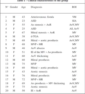
this image view modality is the excellent acoustic window obtained when the LA is dilated by associated mitral re-gurgitation, which thus helps visualize the morphology of the mitral valve components. A example of MVP is shown in figure 1.
Mitral regurgitation – In five patients, 3D dynamic imaging allowed for a better visualization and understan-ding of the jet direction and size projection than could be obtained by 2DE with color flow mapping alone. Mitral regurgitation is seen in figure 2, where a central jet is clearly seen on the atrial perspective.
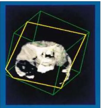
Six patients were studied for evaluation of different kinds of mitral and aortic prostheses (7 mechanical and one biological). 3DE images did not provide additional information when compared with those obtained by 2DE, because 3DE disk evaluation was not adequate, even after using different thresholds with brightness and opacity adjustments, due to the excess of echoes that jeopardized the correct analysis. Figure 3 shows disk prosthesis in systole.
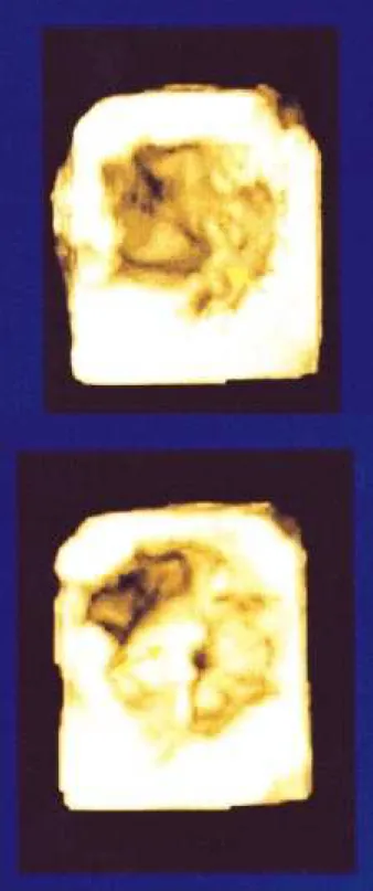
Aortic stenosis – 3D reconstruction of aortic valve was obtained in three patients and the aortic cusps were seen from above. In the dynamic mode, valve opening and closing could be observed. A stenotic aortic valve in diastole can be seen in figure 4.
Table I – Clinical characteristics of the group
No Gender Age Diagnosis ROI
1 M 43 Arteriovenous fistula VM
2 M 23 ASD SIA
3 F 55 Ao stenosis + AoR AoV, MV
4 F 25 ASD AS, AoV
5 F 67 Mitral stenosis + AoR MV
6 M 38 d-TGA AoV, MV
7 M 68 Mitral + aortic prosthesis AoV, MV
8 F 68 MVP + MR MV
9 M 46 AoV abscess AoV
10 F 51 IE of the MV + Ao prosthesis MV
11 F 60 AoV thickening AoV
12 M 60 Mitral prosthesis MV
13 M 75 MVP MV
14 F 60 Mitral + aortic prosthesis MV
15 F 43 Aortic stenosis AoV
16 F 76 Mitral prosthesis MV
17 M 72 MVP + MR MV
18 F 69 Ao prosthesis + MV thickening AoV, MV
19 F 75 Aortic stenosis AoV
20 M 50 IE + AoR AoV
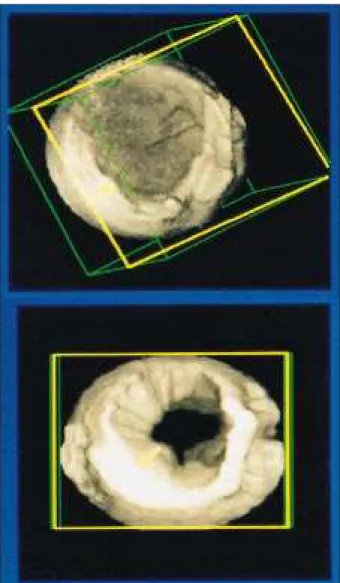
Atrial septal defect – In two patients, 3D recons-truction of the defect was performed and direct view of its size and geometry was obtained, as well as its relationships to other cardiac structures, which could be visualized from both sides of the atrial septum. It was also possible to defect systo-diastolic variation in the size of the defect, which could be visualized only when 3DE was used.
Figure 5 shows a large atrial septal defect from the perspective of the left atrium. Right atrium could be seen through the large defect.
Infective endocarditis – Three patients with infective endocarditis were studied and in one (case 9), a better ana-tomical definition of an aortic perivalvular abscess was obtained. In the two remaining patients, vegetation had already been identified by 2DE.
Fig. 1 – 3D reconstruction in a patient with mitral valve prolapse: A) mitral valve seen from the ventricular perspective. B) systolic prolapse of the anterior leaflet of the mitral valve seen from the atrial perspective, towards the observer.
Discussion
3DE may become the best method for studying car-diac anatomy and pathology. In the last few years, 3D re-construction has been performed using different methods for image acquisition. Up to now, conventional 2DE has always been used for 3D reconstruction.
Rotational acquisition technique offers advantages during a multiplanar transesophageal study due to the
Fig. 4 - 3DE reconstruction of the aortic valve. The stenotic aortic valve is seen in diastole.
Fig. 3 - 3D reconstruction of a mechanical aortic prosthesis: A) opened disk in systole can be seen. B) closed disk is seen in diastole.
excellent quality of the obtained image when the transe-sophageal approach is used 5-9. When the region of
inte-rest is centralized, the transducer is held in a stable posi-tion and then the motor makes it spin around the axis. This technique is relatively easy to perform but there is a lear-ning curve, since the apparatus coupled to the transducer is large and heavy.
TEE, specially with a multiplanar transducer, has be-come a widely used procedure in many centers, because it can be easily employed in different clinical situations. Because the esophagus is posterior to the heart, it is a stable site for placing the multiplanar transducer, and this provides advantages for rotational acquisition of good quality images, when compared with other methods.
The greatest 3D reconstruction advantage consists of the possibility of visualizing structures in unique pers-pectives, due to the sections that can be obtained in any desired plane.10 It is very helpful in the evaluation of valvar
structure (subvalvular apparatus, valvular ring and leaflet thickening which can be visualized in different sections). Defect size and magnitude in different points and angles from those available in 2DE can be obtained in a perspec-tive similar to open heart surgery.
In addition, real time images offer the opportunity to better reproduce valvar movement, providing further information about leaflets mobility, commissures and valvar orifice size. Different aspects from different diseases can be appreciated, when compared with the still heart during surgery 11,12.
1. Martin RW, Bashein G, Zimmer R, Sutherland J. An endodcopic microma-nipulator for multiplanar transesophageal imaging. Ultrasound Med Biol 1986; 12: 965-75.
2. Martin RW, Bashein G, Detmer PR, Moritz WE. Ventricular volume measurement from a multiplanar transesophageal ultrasonic imaging system: an in vitro study. IEEE Trans Biomed Eng 1990; 37: 442-8.
3. Martin RW, Bashein G. Measurement of stroke volume with three-dimensional transesphageal ultrasonic scaning: comparison with thermodilution measu-rement. Anesthesiology 1989; 70: 470-6.
4. Salustri, A, Becker AE, Herwerden L, Vletter, WB, Cate FJT, Roelandt JRT. Three-dimensional echocardiography of normal and pathologic mitral valve: a comparison with two-dimensional transesophageal echocardiography. J Am Coll Cardiol 1996; 27: 1502-10.
5. Sugeng L, Cao QL, Delabays A, Pandian NG, et al. Three-dimensional echocardiographic evaluation of aortic disorders with rotational multiplanar imaging:experimental and clinical studies. J Am Soc Echocardiograpfy 1997; 10: 120-32.
6. Roelandt JRTC, ten Cate FJ, Vletter Wletter WB, et al. Ultrasonic dynamic three-dimensional visualization of the heart with a multiplane transesophageal imaging transducer. J Am Soc Echocardiogr 1994; 7: 217-29.
References
7. Menzel T, Kahaly SM, Kolsch B, Kupferwasser I, Meyer J, et al. Quantitative assessment of aortic stenosis by three-dimensional echocardiography. J Am Soc Echocardiography 1997; 10: 215-23.
8. Salustri A, Roelandt J. Three dimensional reconstruction of the heart with rotational acquisition: methods and clinical applications. B Heart J 1995; 13(suppl 2) : 10-15.
9. Ghosh A, Nanda NC, Maurer G. Three-dimensional reconstruction of echo-cardiographic images using the rotation method. Ultrasound Med Biol 1982; 8: 655 - 661.
10. Pandian NG, Roelandt J, Nanda N, Sugeng L, Cao Q, Azevedo J, et al. Dynamic Three-dimensional echocardiography: methods and clinical potential. Echocardiography 1994; 11: 237-59.
11. Chen Q, Nosir YFM, Vletter WB, Kint PP, Salustri A, Roelandt JRT. Accurate assessment of mitral valve area in patients with mitral stenosis by three-dimensional echocardiography. J Am Soc Echocardiography 1997; 10: 133-40.
12. Levine RA, Handshumaker MD, Sanfilippo AJ, et al. Three-dimensional echocardiographic reconstruction of the mitral valve, with implication for diagnosis of mitral valve prolapse. Circulation 1989; 80: 589-98.
Fig. 5 – Section at the level of atrial septum helped to define atrial septal defect and its relationship with other structures.
Reconstruction image quality, considered adequate when complete deep view of the aimed structure is obtai-ned, depends on the quality of 2DE images gathered during acquisition, which in turn, depends on the transducer stability, the patient’s respiration and heart rate. Due to this fact, patients on atrial fibrillation, when it is difficult to obtain ECG gating, have been excluded, as well as those with respiratory distress, which greatly prolongs image acquisition time.
Several factors are crucial for 3D reconstruction. Selection of gains and threshold in post-processed image are major steps for volume-rendered imaging. When not properly adjusted, they may result in artifacts and limit diagnosis.
Another major point needs to be emphasized: the possibility of the patient moving the transducer during acquisition. This limitation has been overcome by providing adequate instruction to the patient to remain still during acquisition.
Finally, the greatest limitation for the 3DE is the amount of time spent for data processing, which is still too long to be used routinely.