Article 4
Is Anti-Suppression the Quest for Visibility?
Eric S. Hussey, OD, Spokane, WashingtonABSTRACT
Vision science defines the fundamental action of the vision system to be the generation of visible percepts. Intermittent central suppression (ICS) is an intermittent, usually alternating, loss of visual sensation, a repetitive loss of that visual percept. A review of the vision science literature on visibility, Troxler’s Perceptual Fading, and the consequences of loss of visibility suggests that loss of visual motion signal causes loss of visibility. The area of lost visibility is then filled in by a cortical computation mechanism. Fixation, binocular saccades, vergence, and sensory perception are all compromised in loss of visibility.
Some of the characteristics of ICS, as well as suspected consequences of ICS, have been difficult to reconcile with the conventional wisdom on suppression. That conventional wisdom suggests that suppression is solely a function of cortical inhibition. Those ICS characteristics and consequences may be better explained if ICS is viewed as a loss of visibility from Troxler’s Fading. Fixation changes and perceptual changes with loss of visibility parallel findings in ICS and the vision-and-dyslexia literature. Research methods to reverse loss of visibility parallel treatment methods for ICS. These parallels suggest that loss of visibility may be diagnosed clinically as ICS, and ICS should be treated with a goal of ensuring bilateral visibility, that is, binocularity.
Keywords: binocularity, dyslexia, intermittent central suppression, suppression, Troxler’s Perceptual Fading, visibility
Introduction
Suppression of vision has been described as neural inhibition, possibly masking, in the cortex.1 Prior reports have suggested flaws in
the conventional wisdom (diplopiaphobia)
concerning suppression.2,3 Described by
Bielschowski in 1943, suppression is a cortical inhibition triggered by the visual cortex’s fear or phobia of the diplopia of strabismus.4
Diplopia precipitates the cortical response of inhibition. If the term diplopiaphobia has any accuracy, it certainly applies most appropriately to early strabismus. Traumatic strabismus in adults apparently does not trigger a cortical diplopiaphobic suppression response. There-fore, diplopiaphobia as a trigger for producing suppression most certainly requires neurology in an early developmental epoch to complete the inhibitory-suppression model.
Other problems with this suppression conventional wisdom follow from our increas-ing knowledge of non-strabismic intermittent central suppression (ICS). This includes the on-and-off predominantly alternating periodicity of ICS, the genesis of ICS after whiplash, and the very normal refractive error profile without significant anisometropia seen in ICS patients.5-7
The pattern of change in ICS suppression periods with therapy, as documented in recent reports, also presents a challenge to suppression conventional wisdom.2,3
could be regarded as designed to achieve simultaneous, bilateral visibility in the central vision. One advantage of using visibility to describe binocular vision is that vision science has studied visibility at least since the 1800s. In 1804, Troxler found that exceedingly stable fixation causes images to disappear; that is, visibility was compromised by that excessively stable fixation, Troxler’s Perceptual Fading.8 What does the vision science tell us
about loss of visibility, beginning with Troxler? One caveat is that experiments on Troxler’s are done monocularly. However, there is still much to learn about loss of visibility from image stabilization and its consequences. In the world of vision science, the fundamental action of the visual system is to generate visible percepts.9 In this context then, binocularity
might be defined as continuous, bilateral, simultaneous visible percepts (without diplopia and therefore with alignment). Conversely, defective binocularity would be viewed as a loss of visibility amounting to a shift toward monocularity, a loss of fundamental visual function. This literature review will look at the vision science literature on visibility and, in summary, look at possible overlaps with the binocularity literature pertaining to ICS and by extension to binocularity in dyslexia.
What does image stabilization mean?
Stabilizing an image on the retina is the steadying of image edges and borders. In Troxler’s (and some later) work, image stabiliza-tion was accomplished by developing very steady fixation.8,10 In other experiments,
non-ocular mechanisms were developed to stabilize images on the retina without training observers to ultra-stable fixation abilities.11
Edge-detect ing cells, which are normally driven and synchronized by contrast borders,11,12
decrease firing with stabilized fixation.13 With
stabilization, perception (visibility) fades within 0.1 sec.,14 possibly a loss of contrast to
the point of disappearance.15 So, in normal
visibility (that is, maintained-without-fading visibility), something (fixational eye movements or stimulus movements) must keep borders and edges moving across edge-detecting cell receptive fields, driving groups of cells in a synchronous fashion.12 Eye or stimulus
movements that are larger than the confines of contrast-edge-detecting-cell receptive fields will sustain visibility.11,16 Image stabilization can
be viewed as the removal of on-off signals from those receptive fields.16 Said differently, visibility
requires visual motion. Experimentally perfect image stabilization requires image movements across the retina to be held below 0.1 arcmin.17
Fixational eye movements keep target edges moving across the retina(s), usually providing the visual motion necessary to keep the image(s) visible. All fixational eye movements are involved: tremor, drifts, and microsaccades (earlier called flicks).11 Fixational
eye movements have predominantly excitatory effects at all levels of the visual system.11
However, ballistic gaze-shifting eye movements (saccades) suppress vision, probably very early in the visual system between the retina and the cortex.18-20 Saccadic suppression of visibility
selectively suppresses motion mechanisms, probably in the magnocellular (M) pathway and probably early in visual processing.20,21 That
saccadic suppression of visibility presumably reduces or maybe eliminates the fast, sweep-of-panorama visual motion that would have to be endured during a non-suppressed saccadic shift in gaze.
Drifts and saccades tend to counteract each other during fixation.22 Drifts produce a
constant global background drifting motion that must be computed out of the central target (local) motion to produce a stable visual world. Drifts differ somewhat between eyes, and that target motion computation occurs prior to the merger of the visual pathways, probably at the level of ganglion cells.23
bursts of neural spikes in ganglion cells, and at the Lateral Geniculate Nucleus (LGN).25 Bursts
of spikes increase in response to motion, not detail, although higher contrast increases the number of spikes in those neural bursts.25
Fixational eye movements have the greatest effect on spike rates and therefore on visibility when the target is most likely to be detected by the M pathway (versus the parvocellular (P) pathway). The increasing spikes are mostly facilitative, not inhibitory, and the enhancement of magnocellular spikes is quadruple that of parvocellular spikes.18
Binocular, or bilateral, fixational movements may have a stronger role in counteracting fading than monocular fixational movements.27 Head
movements also contribute to maintaining visibility.28 Visibility can be maintained with
head rotation, and all fixational eye movements increase in speed relative to the target when the head is free to move. Drifts and tremor happen simultaneously and are sufficient to maintain visibility centrally, but they may not be sufficient for visibility in the periphery.27 Tremor
alone increases visibility centrally only when the tremor surpasses 0.3 minutes of arc, the inter-cone spacing, and when cones receive complete on-off exposure,15 that neural activity
being early in the visual system.9 Although
microsaccades may correlate strongly to visibility in controlled experiments, in natural conditions they apparently have no special role and actually seldom happen during reading.28
Fixational eye movements support
visibil-ity through retinal image changes,29 but
stimulus motion can also keep the image visible.27,30 An especially strong form of image
motion stimulus is on-off flicker.1,25,31 In the
real visual world, visual stimuli turn on and off several times per second, when viewed from the standpoint of receptive fields.32
The onset response to flashes (visual flicker) may be seven times as strong as the neural response to the same stimulus moving across a receptive field.25
In sum, visual motion is necessary for sus-taining visibility of the retinal image, whether through fixational eye movements, head movements, or image motion (including stimulus flicker). The operative neural signal supporting visibility is visual (sensory), not motor.11
Image stabilization reduces the motion signal that keeps visibility intact. Transient-neuron responses are more import ant to visibility than sustained-neuron responses.32 Fixational eye
movements have the greatest effect on visibility when the target is most likely to be detected by M pathway neurons.18
Importantly, all of these image motion visibility-sustaining effects assume unimpaired neurological motion sensitivity. Approached from the opposite side – not from sustaining but from losing visibility – suppose we ask how much impairment in motion sensitivity is needed to trigger loss of visibility? If we can extrapolate from the loss of visibility during a ballistic gaze-shifting saccade, a drop in magnocellular spike rate of about 20% is enough.18 Visual motion is, indeed, the
“on-switch” for visibility, and a relatively small loss in neurological motion signal is enough to flip that switch to “off.”
Loss of Visibility and its Consequences
Loss of the motion signal carried in transient-response neurology causes loss of visibility. We can speculate on the loss of that motion signal and its consequences to binocularity such as loss of binocular signal convergence and binocular facilitation for fusion and stereopsis,2,3,26 or we can again
follow the vision science on Troxler’s and see what happens during a lack-of-motion loss of visibility.
During a Troxler’s fade loss of visibility, decreased motion feedback may accompany that loss of visibility, changing fixational movement dynamics as feedback for fixation is compromised.33 Microsaccade rates reduce,
off target are larger, presumably because only larger target displacements trigger correction.15,22,24 Gaze inaccuracy increases by
a factor of four when fixation loses a visible marker, possibly with some limitation in error contributed by peripheral vision.22 Fixation
just gets sloppy during a Troxler’s fade loss of visibility.
Binocular (perhaps more accurately when discussing loss of visual sensation, “bilateral”) eye aiming might also be affected. Saccade amplitudes become more unequal immediately after loss of binocular vision. Therefore, binocular vision is essential for keeping the amplitudes of binocular saccades as equal as possible.27 Disjunctive (vergence)
eye movements are corrected during their course on the basis of continuous disparity information, the sensory system being capable of disparity discriminations even less than a minute of arc.34 The necessary matching
of bilateral features may happen prior to actual depth percept processing.35 Although
vergence during Troxler’s fading has not been studied, mechanical interference in one eye’s stimulus during binocular saccade recording has been studied, showing vergence errors that are not corrected during inter-saccadic fixation periods.36
Target movement strengthens the probability that a stimulus will dominate and also prevents fading. Therefore, lack of motion signal allows the opposite eye, the eye with retinal image changes, to dominate29,30,37 and
allows fading as above. The lack-of-motion-signal-triggered loss of visibility (Troxler’s fade), as well as the opposite effect, the motion-triggered domination of one eye over the other, apparently occurs at early stages in the visual system, possibly at the level of the LGN.25,30,38,39 At a sensitive developmental
period, this could provide the mechanism for a subcortical monocular suppression.26 If so, the
suppression at a lower level in the visual system would influence processing at subsequent
stages,39 conceivably contributing to the
cortical (interocular) inhibition of strabismic suppression26,40 and positioning amblyopia
as secondary to loss of binocularity,41 but
also affecting the target velocity and position comparisons made centrally that control vergence.34 Therefore, a
decreased-motion-signal fade of visibility, allowing the opposite eye to dominate and interfering with vergence-control velocity and position comparisons, might easily cause vergence errors and non-correction of vergence errors similar to those
shown in Collewijn et al’s36 experimental
conditions and results. Loss of visibility sets the stage for sloppy fixation, unequal saccades, and erroneous vergence.
If this loss of visibility from lack of motion signal does, as the visibility literature suggests, impair accurate fixation and vergence, then any abrupt restoration of visibility would reap the harvest of these inaccuracies: Any errors in accurate fixation and vergence during the loss of visibility would require correction when both eyes are seeing simultaneously.2,3 But what of
perception during the loss of visibility? When visibility is lost, is the brain left with intact perception from one side to be combined with a black “patch” from the other?
Filling-in as a Consequence of Loss of Visibility
During a Troxler’s loss of visibility, the faded area - the area that has lost visibility - is filled in with surround, apparently by the same process that fills in the natural blind spot.39 Therefore,
loss of visibility does not equate to positive scotoma. Just as most people don’t notice their natural blind spot, and a specific search task such as a visual field test must be used to define its boundaries, perceptual filling-in makes it less likely that a monocular loss of visibility will reach a conscious level.10,13,39 This
original percept. Since it is an active process, it is possible that measurements of cortical activity in strabismus may include this filling-in neural activity.
The perceptual filling-in computation occurs beyond the primary visual cortex.13 Perceptual
filling-in can create rivalry,13 suggesting cortical
feedback down to lower levels where rivalry is thought to occur, possibly at the LGN.43-46
Rivalry between the side with visibility versus the side without visibility but with perceptual filling-in suggests the possibility that a loss of visibility is not some sort of a mild, essentially innocuous loss of attention but could actually produce visual confusion from percept changes produced by that rivalry.
This possibility of and the consequences of filling-in occurring in a lost-visibility (i.e., suppressed?) visual area is ignored in the binocularity literature. Other disparate effects that might be involved with a loss of visibility are similarly unknown. For example, if lost visibility takes away retinotopic frames of reference or spatial updates, both visual attention and visual spatial maps might be affected.47,48 The consequences of a loss of
visibility may be much more far reaching than previously addressed.
What does the Vision Science Tell Us About Ways to Ensure Visibility?
The vision science primarily addresses what stimuli reverse a Troxler’s Fade loss of visibility. Perhaps, though, we can extrapolate from what reverses fading to what might continuously maintain rather than restore visibility. With reversal of perceptual fading projected into the possibility of sustained visibility, we would also expect to reverse or to eliminate some of the oculomotor effects of loss of visibility as well as eliminating any consequences of perceptual filling-in.
Throughout the discussion of loss of visibil-ity, motion is key to reversing the fade and restoring visibility. Transient-neuron
respons-es associated with onset and offset of a stimulus drive visibility. When viewed from the standpoint of receptive fields, real world stimuli turn on and off several times per second.32 If
we can define a strong motion stimulus that provides onset/offset for the visual neurology, visibility should be facilitated.
As discussed above, a particularly strong motion stimulus is on-off flicker. On-off visual
flicker equals motion,31 keeping targets
visible.49 Neural responses to onset of flashing
bars are seven times larger at the LGN and at V1 than responses to the same stimulus bars whose edges are moved across receptive fields by microsaccades, rather than being flashed.11
“Flash suppression” experiments use a single-target flicker event to override the other side’s visual stimulus through that strong response to flicker, suppressing the non-flickered side. Flash suppression probably occurs at a lower visual area.50 Similarly, masking can override
the opposite image. However, masking apparently is cortical and works by inhibiting neural on- and off-response.32
Stringing a series of flicker-flash events together at the correct rate will override the other eye’s percept for an extended period,37,51,52 called continuous flash
suppres-sion. Continuous flash suppression (CFS) rapidly updates a complex stimulus image37 and is
facilitative to the “dominant” signal.51 Rather
than decreasing inhibition, CFS increases dominance time of the flashing stimulus but does not decrease the duration of the period of visibility when the “suppressed side” shows. The sustained “barrage of transients” is responsible for CFS’s potency in keeping one side dominant, and at five flashes the neurology is being driven, creating neural learning.51
Although a flash rate between 3 and 10 Hz will reliably suppress a salient image on the other side,51 5 Hz is accepted as the strongest driving
rate.52 That 5 Hz flash rate is a 100 msec square
produces,51 providing a steady stream of
onset-offset transient (motion) signals.
Both spatial and temporal profiles of the strongest CFS stimulus suggest that the M pathway is primarily involved, supporting the idea that CFS uses a strong sustained, apparently additive motion stimulus to drive visibility in the early visual system.52 If we go
back to our original definition of binocularity as both eyes having intact visibility of central images that are combined into one percept without loss of visibility of either central image, the only way to assure that a potentially asymmetric bilateral vision system is equally driven to facilitate bilateral simultaneous percept is to alternately drive the two sides with a strong CFS-style stimulus, as paradoxical as that might seem. It would follow that bilateral visibility should be facilitated by alternating flashing targets at 5 Hz, a strong CFS motion stimulus, now applied bilaterally.
Although this concept has not been studied by vision science, the concept matches clinical experience.3 Further, the clinical experience
of changing intermittent central suppression (ICS) with 5 Hz alternation parallels CFS since CFS increases dominance time without decreasing suppressed time on the other side, perhaps partially explaining post-therapy changes in ICS suppression periods versus binocular periods.2,3
If 5 Hz alternation can facilitate bilateral simultaneous visibility (binocularity), we would expect bilateral simultaneous visibility to change what happens during a Troxler’s fade toward more “normal” oculomotor behavior. Therefore, fixational eye movements should normalize to reduce the sloppiness that occurs during fading. Bilateral saccades should return to being symmetrical, and the suspected vergence errors should decrease. Those changes should improve stability of perception
just by reducing inaccuracies in oculomotor behavior.
At this point, we might reconsider the intermittency of ICS, defined by its typical 2- to 3-second on-off cycle, but change the language of the intermittency from “on-off cycle” to “normal-visibility/loss-of-visibility cycle.” If we then apply the lessons from perceptual fading to a normal-visiblity/loss-of-visibility cycle, not only is oculomotor behavior compromised in a number of ways by losses of visibility, but during the loss-of-visibility periods, some rivalry can occur to interfere with the detail that remains visible on the non-suppressed/ normal-visibility side. Obviously, if there is any accuracy to that constellation of fade-effects, something requiring constancy of detail like reading could be affected.
Are There Commonalities Between Loss of Visibility, ICS, Reading Problems, and Anti-suppression Treatment?
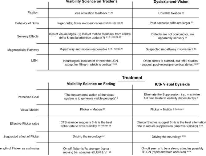
Table 1 shows a listing of effects of loss of visibility from the vision science versus some of the vision-and-dyslexia research. The visibility science, as discussed above, suggests oculomotor errors during loss-of-visibility fades. The vision-and-dyslexia research also supports oculomotor errors in “dyslexia.”53 Loss
of visibility through a Troxler’s fade is sensory; some of the defects in “dyslexia” are sensory.54
The magnocellular pathway is intimately involved in Troxler’s perceptual fading and has been implicated in “dyslexia.”55 Further, as
in Troxler’s, that defect is thought to be post-retinal and pre-cortical.56,57
On the treatment side, assuming (for example) liquid crystal alternation to reduce suppression and CFS have some actions in common, the visibility research suggests that a strong motion stimulus like 5 Hz flicker will drive the visual neurology and restore or retain visibility,51 just as 5 Hz alternation reduces
suppression.3 Since a 5 Hz CFS stimulus of five
flashes (that is, one second of continuous 5 Hz
flashing) creates neural learning,51 permanent
neural change should follow from sustained use, paralleling clinically-testable decreases in suppression with sustained 5 Hz alternation in a therapy regimen.3 Further, a change from
Troxler’s fading and loss-of-visibility to constant bilateral visibility (elimination of ICS?) would imply reversal of loss-of-visibility oculomotor errors, presumably meaning improved fixation stability as well as normal visual sensation rather than perceptual filling-in – all changes that would normalize visual perception. Normalized visual perception would support normalized reading behavior, paralleling the improvements in reading as reported in the ICS literature.3
Conclusions
At least for non-strabismic, non-amblyopic ICS, Troxler’s perceptual fading is a likely mechanism explaining suppression. A 20% decrease in magnocellular spike activity may be enough to cause perceptual fading that would be clinically-diagnosed ICS. Further, ICS, and a Troxler’s fade, is likely to interfere with detail-intensive tasks such as reading. Correcting ICS, whether by more traditional means or with electronic rapid alternate occlusion at 5 Hz, establishes simultaneous bilateral visible percepts, also known as binocularity, which should improve visual stability and accuracy with some positive effects on detail-intensive tasks such as reading.
References
1. Schor C, Terrell M, Peterson D. Contour interaction and temporal masking in strabismus and amblyopia. J Optom Physiol Optics 1976;53:217-23. http://bit.ly/1ETg7FJ
2. Hussey ES. Increases in binocularity periods with treatment of intermittent central suppression contradict suppression as solely inhibitory. Accepted for publication, in press.
3. Hussey ES. Remote treatment of intermittent central suppression improves quality of life measures. Optometry 2012;83:19-26. http://bit.ly/1DZ3YLn
5. Hussey ES. Temporal characteristics of intermittent central suppression. J Behav Optom 2002;13:149-52. http://bit. ly/1DZ4h8X
6. Hussey ES. Intermittent central suppression: A missing link in reading problems? J Optom Vis Devel 1990;21:11-6. http://bit. ly/1KGJY2o
7. Hussey ES. Intermittent central suppression caused by cervical trauma (whiplash). J Behav Optom 1997;8:31-6. http://bit. ly/1KGJY2o
8. Troxler D. Uber das Verschwinden gegebener Gegenstande innerhalb unseres Geisichtskreises. In: Himly K, Schmidt JA, eds. Ophthalmische bibliothek. Jena: Fromann, 1804:51-0.
9. Martinez-Conde S, Macknik SL, Hubel DH. The role of fixational eye movements in visual perception. Nature Reviews/ Neuroscience 2004;5:229-40. http://bit.ly/1uwN7jp
10. Troncoso XG, Macknik SL, Martinez-Conde S. Microsaccades counteract perceptual filling-in. J Vis 2008;8(14):1-9. http:// www.journalofvision.org/content/8/14/15
11. Ditchburn RW. Eye movements in relation to retinal action. Optica Acta 1955;1:171-6. http://bit.ly/1KGKbCN
12. Greschner M, Bongard M, Rujan P, Ammermuller J. Retinal ganglion cell synchronization by fixational eye movements improves feature estimation. Nat Neurosc 2002;5:341-7. http://bit.ly/1M7grTa
13. Komatsu H. The neural mechanisms of filling-in. Nature Reviews/Neuroscience 2006;7:220-31. http://bit.ly/1M7guP0
14. Olveczky BP, Baccus SA, Meister M. Segregation of object and background motion in the retina. Nature 2003,423:401-8. http://bit.ly/1Ac0iqX
15. Poletti M, Rucci M. Eye movements under various conditions of image fading. J Vis 2010;10(3):1-18. http://bit.ly/1Dd20Zy.
16. Ditchburn RW, Fender DH, Mayne S. Vision with controlled movements of the retinal image. J Physiol 1959;145:98-107. http://1.usa.gov/1C8LLOH
17. Ditchburn RW. What is psychophysically perfect image stabilization? Do perfectly stabilized images always disappear? comment. J Optical Society Am 1987;4:405-6. http://bit.ly/16HPZyt
18. Reppas JB, Usrey WM, Reid RC. Saccadic eye movements modulate visual responses in the lateral geniculate nucleus. Neuron 2002;35:961-74. http://bit.ly/1Dd2aAk
19. Shioiri S, Cavanaugh P. Saccadic suppression of low-level motion. Vis Res 1989;29:915-28. http://bit.ly/1DUTInA
20. Thilo KV, Santoro L, Walsh V, Blakemore C. The site of saccadic suppression. Nat Neurosc 2004;7:13-4. http://bit.ly/1vesKCu
21. Burr DC, Morrone MC, Ross J. Selective suppression of the magnocellular visual pathway during saccadic eye movements. Nature 1994;371:511-3. http://bit.ly/1M7h5Af
22. Cherici C, Kuang X, Poletti M, Rucci M. Precision of sustained fixation in trained and untrained observers. J Vis 2012;12(6):1-16. http://bit.ly/1ICoFhr, doi:10.1167/12.6.31.
23. Buckthought A, Mendola JD. A matched comparison of binocular rivalry and depth perception with fMRI. J Vis 2011;11(6);1-15. http://bit.ly/1y2KeIl, doi:10.1167/11.6.3.
24. Kagan I, Gur M, Snodderly DM. Saccades and drifts differentially modulate neuronal activity in V1: Effects of retinal image motion, position, and extraretinal influences. J Vis 2008;8(14):1-25. http://bit.ly/1C3AUlo, doi:10.1167/8.14.19.
25. Martinez-Conde S, Macknik SL, Hubel DH. The function of bursts of spikes during visual fixation in the awake primate lateral geniculate nucleus and primary visual cortex. PNAS 2002;99:13920-5. http://bit.ly/1zq9LW3
26. Sengpiel F, Jirman K, Vorobyov V, Eysel UT. Strabismic suppression is mediated by inhibitory interactions in the primary visual cortex. Cerebral Cortex 2006;16:1750-8. http:// bit.ly/175nJag
27. Martinez-Conde S, Macknik SL, Troncoso XG, Dyar TA. Microsaccades counteract visual fading during fixation. Neuron 2006;49:297-305. http://bit.ly/16LEFBN
28. Collewijn H, Kowler E. The significance of microsaccades for vision and oculomotor control. J Vis 2008;8(14):1-21. http:// bit.ly/1DMjjPZ, doi:10.1167/8.14.20.
29. van Dam LCJ, van Ee R. Retinal image shifts, but not eye movements per se cause alternations in awareness during binocular rivalry. J Vis 2006;6:1172-9. http://bit.ly/1xYOIyg doi:10.1167/6.11.3
30. Arnold DH, Law P, Wallis TSA. Binocular switch suppression: A new method for persistently rendering the visible “invisible.” Vision Research 2008;48:994-1001. http://bit.ly/1Fr2Txl
31. Wallis TSA, Arnold DH. Motion-induced blindness is not tuned to retinal speed. J Vision 2008;8(2):1-7. http://bit.ly/1wL0Sq6, doi:10.1167/8.2.11. http://bit.ly/1wL0Sq6
32. Macknik SL, Livingstone MS. Neuronal correlates of visibility and invisibility in the primate visual system. Nat Neurosc 1998;1:144-9. http://bit.ly/1Dd2Lls
33. Engbert R, Mergenthaler K. Microsaccades are triggered by low retinal image slip. PNAS 2006;103(18):7192-7. http://bit. ly/16HS9y2
34. Rashbass C, Westheimer G. Disjunctive eye movements. J Physiology 1961;159:339-60. http://1.usa.gov/1AmKwEx
35. Masson GS, Busettini C, Miles FA. Vergence eye movements in response to binocular disparity without depth perception. Nature 1997;389:283-6. http://bit.ly/1KGLGB2
36. Collewijn H, Erkelens CJ, Steinman RM. Binocular Co-ordination of human horizontal saccadic eye movements. J Physiol 1988;404:157-82. http://bit.ly/1Dy5dTZ
37. Nichols DF, Wilson HR. Effect of transient versus sustained activation on interocular suppression. Vis Res 2009;49:102-14. http://bit.ly/1IFQf2M
38. Kotulak JC, Schor CM. The accommodative response to subthreshold blur and to perceptual fading during the Troxler phenomenon. Perception 1986; 15:7-15. http://bit.ly/1Ac2Sx8
39. Lou L. Selective peripheral fading: how attention leads to loss of visual consciousness. http://bit.ly/1tWDbdc. Last accessed September 21, 2004. http://bit.ly/1tWDbdc
41. Hess RF, Thompson B, Black JM, Maehara G, et al. An iPod treatment of amblyopia: An updated binocular approach. Optometry 2012;83:87-94. http://bit.ly/1zQc5JE
42. Magnussen S, Spillmann L, Sturzel F, Werner JS. Filling-in of the foveal blue scotoma. Vision Res 2001;41:2961-7. http://bit. ly/1y0FY6a
43. Haynes J-D, Deichmann R, Rees G. Eye-specific suppression in human LGN reflects perceptual dominance during binocular rivalry. Nature 2005;438:496-9. http://1.usa.gov/1zQcgEM
44. Wunderlich K, Schneider KA, Kastner S. Neural correlates of binocular rivalry in the human lateral geniculate nucleus. Nat Neurosc 2005;8:1595-1602. http://bit.ly/1uwQiaP
45. Lee S-H, Blake R. V1 activity is reduced during binocular rivalry. J Vis 2002;2:618-26. http://bit.ly/1C3BCim doi 10:1167/2.9.4. http://bit.ly/1C3BCim
46. Knapen T, van Ee R, Blake R. Stimulus motion propels traveling waves in binocular rivalry. PLoS ONE 2007;2(8):e739. doi:10.1371/ journal.pone.0000739. http://bit.ly/1CMMrct
47. Golomb JD, Chun MM, Mazer JA. The native coordinate system of spatial attention is retinotopic. J Neuroscience 2008;28:10654-62. **J Neuroscience currently unavailable
48. Golomb JD, Pulido VZ, Albrecht AR, Chun MM, Mazer JA. Robustness of the retinotopic attentional trace after eye movements. J Vis 2010;10(3):1-12. http://bit.ly/1KyBLAk doi: 10.1167/10.3.19. http://bit.ly/1KyBLAk
49. Simons S, Lleras A, Martinez-Conde S, Slichter D, et al. Induced visual fading of complex images. J Vis 2006;6:1093-1101. http://bit.ly/1DMjYAM doi: 10.1167/6.10.9
50. Brascamp JW, Knapen THJ, Kanai R, van Ee R, van den Berg AV. Flash suppression and flash facilitation in binocular rivalry. J Vis 2007;7(12):1-12. http://bit.ly/1Dy1zdH doi: 10.1167/7.12.12
51. Tsuchiya N, Koch C, Gilroy LA, Blake R. Depth of interocular suppression associated with continuous flash suppression, flash suppression and binocular rivalry. J Vis 2006;6:1068-78. http://bit.ly/1KyBWvh doi: 10.1167/6.10.6
52. Yang E, Blake R. Deconstructing continuous flash suppression. J Vis 2012;12(3):1-14. http://bit.ly/1y2KNlx doi: 10.1167/12.3.8
53. Jainta S, Kapoula Z. Dyslexic children are confronted with unstable binocular fixation while reading. PLoS One 2011;6(4):e18694. doi:10.1371/ journal.pone.0018694. http:// bit.ly/1CaSVwo
54. Yang Q, Kapoula Z. Binocular coordination of saccades at far and at near in children and adults. J Vis 2003;3:554-61. http:// bit.ly/1IdpAGO doi 10:1167/3.8.3
55. Bucci MP, Bremond-Gignac D, Kapoula Z. Poor binocular coordination of saccades in dyslexic children. Graefes Arc Clin Exp Ophthalmol 2008;246:417-28. doi 10.1007/s00417-007-0723-1. http://bit.ly/1zQd6Bx
56. Demb JB, Boynton GM, Heeger DJ. Functional magnetic resonance imaging of early visual pathways in dyslexia. J Neuroscience 1998;18:6839-951. **J Neuroscience currently unavailable
57. Kirby JA, Blythe HI, Drieghe D, Liversedge SP. Reading text increases binocular disparity in dyslexic children. PLoS One 2011;6(11):e27105. doi:10.1371/journal.pone.0027105. http:// bit.ly/1uwRf2I
58. Yo C, Wilson HR. Peripheral temporal frequency channels code frequency and speed inaccurately but allow accurate discrimination. Vision Research 1993;33:33-45. http://bit. ly/1AQ7j1K
59. Hussey ES. Correcting intermittent central suppression improves binocular marksmanship. J Military Med 2007;172:414-7. http://bit.ly/1Dd3K5a
Correspondence regarding this article should be emailed to Eric S. Hussey, OD, at [email protected]. All statements are the author’s personal opinion and may not reflect the opinions of the representative organizations, ACBO or OEPF, Optometry & Visual Performance, or any institution or organization with which the author may be affiliated. Permission to use reprints of this article must be obtained from the editor. Copyright 2015 Optometric Extension Program Foundation. Online access is available at www.acbo.org.au,
www.oepf.org, and www.ovpjournal.org.