DOI: 10.1590/0004-282X20150106
ARTICLE
Atypical and anaplastic meningiomas in a
public hospital in São Paulo State, Brazil
Meningiomas atípicos e anaplásicos em um hospital público de São Paulo, Brasil
Benedicto Oscar Colli1, Carlos Gilberto Carlotti Junior1, João Alberto Assirati Junior1,Vicente de Paulo
Martins Coelho Junior1, Luciano Neder2
Meningiomas constitute 13 to 26% of all intracranial tu-mors1,2,3,4,5 Cushing and Eisenhardt4, described meningiomas
with more aggressive histopathological characteristics and
worst clinical prognosis and classiied them as malignant
forms. Later, D`Arrigo et al.6 described another subgroup
of meningiomas, atypical, characterized by slower growth,
but, with high recurrence rates. hese tumors are included
in grades II and grade III of the World Health Organization
(WHO) classiication1,7; they can originate from malignant
pro-gression of a benign meningioma that accumulates mutations.
Atypical and anaplastic meningiomas account for 3.0-7.2% and 0.4-3.7%1,3,5,8,9, respectively, of intracranial meningiomas. Female
predominance is less marked and there is even male predomi-nance among them2,8,9,10, and they are more common in the
ce-rebral convexities2,9,10. Although surgery is considered the
pri-mary treatment for these forms10,11, the high recurrence rates
requires other therapeutic modalities, such as radiation
thera-py, and chemotherapy. his study aimed to identify factors that
inluence the clinical outcome of patients with atypical and an
-aplastic meningiomas treated at our institution.
1Universidade de São Paulo, Faculdade de Medicina de Ribeirão Preto, Hospital das Clínicas, Departamento de Neurocirurgia, Ribeirao Preto SP, Brazil; 2Universidade de São Paulo, Faculdade de Medicina de Ribeirão Preto, Hospital das Clínicas, Departamento de Patologia, Ribeirao Preto SP, Brazil.
Correspondence: Benedicto Oscar Colli; HCFMRP-USP, Divisão de Neurocirurgia, Departamento de Cirurgia, Campus Universitário USP; 14048-900 Ribeirão Preto SP, Brasil; E-mail: [email protected]
Conflict of interest: There is no conlict of interest to declare.
Received 05 November 2014; Received in inal form 22 April 2015; Accepted 13 May 2015.
ABSTRACT
Atypical/anaplastic (World Health Organization (WHO) grades II and III) are less common and have poorer outcomes than benign meningiomas. This study aimed to analyze the outcome of patients with these tumors. Method: Overall/recurrence-free survivals (RFS) and the Karnofsky Performance Scale of 52 patients with grades II (42) and III (9) meningiomas surgically treated were analyzed (uni/multivariate analysis). Results: Total/subtotal resections were 60.8%/35.3%. Patients <60 years-old and grade II tumors had longer survival. Grade II tumors, total resection and de novo meningioma had better RFS (univariate analysis). Patients >60 years-old, de novo meningioma and radiotherapy had longer survival and patients <60 years-old and with grade II tumors had longer RFS (multivariate analysis). Recurrence rate was 51% (39.2% Grade II and 66.7% Grade III). Operative mortality was 1.9%. Conclusion: Age <60 years-old, grade II tumors and de novo meningiomas were the main predictors for better prognosis among patients with grades II and III meningiomas.
Keywords: atypical and anaplastic meningiomas, surgical treatment, extent of resection, survival curves, recurrence survival curves.
RESUMO
Meningiomas atipicos/anaplásticos (graus II e III da World Health Organization (WHO)) são menos comuns e tem prognóstico pior que os benignos. Este estudo visa analisar o prognóstico de pacientes com estes tumores. Método: Sobrevida/sobrevida livre de doença (SLD) e índice de Karnofsky de 52 pacientes com meningiomas graus II (42) e III (9) tratados cirurgicamente foram avaliados (análises uni/multivariada). Resultados: Pacientes <60 anos e com tumores grau II tiveram sobrevida mais longa. Tumores grau II , ressecção total e meningioma de novo tiveram melhor SLD (análise univariada). Pacientes >60 anos, meningioma de novo e radioterapia tiveram sobrevida mais longa e, pacientes <60 anos e com tumores grau II tiveram SLD mais longa (análise multivariada). Recidiva ocorreu em 51% (39.2% Graus II e 66,7% Graus III). A mortalidade operatória foi 1,9%. Conclusão: Idade <60 anos, meningiomas grau II e de novo foram preditores de melhor prognóstico entre pacientes com meningiomas graus II/ III.
METHOD
Patient population
his study was a retrospective review of the medical
records of 52 consecutive patients with WHO Grade II or Grade III meningiomas who underwent surgery from 1984 to 2013 at the Division of Neurosurgery, Department of Surgery, Hospital das Clinicas, Ribeirão Preto Medical
School - University of São Paulo (HCFMRP-USP). he study
was approved by the Ethics in Research Committee of the
HCFMRP-USP (Nº 736.988, 21/07/14). hese patients com
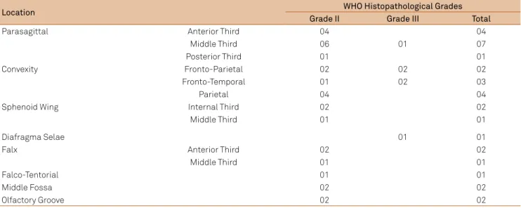
-prised 10% (52 of 522) of patients with intracranial me-ningioma treated in the same period and 75% of the cases were operated on by the senior author (BOC). Diagnosis was performed using CT and/or MR imaging of the head. Tumors were more frequently located in the parasagittal region and on the convexity (Table 1). Tumors were
clas-siied according to the 2007 histopathological WHO cri
-teria1,7, (slides of patients operated in the irst years were
reviewed). Forty-three (82.7%) patients had Grade II me-ningiomas (37 atypical, 3 clear cell and 3 chordoid sub-types) and nine (17.3%) had Grade III (7 anaplastic, 1 pap-illary and 1 rhabdoid) meningiomas. Nine (17.3%) patients had multiple meningiomas (six with two, one with three
and two with ive tumors). One patient with multiple me
-ningiomas had neuroibromatosis type 2. Forty-three
(82.7%) were de novo meningiomas and nine (17.3%) were tumors with malignant progression. Among the later, four patients progressed from grade I to atypical grade II (0.8% of grade I meningiomas) after recurrence (at 39, 45, 84, and
195 months); the earlier, irst biopsies of these 4 patients had focal areas of atypia not suicient to be considered
grade II. Two patients with grade I (0.4% of grade I menin-giomas) and three with atypical grade II tumors progressed (7% of grades II meningiomas) to grade III after recurrence (at 4, 5, 12, and 26 months).
Clinical findings
he most important demographic characteristics of pa
-tients are presented in Table 2 and Figure 1. here was a female
predominance for all patients (1.3:1) and for patients with Grade II (1.5:1), and a slight male predominance among patients with
Grade III tumors (0.8:1). he sex distribution was similar for pa
-tients with Grade II and Grade III tumors (p = 0.8732).
Age ranged from 16 to 89 years old (mean = 54.77 ± 15.91). Patients with Grade II were signiicantly younger than were
patients with Grade III tumors (means: 53.86 vs 75.22 months,
p = 0.0015, Kruskal-Wallis test, with Dunn’s multiple com
-parison test); the same was observed for females (means:
51.96 vs 73.75 years, p = 0.0007, Kruskal-Wallis test, with Dunn´s
multiple comparison test) and for males (means: 47.56 vs 76.40
years, p = 0.0034, Kruskal-Wallis test, with Dunn´s multiple
comparison test). Follow-up ranged from 3 to 257 months
(mean = 66.02 ± 59.96; median = 44.50 months).
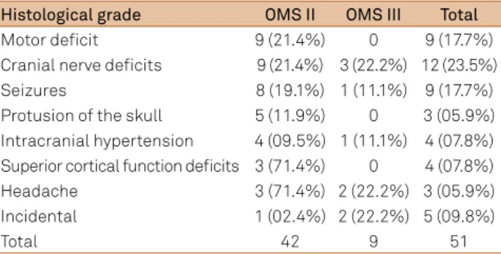
he main clinical signs and symptoms at admission are
summarized in Table 3. he most frequent were motor dei
-cits, cranial nerve palsies and seizures.
Two male patients with grade II (atypical) meningiomas ( falcine and petrous) underwent previous whole brain radio-therapy and chemoradio-therapy (one 27-year-old for treatment of a medulloblastoma 8 years earlier; and another 23-year-old for lymphoid leukemia twice, 14 and 3 years earlier).
Management of the disease
Surgical Treatment:Surgery was performed using micro-surgical techniques, with the aims of the most extensive safe resection possible and avoidance of new or increased
neu-rological deicits. he extent of resection was assessed mac
-roscopically during surgeryand postoperatively using CT or MR imaging 48 hours and 6 months after operation catego-rized as radical removal (no evidence or doubt about residual tumor in the MR image); subtotal (resection >90%) and par-tial (resection was >90%).
Table 1. Location of grades II and III meningiomas in 50 patients operated on.
Location WHO Histopathological Grades
Grade II Grade III Total
Parasagittal Anterior Third 04 04
Middle Third 06 01 07
Posterior Third 01 01
Convexity Fronto-Parietal 02 02 02
Fronto-Temporal 01 02 03
Parietal 04 04
Sphenoid Wing Internal Third 02 02
Middle Third 01 01
Diafragma Selae 01 01
Falx Anterior Third 02 02
Middle Third 01 01
Falco-Tentorial 01 01
Middle Fossa 02 02
Twenty (36.5%) patients (16 with Grade II and 4 with Grade III meningiomas) underwent adjuvant radiotherapy. Nineteen were submitted to external beam fractionated ra-diotherapy and one to conformational rara-diotherapy (doses: 4,500 to 6,000 cGy). Twelve patients were treated after the
irst surgery (all Grade II) and 8 after the irst recurrence.
Functional outcome
Functional outcomes were compared between patients with histopathological Grades II vs III tumors and between
atypical and other subtypes of Grade II tumors. he preopera
-tive, postoperative ( irst 10 days) and the follow-up outcome were analyzed using the Karnofsky Performance Scale (KPS)
for 48 patients. Patients were classiied into one of three
functional status categories: 1) Normal function or minimal
symptoms and ability to work (KPS 80-100), 2) Independent
but not able to work (KPS 70), and 3) Moderate or severe dis
-ability (KPS <70. For patients with tumor recurrence and clin
-ical deterioration, the highest KPS score obtained during the
follow-up evaluations was used. Two patients did not attend
the 6 months follow-up and did not receive a KPS score at
this time.
Survival analysis was performed using Kaplan-Mayer over
-all survival (censoring event: death), and recurrence-free sur-vival (RFS) curves (censoring event: recurrence) and rates in relation to sex, age, histopathological grade, and extent of re-section, use of radiation therapy, and malignant progression.
Statistical analysis
Statistical analyses was performed using the Chi-square
and Fisher’s exact tests to compare proportions, the
Kruskall-Wallis non-parametric test and the analysis of variance (ANOVA) to compare medians, and the log-rank test to com-pare overall and RFS curves using the Graph Pad PRISM (ver-sion 3.0; Graph Pad Software Inc. San Diego, CA). Multivariate analysis for selected clinical variables was performed us-ing Cox-Regression (SPSS-Version 21.0, IBM Corporation, Armonk, NY). An α-error probability not exceeding 5% was
considered signiicant for two-tailed probability tests.
10 8 6 4 2 0 2 4 6 8 10
11-20 21-30 31-40 41-50 51-60 61-70 71-80 81-90
0
9 7 5 3 1 1 3 5 9 9
Figure 1. Age and sex distribution of WHO grades II and III meningiomas based on 50 cases treated at the Hospital das Clínicas, Ribeirão - Preto Medical School - University of São Paulo, Ribeirão Preto, São Paulo, Brazil.
Table 2. Summary of demographic data in 50 patients with WHO grades II and III meningiomas.
Characteristic Grade II
Meningiomas atypical
Grade II Meningiomas other
Grade II Meningiomas total
Grade III
Meningiomas Total
Sex
Female 22 5 27 4 31
Male 15 1 16 5 21
Female/male 1.5:1 5:1 1.7:1 0.8:1 1.5:1
Age (yrs)* 53.95 ± 14.70 53.33 ± 21.32 53.86 ± 15.47 75.22 ± 7.546 54.77 ± 15.91
Age range (yrs) 17-76 25-81 16-81 31-89 16-89
Female 25-64 65-68 25-68 69-81 25-81
Male 16-62 65-65 16-25 67-89 16-89
Follow-up (months)* 66.59 ± 59.85 59.17 ± 41.00 65.23 ± 57.56 78.25 ± 74.52 69.78 ± 74.20 * Values presented as the means ± standard deviation.
Table 3. Initial signs and symptoms in 51 patients with grades II and III meningiomas.
Histological grade OMS II OMS III Total
Motor deicit 9 (21.4%) 0 9 (17.7%)
Cranial nerve deicits 9 (21.4%) 3 (22.2%) 12 (23.5%)
Seizures 8 (19.1%) 1 (11.1%) 9 (17.7%)
Protusion of the skull 5 (11.9%) 0 3 (05.9%) Intracranial hypertension 4 (09.5%) 1 (11.1%) 4 (07.8%) Superior cortical function deicits 3 (71.4%) 0 4 (07.8%)
Headache 3 (71.4%) 2 (22.2%) 3 (05.9%)
Incidental 1 (02.4%) 2 (22.2%) 5 (09.8%)
RESULTS
Summary of surgical treatment
hirty-ive (67.3%, 29 Grade II/6 Grade III) patients un
-derwent radical tumor resection, fourteen (26.9%, 10 Grade II/4 Grade III) had subtotal and three (5.8%, Grade II) had partial resection.
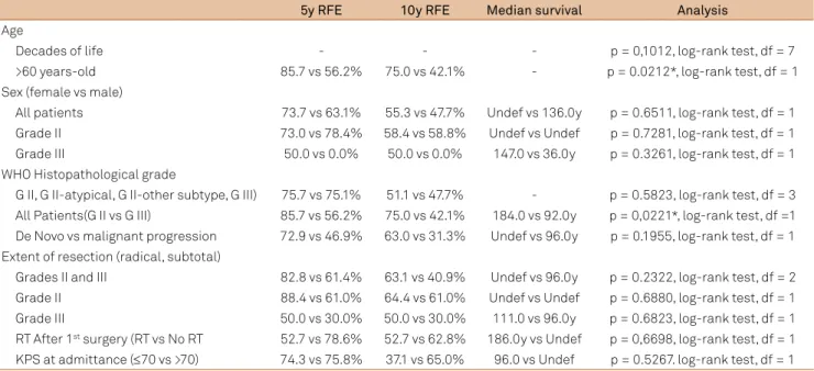
Survival:Tables 4 (univariate analysis) and 5 (multivariate analysis) presents the results of the analysis of factors evaluat-ed for an association with the overall survival of patients with
WHO grades II and III meningiomas. Patients <60-years-old
survived longer than did patients >60-year-old (univariate - Figure 2, and multivariate analysis), and patients with grade II tumors survived longer than did patients with grade III tu-mors (univariate analysis - Figure 3). Histological progres-sion, radiotherapy and recurrence were predictors for longer survival (multivariate analysis).
Recurrence:he results of the analysis of factors that can inluence the RFS of patients with WHO grades II and
III meningiomas are presented in Tables 4 (univariate) and
5 (multivariate). he recurrence rates during follow-up
were 50% (26/52 patients), 46.5% (20/43 patients) and 66.7% (6/9 patients), respectively, in all patients, in patients
with WHO Grade II and with III meningiomas. Among
pa-tients with Grade II tumors, 16 (80%) recurred in the irst
5 years and the other 4 (20%) recurred after 10 years. All six recurrences in patients with Grade III meningiomas
oc-curred in the irst 3 years.
RFS was longer for patients with grade II than with grade III meningiomas (univariate - Figure 4, and multivariate analysis), for patients who underwent total resection than with subtotal resection, for patients with de novo than in patients with malignant transformation (univariate analy-sis - Figures 5 and 6), and for patients >60-years-old (multi-variate analysis).
Mortality and morbidity
One patient (grade II) died in the irst postoperative
month after surgery, due to pulmonary embolism. Two pa-tients (grades II and III) died, respectively, three and two months after surgery due to pulmonary infection and 13 pa-tients died during the follow-up period (operative mortality: 2 [3.9%] patients; surgery related mortality: 3 [5.8%] patients; and overall mortality: 16 [30.8%] patients). During follow-up,
nine deaths occurred due to progression of Grades II ( ive pa
-tients) and III ( four pa-tients) tumors and four deaths were not related to tumor.
Transient (5 [9.6%] patients) or permanent (6 [11.5%] pa-tients) neurological postoperative complications occurred in 11 (21.2%) patients (10 with Grade II and one with Grade III
tumors). he most frequent complication were hemiparesis
in six (11.5%) patients ( four transient [75%] and two [25%]
permanent), and cranial nerve deicits in 3 (5.8%) patients
(II/III/IV/VI in one; V/VII in the second; and V/VI/VII in the
third patient ). Two patients had deep venous thrombosis, one of them developed pulmonary embolism. Another pa-tient had osteomyelitis.
0 50 100 150 200 250
0 25 50 75 100 125
Grade II
Grade III
Time (Months)
Pr
opor
ti
on
of
Su
rv
iv
al
Figure 4. Recurrence-free survival curve for patients with WHO grades II and III. Signiicant difference (p = 0.0030, df = 1, log-rank test ).
0 50 100 150 200 250
0 25 50 75 100 125
Time (Months)
Pr
opor
ti
on
of
Su
rv
iv
al
Grade II Grade III
Figure 3. Survival curves for patients according WHO histological grades. Signiicant difference (p = 0.0034, df = 1, log-rank test).
0 100 200 300
0 25 50 75 100 125
00-60 years 61-90 years
Pr
obab
ilit
y
of
Su
rv
iv
al
Time (Months)
Postoperative functional outcome
Preoperative, immediate postoperative and follow-up
functional disabilities assessed by KPS are presented in
Table 6. he proportions of patients in the functional cat
-egories were similar in all evaluations (p = 0.5107 for all patients, p = 0.4694 for patients with Grade II tumors, and p = 0.9056 for patients with Grade III tumors). Preoperatively
12 (27.9%) patients had KPS scores ≤ 70, and postopera
-tively 14 (32.6%) had similar KPS scores (p = 0.5059). hree
(5.9%) patients (two with Grade II and one with Grade III)
experienced postoperatively deterioration (KPS score ≤70) and one Grade III (1.9%) experienced improvement (KPS
≥80). Assessment at 6 months identiied 5 additional pa
-tients ( four Grade II and one Grade III) with KPS score
≤70. here was no signiicant diferences between the over
-all survival and RFS curves for patients with KPS score ≤70 and with KPS >70 on admission (respectively, p = 0.5107 and p = 0.4719) Tables 4 and 5.
Table 4. Summary of the analysis of survival curves according factors that can inluence the overall survival of patients with WHO grades II and III meningiomas.
5y RFE 10y RFE Median survival Analysis
Age
Decades of life - - - p = 0,1012, log-rank test, df = 7
>60 years-old 85.7 vs 56.2% 75.0 vs 42.1% - p = 0.0212*, log-rank test, df = 1
Sex (female vs male)
All patients 73.7 vs 63.1% 55.3 vs 47.7% Undef vs 136.0y p = 0.6511, log-rank test, df = 1 Grade II 73.0 vs 78.4% 58.4 vs 58.8% Undef vs Undef p = 0.7281, log-rank test, df = 1 Grade III 50.0 vs 0.0% 50.0 vs 0.0% 147.0 vs 36.0y p = 0.3261, log-rank test, df = 1 WHO Histopathological grade
G II, G II-atypical, G II-other subtype, G III) 75.7 vs 75.1% 51.1 vs 47.7% - p = 0.5823, log-rank test, df = 3 All Patients(G II vs G III) 85.7 vs 56.2% 75.0 vs 42.1% 184.0 vs 92.0y p = 0,0221*, log-rank test, df =1 De Novo vs malignant progression 72.9 vs 46.9% 63.0 vs 31.3% Undef vs 96.0y p = 0.1955, log-rank test, df = 1 Extent of resection (radical, subtotal)
Grades II and III 82.8 vs 61.4% 63.1 vs 40.9% Undef vs 96.0y p = 0.2322, log-rank test, df = 2 Grade II 88.4 vs 61.0% 64.4 vs 61.0% Undef vs Undef p = 0.6880, log-rank test, df = 1 Grade III 50.0 vs 30.0% 50.0 vs 30.0% 111.0 vs 96.0y p = 0.6823, log-rank test, df = 1 RT After 1st surgery (RT vs No RT 52.7 vs 78.6% 52.7 vs 62.8% 186.0y vs Undef p = 0,6698, log-rank test, df = 1 KPS at admittance (≤70 vs >70) 74.3 vs 75.8% 37.1 vs 65.0% 96.0 vs Undef p = 0.5267. log-rank test, df = 1 RFE: recurrence-free estimate; Undef: undeined; WHO: World Health Organization; y: year; * signiicant difference.
Table 5. Summary of the analysis of factors that can inluence the overall recurrence-free survival of patients with WHO grades II and III meningiomas.
5y RFE 10yr RFE Median
Survival Analysis
Recurrence/Regrowing rate GII vs G III (46.5% vs 66.7%) - - - p = 0.4654, Fisher’s exact test Age
Decades of life - - - p = 0.7799, log-rank test, df = 7
>60 years-old 45.9 vs 53.0% 25.5 vs 12.9% 48.0 vs 84.0y p = 0.9469, log-rank test, df = 1 WHO Histopathological grade
Grades (II vs III) 55.1% vs 0.0% 32.1 vs 0.0% 78.0 vs 15.0y p < 0.0030*, log-rank test, df = 1 AtypicaI vs other subtypes G II 52.1 vs 10.5% 26.0 vs 32.5% 72.0 vs Undef p < 0.2033, log-rank test, df = 1 De Novo vs Malignant progression 51.1 vs 0.0% 31.8 vs 0.0% 72.0 vs 15.0y p = 0.0010*, log-rank test, df = 1 Sex
All patients, 53.7 vs 32.2% 25.6 vs 32.2% 72.0 vs 45.0% p = 0.7396, log-rank test, df = 1 Patients with grade II 64.3 vs 32.5% 30.6 vs 32.5% 78.0 vs 45.0y p = 0.5612, log-rank test, df = 1 Patients with grade III 0.0 vs 0.0% 0.0 vs 0.0% 15.0 vs 29.0y p = 0.7675, log-rank test, df = 1 Extent of resection (radical, subtotal)
All patients 65.4 vs 33.0% 46.5 vs 0.0% - p = 0.011*9 log-rank test, df = 1
DISCUSSION
Epidemiology
he wide range in the relative frequency of atypical (3.6
and 7.5%) and anaplastic meningiomas (0.4 to 2.8%)1,2,3,5,8,9,10,11,
can be explained by variable pathological criteria for their
classiication. However, the use of the WHO criteria im
-proved comparisons among more recent series. We found 8.2% of patients with grade II and 1.7% with grade III (10% of the patients with meningiomas surgically treated), data similar to the recent series. Benign meningiomas predomi-nates in patients of female sex; in contrast, gender distribu-tion has been reported similar or even with male predomi-nance among patients with Grades II/III2,8,12,13,14,15. Our results
kept this tendency for patients with grade II and III tumor. Atypical meningiomas have been reported to occur after cra-nial irradiation for other tumors or conditions16,17. We
ob-served two patients (3.8%) with radiation-induced grade II (atypical) meningiomas.
Histopathological features
Several histopathological classiications were proposed
for more aggressive meningiomas2,18, however, these classii
-cations are potentially subjective. Perry et al.19,20, elaborated
a simple and reproducible grading scheme for meningiomas
(he Mayo Clinic Scheme), based primarily on histological features in patients who underwent total resection. hey
deined the following criteria for the diagnosis of 1) atypi
-cal meningiomas: (≥4 mitoses/10 HPF, ≥2.5/mm2) or at least
three of the following features: sheeting, macronuclei, small
cell formation, hypercellularity (≥53 nuclei/HPF, ≥118/mm2), and brain invasion; 2) anaplastic meningiomas (≥20 mitotic
igures/10 HPF, ≥12.5 mm2), or focal or difuse loss of menin
-gothelial diferentiation (carcinoma-, sarcoma-, or melano
-ma-like appearance).
he WHO classiications1,7 incorporated the Mayo
Clinic criteria for grading atypical and anaplastic meningi-omas (proliferation index, brain invasion and mitotic activ-ity), and emphasized more strict and objectives histologi-cal criteria. Grade II tumors included atypihistologi-cal tumors with three or more of the following features: increased mitotic
activity (≥4 mitoses/10 HPF), increased cellularity, small
cells with a high nuclear:cytoplasmic ratio, prominent nucleoli, uninterrupted patternless or sheet-like growth, and foci of necrosis. Grade II also includes clear cell and chordoid subtypes. Grade III tumors had obvious malig-nant cytology resembling that of carcinoma, melanoma or high-grade sarcoma or a markedly elevated mitotic index
(≥20 mitoses /10 HPF). Grade III also includes rhabdoid
and papillary subtypes.
0 50 100 150 200 250
0 25 50 75 100 125
De Novo Progression
Time (Months)
Pr
obabi
li
ty
of
Su
rv
iv
al
Figure 6. Recurrence-free survival curves for patients with de novo and for patients with progression meningiomas. Signiicant difference (p = 0.0010, df = 1, log-rank-test.
0 50 100 150 200 250
0 25 50 75 100 125
Total Subtotal
Time (Months)
Probability of Survival
Figure 5. Recurrence-free survival curves for patients according the extent of resection. Signiicant difference (p = 0.0192, df = 2, log-rank-test.
Table 6. Preoperative, postoperative, and follow-up functional status*.
Functional status KPS Score
WHO Histopathological Grade
Preoperative Immediate Postoperative Follow-Up
Grade II Grade III Total Grade II Grade III Total Grade II Grade III Total
No. of patients 43 09 52 43 09 52 39 08 47
Normal/minimal symptoms & working
80-100 31 (72.1%) 06 (66.7%) 37 (71.1%) 29(67.4%) 6 (66.7%) 35 (67.3%) 31 (79.5%) 6 (66.7%) 37 (78.7%) Independent, not working 70 12 (27.9%) 03 (33.3%) 15 (28.9%) 14 (32.6%) 2 (22.2%) 16 (30.8%) 08 (20.5%) 2 (22.2%) 10 (21.3%)
Moderate or severe disability <70 1 (11.1%) 1 (1.9%)
Histopathological progression of meningiomas from Grade I to II and from Grade II to III has been reported in 0.16 to 2% of cases8,13,15,18,19,21. We found 9 patients (1.7% of all
meningiomas and 17.3% of Grades II and III meningiomas) that had tumor progression at time of their recurrence. All patients progressing from Grade I to II had focal atypia in
the irst biopsy slides not considered suicient to classify
them initially as atypical. his observation should be con
-sidered at least as an alert: patients with Grade I meningio-mas with focal atypia deserve close follow-up imaging look-ing for recurrence.
Some authors postulated that genetic changes in menin-giomas progress stepwise, from grade I to III1,22,23. his is sup
-ported by histopathological progression of benign meningio-ma at recurrence11. However, other authors15 genetic changes
in tumor cells already present in the biopsies of benign
re-current progressing tumors, a inding inconsistent with the
stepwise progression.
Prognostic factors
In addition to the histopathological features, several other
factors were reported to inluence the prognosis of the patients
with atypical and anaplastic meningiomas. Age at diagnosis is a recognized prognostic factor for patients with intracranial tu-mors, including grades II and III meningiomas8,10,12,14,19. Among our patients, age <60-years-old was also a prognostic factor for
overall survival, but not for RFS. Although some authors report that female sex is an unfavorable prognostic factor2, more evi-dence indicates that gender does not inluence prognosis of
patients with grades II and III meningiomas10,12,13,14, including
our results. Some authors21 found that patients with Grade II
tumors progressing to Grade III have shorter overall survival than patients with “de novo” grade II meningioma and our
re-sults support these indings.
Extent of resection is considered a strong prognostic fac-tor in several studies. Recurrence rate is lower for patients submitted to total (17%) compared with subtotal (87%) re-section3. Survival is longer in patients that had Simpson 1
re-section13, and RFS is longer in patients with total resection3. Total or subtotal resections did not inluence the survival of
our patients and the RFS was longer for patients with total resection only on univariate analyis.
Molecular markers were also reported to be related to prognosis. Poor prognosis may be associated with a high MIB-1 labeling index. However, this index ranges overlap considerably for benign, atypical, and anaplastic meningio-mas11,19,20, and it is only valuable to evaluate tumors with
bor-derline atypia. In such a case, an index of ≥ 4.2% would clas
-sify the tumor as atypical11,19,20.
Surgical treatment
Surgery is the primary treatment for atypical and
anaplas-tic meningiomas aiming a deinitive diagnosis, reducing any mass efect and alleviating symptoms. Complete resection is
the goal and the involved dura mater and bone should also be resected to prevent recurrence. Repeated surgery should be considered in cases where subtotal resection is required to
avoid neurological deicits, such as for tumors of the cranial
base and tumors with dense cortical adherence11.
Radiation therapy
Conventional fractionated radiation therapy
Results of fractionated radiation therapy for atypical and
anaplastic meningiomas are diicult to evaluate because of
the variation of histopathology, the small number of cases and other variables, such as extent of resection and brain in-vasion11. he 5-year survival rate for patients with atypical
and anaplastic meningiomas was found to be 58% and the 5-year RFS rate was 48%24, the 2-year RFS rate was 89% for
irradiated patients compared to 50% for patient not
irradiat-ed. he overall survival rate was better for patients submitted
immediately to radiation therapy3. herefore, the consensus
favors fractionated radiation therapy for patients submitted to total or subtotal resection of atypical and anaplastic me-ningiomas11, even though this recommendation is not sup-ported by prospective controlled trials. We ind better overall
survival favoring patients submitted to fractionated
radia-tion therapy on multivariate analysis but no diference was
found between the RFS curves.
Stereotatic radiosurgery
Some authors have reported good results using stereo-tactic radiosurgery to treat benign, atypical and anaplastic tumors, 10-year survival rates of 59% for atypical and of 59 and 0% for anaplastic meningiomas, and 5-year RFS rates of 83 and 72% for atypical and anaplastic meningiomas, respectively25. However, another report did not support
these favorable results for stereotatic radiosurgery show-ing a very poor result for anaplastic lesions26. Tumor size
is a limiting factor for stereotactic radiosurgery25,27; lesions
>3 cm in size do not respond to this treatment25 and
radia-tion necrosis occurs in 23% of the cases, with a few cases requiring surgery27.
Based on the fairly good results reported in the literature and because complications are considered uncommon11,
ex-cept when radiosurgery is administered after conformal ra-diation therapy26, stereotactic radiosurgery probably could
be administered to patients with nodular residual atypical or anaplastic meningiomas along with fractionated radiation therapy to the tumor bed, even though no prospective con-trolled studies support this approach11.
CHEMOTHERAPY
vincristine, were used to treat unresectable benign, atypical and anaplastic meningiomas and produced tumor shrinking
or stabilization in most cases in preliminary studies. he ini
-tial results were not reproduced and none of these agents has shown convincing results in the treatment of atypical and an-aplastic meningiomas11.
Molecular biology suggested new options for the man-agement of atypical and anaplastic meningiomas. Vascular Endothelium Growth Factor (VEGF) and Platelet-Derived Growth Factor (PGDF)-A and B and PDGF-β-receptor cause increased cell division and tumor proliferation28, and their
ex-pression are increased in atypical and in anaplastic meningi-omas29. Based on this knowledge, inhibitors of VEGF or VEGF
receptors (vandetanib, vatalanib, bevacizumab, AEE788, and IMC-1C11), and VEGF receptor antagonists (bevacizumab and erlotinib), are available and some of them are being test-ed in phase II trials11.
Atypical and anaplastic meningiomas are distinct enti-ties with poorer prognosis than benign meningiomas when treated with the current therapeutic options. Objective
clas-siication systems to grade meningiomas, which were im
-proved by the Mayo Clinic scheme followed by the WHO grading system, which was based on the former, allow better
comparison between diferent series. he algorithm for treat
-ment of atypical and anaplastic meningiomas proposed by
Perry et al.20, and modiied by Modha & Gutin11 is a
reason-able option for treating these patients (Figure 7).
In conclusion, in this study we found that age under sixty-years old, grade II tumors, de novo meningiomas and total resection were main predictors for better prognosis fac-tors among patients with atypical and anaplastic
meningio-mas. he treatment of atypical and anaplastic meningiomas
remains a challenge for neurosurgeons, and new strategies should be developed to reduce recurrence and improve the prognosis of patients harboring these tumors.
References
1. Louis DN, Scheithauer BW, Budka H, von Deimling A, Kepes JJ. Meningiomas. In: Kleihues P, Cavenee WK, eds. World Health Organization Classiication of tumours Pathology and genetics of tumours of the nervous system. Lyon: IARC; 2000. p. 175-84. 2. Mahmood A, Caccamo DV, Tomecek FJ, Malik GM. Atypical and
malignant meningiomas: a clinicopathological review. Neurosurgery. 1993;33(6):955-63. doi:10.1227/00006123-199312000-00001
3. Dziuk TW, Woo S, Butler EB, Thornby J, Grossman R, Dennis WS, et al. Malignant meningioma: an indication for initial aggressive surgery and adjuvant radiotherapy. J Neurooncol. 1998;37(2):177-88. doi:10.1023/A:1005853720926
4. Cushing H, Eisenhardt L. Meningiomas: their classiication, regional behaviour, life history, and surgical results. Springield: Charles C. Thomas; 1938.
5. Pourel N, Auque J, Bracard S, Hoffstetter S, Luporsi E, Vignaud JM et al. Eficacy of external fractionated radiation therapy in the treatment of meningiomas: a 20-year experience. Radiother Oncol. 2001;61(1):65-70. doi:10.1016/S0167-8140(01)00391-7
6. D’Arrigo B, Morello G, De Divitiis E, Cucciniello B. [Atypical meningiomas. Histopathological considerations on 13 personal cases]. Rass Neuropsichiatr. 1966;19(3):577-90. Italian. 7. Louis DN, Ohgaki H, Wiestler OD, Cavenee WK, Burger PC,
Jouvet A et al. The 2007 WHO classiication of tumours of the central nervous system. Acta Neuropathol. 2007;114(2):97-109. doi:10.1007/s00401-007-0243-4
8. Duntze J, Metellus P, Litre CF, Eap C, Theret E, Colin P. [Management of WHO grade II and II meningiomas: retrospective study of
surgical series of 36 cases at a single institution]. Neurochirurgie. 2012;58(5):275-81. French. doi:10.1016/j.neuchi.2012.01.006 9. Kane AJ, Sughrue ME, Rutkowski MJ, Shangari G, Fang S,
McDermott MW et al. Anatomic location is a risk factor for atypical and malignant meningiomas. Cancer. 2011;117(6):1272-8. doi:10.1002/cncr.25591
10. Cornelius JF, Slotty PJ, Steiger HJ, Hänggi D, Polivka M, George B. Malignant potential of skull base versus non-skull base meningiomas: clinical series of 1,663 cases. Acta Neurochir (Wien). 2013;155(3):407-13. doi:10.1007/s00701-012-1611-y
11. Modha A, Gutin PH. Diagnosis and treatment of atypical and anaplastic meningiomas: a review. Neurosurgery. 2005;57(3):538-50. doi:10.1227/01.NEU.0000170980.47582.A5
12. Rosenberg LA, Prayson RA, Lee J, Reddy C, Chao ST, Barnett GH et al. Long-term experience with World Health Organization grade III (malignant) meningiomas at a single institution. Int J Radiat Oncol Biol Phys. 2009;74(2):427-32. doi:10.1016/j.ijrobp.2008.08.018
13. Palma L, Celli P, Franco C, Cervoni L, Cantore G. Long-term prognosis for atypical and malignant meningiomas: a study of 71 surgical cases. J Neurosurg. 1997;86(5):793-800. doi:10.3171/jns.1997.86.5.0793
14. Pasquier D, Bijmolt S, Veninga T, Rezvoy N, Villa S, Krengli M et al. Atypical and malignant meningioma: outcome and prognostic factors in 119 irradiated patients. A multicenter, retrospective study of the Rare Cancer Network. Int J Radiat Oncol Biol Phys. 2008;71(5):1388-93. doi:10.1016/j.ijrobp.2007.12.020
Consider RT (SRS/FRT) MIB-1 Index
Absent
Brain Invasion Present Post-operative SRS/FRT Total Subtotal
-Atypical Meningiomas -Anaplastic Meningiomas Extent of Resection
<4.2%
Watch
15. Al-Mefty O, Kadri PA, Pravdenkova S, Sawyer JR, Stangeby C, Hussain M. Malignant progression in meningioma: documentation of a series and analysis of cytogenetic indings. J Neurosurg. 2004;101(2):210-8. doi:10.3171/jns.2004.101.2.0210
16. Hug EB, Devries A, Thornton AF, Munzenride JE, Pardo FS, Hedley-Whyte ET, et al. Management of atypical and malignant meningiomas: role of high-dose, 3D-conformal radiation therapy. J Neurooncol. 2000;48(2):151-60. doi:10.1023/A:1006434124794
17. Al-Mefty O, Topsakal C, Pravdenkova S, Sawyer JR, Harrison MJ. Radiation-induced meningiomas: clinical, pathological, cytokinetic, and cytogenetic characteristics. J Neurosurg. 2004;100(6):1002-13. doi:10.3171/jns.2004.100.6.1002
18. Jääskelainen J, Haltia M, Servo A. Atypical and anaplastic meningiomas: radiology, surgery, radiotherapy, and outcome. Surg Neurol. 1986;25(3):233-42. doi:10.1016/0090-3019(86)90233-8 19. Perry A, Stafford SL, Scheithauer BW, Suman VJ, Lohse
CM. Meningioma grading: an analysis of histologic parameters. Am J Surg Pathol. 1997;21(12):1455-65. doi:10.1097/00000478-199712000-00008
20. Perry A, Scheithauer BW, Stafford SL, Lohse CM. “Malignancy” in meningiomas: a clinicopathologic study of 116 patients with grading implications. Cancer. 1999;85(9):2046-56. doi:10.1002/(SICI)1097-0142(19990501)85:9<2046::AID-CNCR23>3.0.CO;2-M
21. Krayenbühl N, Pravdenkova S, Al-Mefty O. De novo versus transformed atypical and anaplastic meningiomas: comparisons of clinical course, cytogenetics, cytokinetics, and outcome.. Neurosurgery. 2007;61(3):495-503. doi:10.1227/01.NEU.0000290895.92695.22
22. Boström J, Meyer-Puttlitz B, Wolter M, Blaschke B, Weber RG, Lichter P et al. Alterations of the tumor suppressor genes CDKN2A (p16(INK4a), p14(ARF)), CDKN2B (p15(INK4b)), and CDKN2C
(p18(INK4c)) in atypical and anaplastic meningiomas. Am J Pathol. 2001;159(2):661-9. doi:10.1016/S0002-9440(10)61737-3 23. Perry A, Banerjee R, Lohse CM, Kleinschmidt-DeMasters BK,
Scheithauer BW. A role for chromosome 9p21 deletions in the malignant progression of meningiomas and the prognosis of anaplastic meningiomas. Brain Pathol. 2002;12(2):183-90. doi:10.1111/j.1750-3639.2002.tb00433.x
24. Goldsmith BJ, Wara WM, Wilson CB, Larson DA. Postoperative irradiation for subtotally resected meningiomas. A retrospective analysis of 140 patients treated from 1967 to 1990. J Neurosurg. 1994;80(2):195-201. doi:10.3171/jns.1994.80.2.0195
25. Kondziolka D, Levy EI, Niranjan A, Flickinger JC, Lunsford LD. Long-term outcomes after meningioma radiosurgery: physician and patient perspectives. J Neurosurg. 1999;91(1):44-50. doi:10.3171/jns.1999.91.1.0044
26. Stafford SL, Pollock BE, Foote RL, Link MJ, Gorman DA, Schomberg PJ et al. Meningioma radiosurgery: tumor control, outcomes, and complications among 190 consecutive patients. Neurosurgery. 2001;49(5):1029-37.
27. Ojemann, SG, Sneed PK, Lardon DA, Gutin PH, Berger MS, Verhey L et al. Radiosurgery for malignant meningioma: results in 22 patients. J Neurosurg. 2000;93 Suppl 3:62-7.
28. Johnson MD, Woodard A, Kim P, Frexes-Steed M. Evidence for mitogen-associated protein kinase activation and transduction of mitogenic signals by platelet-derived growth factor in human meningioma cells. J Neurosurg. 2001;94(2):293-300. doi:10.3171/jns.2001.94.2.0293