ARTICLE
Comparison of functional electrical
stimulation associated with kinesiotherapy
and kinesiotherapy alone in patients with
hemiparesis during the subacute phase of
ischemic cerebrovascular accident
Comparação da estimulação elétrica funcional associada à cinesioterapia
com a cinesioterapia isolada em pacientes com hemiparesia na fase
subaguda por acidente vascular cerebral isquêmico
Paulo Cesar Modesto1, Fernando Campos Gomes Pinto2
he cerebral vascular accidents (CVA) are divided, according to their anatomical and pathological features, into subarachnoid and intraparenchymatous hemorrhages; and as ischemic (ICVA), caused by primary vascular disease, thrombosis or embolism
and hemorrhagic (HCVA); divided, respectively1,2. Approximately
75% of CVA are ischemic and 25% are hemorrhagic2.
he acute stage of the disease is characterized by a state of laccidity, arelexia, hemiplegia, hemi-anesthesia and
1Physical therapist, Master’s Degree Student at Hospital do Servidor Público Estadual (IAMSPE-SP), São Paulo SP, Brazil;
2Physician, Neurosurgeon, Doctor of Neurosurgery at Universidade de São Paulo (USP); Post-graduate Advisor of IAMSPE-SP, São Paulo SP, Brazil.
Correspondence: Paulo Cesar Modesto; Rua Corcovado 134 / bloco 54 / apto 28; 05038-040 São Paulo SP - Brasil; E-mail: [email protected]
Conlict of interest: There is no conlict of interest to declare.
Received 22 August 2012; Received in inal form 11 October 2012; Accepted 18 October 2012.
ABSTRACT
Objective: To compare the functional electrical stimulation associated with functional kinesiotherapy alone in patients after ischemic cere-brovascular accident. Methods: The study included 20 patients who were divided into two groups: Group I (GI): functional electrical stimula-tion plus funcstimula-tional kinesiotherapy and Group II (GII): funcstimula-tional kinesiotherapy. We evaluated active and passive range of mostimula-tion, in knee lexion and extension muscle strength, activities of daily living and quality of life. The evaluations were conducted in the pretreatment period, after 10 sessions and after 20 physical therapy sessions. Results: There was a signiicant improvement in all variables studied for both groups. However, signiicant improvements for the sub-items functional capacity and social aspects were seen only in the patients treated with associated functional electrical stimulation and kinesiotherapy. Conclusion: Although both groups of patients improved with the treat-ment, the association of functional electrical stimulation and kinesiotherapy showed superiority in two quality of life items, in the sub-items functional capacity and social aspects.
Key words: electric stimulation, stroke, paresis, physiotherapy.
RESUMO
Objetivo: Comparar a estimulação elétrica funcional associada à cinesioterapia com a cinesioterapia funcional isolada no membro inferior de pacientes em fase subaguda após acidente vascular cerebral isquêmico. Método: Participaram do estudo 20 pacientes divididos em 2 grupos: Grupo I (GI): eletroestimulação funcional mais cinesioterapia funcional e Grupo II (GII): cinesioterapia funcional. Foram avaliadas as amplitudes de movimento ativo e de movimento passivo em lexão e extensão do joelho, a força muscular, as atividades da vida diária e a qualidade de vida. As avaliações foram realizadas nos períodos pré-tratamento, após 10 e após 20 sessões de isioterapia. Resultados: Houve melhora signiicativa em todas as variáveis estudadas para ambos os grupos. Contudo, melhorias signiicativas para os subitens capacidade funcional e aspectos sociais foram vistos apenas nos pacientes tratados com a estimulação elétrica funcional associada à cinesioterapia. Conclusão: Os dois grupos de pacientes melhoraram com o tratamento, mas a associação da estimulação elétrica funcional à cinesioterapia mostrou superioridade nos subitens capacidade funcional e aspectos sociais da qualidade de vida.
Palavras-Chave: estimulação elétrica, acidente vascular cerebral, paresia, isioterapia.
cognitive alterations, which are the result of cerebral hypoxia and in accordance with the location of the lesion. he dura-tion of this stage is usually brief and may inish in approx-imately 15 to 30 days, when the clinical course is modiied and the individual then goes to a subacute stage, which last 1–3 months and can reach up to 6 months3-6. In most cases,
the return of function occurs spontaneously in one to three months after the CVA, reaching a plateau from six months to one year after the injury, although some patients exhibit sub-stantial recovery at the later stages7-9.
Functional electrical stimulation (FES) is a resource used by physical therapy that enables the transmission of electrical signals to the muscles, facilitating movement10,11. It consists of functional
electrical stimulation of a muscle deprived of normal control to produce a functionally useful contraction. his stimulation de-polarizes the motor nerve, producing a synchronous response in all motor units of the stimulated muscle, improving throphism11.
Its mechanism of action is closely linked to the facilitation of phys-iological mechanisms of striated muscle (muscle contraction), al-lowing selective and repetitive aferent input to the CNS, activat-ing not only the local muscles, but also relex mechanisms that are necessary for reorganization of the motor activity10,11.
he present study aimed to compare the FES associated with functional kinesiotherapy (FK) and functional kinesio-therapy alone in patients with sequelae of ICVA with hemi-paresis in the subacute phase.
METHODS
he study included 20 patients of both genders with hemipa-resis in the subacute phase after an ICVA. he sample consisted of 11 female and 9 male patients, with a mean age in Group I (GI) of 66.70 and 66.90 years in Group II (GII). he project was ap-proved by the Committee of Ethics in Research of the Hospital do
Servidor Público Estadual, São Paulo SP, Brazil, and the free and
informed consent form was signed by all patients.
Inclusion criteria
Adult literate patients of both genders, aged 18 years and older, who were treated between July 2011 and January 2012 with ischemic CVA in the middle cerebral artery terri-tory, with clinical and imaging diagnosis and within 90 days since the onset of the condition, with no structural deformi-ties were included in the study.
Exclusion criteria
Patients with pacemakers, vascular alterations and sensi-tivity impairment in hemiparetic lower limb were excluded.
Matching
Study patients, whose score was lower than 85 in the ini-tial Barthel Index (pretreatment assessment) were selected for the comparative analysis between the groups (study and
control) to allow comparison of health-related quality of life (QOL) results from the patient’s perspective and the perfor-mance of activities of daily life assessed by physical therapists in patients treated with FES+FK (GI) and patients treated with FK alone (GII).
Evaluations
Muscle strength (MS) assessed by Daniels numerical grading system12.
Barthel index (BI) is a scale that assesses functional ca-pacity in activities of daily living and the degree of depen-dence from the perspective of the examiner.
Quality of Life Questionnaire-SF-36.
Joint range of motion (ROM): joint range of motion (ROM) assessment was performed using a goniometer for the assessment of posterior and anterior ROM of the hemi-paretic lower limb in knee extension and lexion. he patient remained supine and the lower limp (LL) was unclothed.
Intervention
he physical therapy intervention program for the groups (GI and GII) consisted of 20 physical therapy sessions twice a week in consecutive weeks, with 60-minute sessions for groups GI and GII, administered by the irst author only.
First phase
Use of FES current; the electrodes were ixed on the thigh of the hemiparetic leg in the quadriceps muscle group (con-sisting of the rectus femoris, vastus lateralis/vastus media-lis/vastus intermedius, knee joint), which promotes contrac-tions in muscles deprived of nervous control.
We used the following stimulation parameters: frequen-cy (F) of 30 to 100 Hz13,14; pulse width (T)=250
microsec-onds13-15, cycle ON/OFF=1/2 (7 seconds of contraction/14
seconds of relaxation); the intensity was the minimum possi-ble to produce an efective and uniform muscle contraction, respecting the voluntary thresholds16-19, as high intensities
cause muscle fatigue18. Each FES session lasted 30 minutes.
Second phase
FK (therapeutic exercises directed at activities of daily liv-ing – ADLs), therapeutic exercises with entertainliv-ing features that encourage interpersonal relationship.
he FK that was administered is based on the guidelines of the American Physical herapy Association. he summary of the exercises and their therapeutic goals are showed in Table 120.
Statistical analysis
displayed as igures. hese were distributed in order to demonstrate the evolution of patients from GI and GII, in the pretreatment period, after 10 sessions and at the end of 20 physical therapy sessions, verifying whether there was a diference between the evaluation periods, between the groups and for the interaction.
RESULTS
General distribution of patients in Groups I and II accord-ing to age and time since injury, are shown in Table 2.
here were no signiicant diferences between groups re-garding age and time since injury.
Table 3 shows gender, afected hemisphere and comor-bidities. here were no signiicant diferences between the groups regarding the parameters of the table.
Quality of life: SF-36
he SF-36 scale is subdivided into eight domains, namely: functional capacity, physical aspect limitation, pain, general health status, vitality, social aspects, emotional aspect limita-tion, and mental health.
Functional capacity
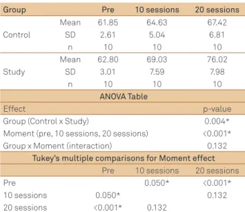
he mean score in the study group ranged from 62.80 (pre) to 69.03 (10 sessions) and 76.02 points (20 sessions) and in the control group, it ranged from 61.85 (pre) to 64.63 points (10 ses-sions) and 67.42 (20 sesses-sions). he GI group showed signiicant improvement after the sessions, compared to GII (p=0.004) and at the time efect (p<0.001) (Fig 1). We have the following hierar-chy as a result: (Pre)<(10 sessions)=(20 sessions) for both groups. Table 4 shows the means and standard deviations of functional capacity.
Fig 1 shows the conidence intervals for each combina-tion of group and time of assessment of the funccombina-tional capac-ity sub-item for both groups.
Social aspects
he mean in the study group ranged from 66.20 to 66.30 (10 sessions) and remained at 66.30 (20 sessions), and in the
control group, it ranged from 63.90 points (pre) to 64.60 (10 ses-sions) and remained at 64.60 (20 ses(10 ses-sions). We observed a sig-niicant diference for both groups (p=0.034) and at the time
Table 1. Functional kinesiotherapy. Physical exercises for lower limbs with therapeutic objective for orthostatic position.
Physical exercises Therapeutic objectives
Passive mobilization, active assisted or active free exercises, of small and large joints of the axial and appendicular skeleton
Maintain or restore normal joint lexibility and prevent clinical complications arising from motor skill reduction
Passive, active assisted, free, and resisted exercises of excursion, stretching and muscle relaxation
Maintain or restore the viscoelastic properties of muscle ibers, preventing muscle shortening, as well as stimulating ideal tissue conditions for best neuromotor performance
Exercises that stimulate postural control, balance reactions and reactions of protection on stable and unstable surfaces in different postures (lying, sitting, on all fours, kneeling, half-kneeling, standing, one-leg support)
Stimulate the development of strategies to maintain balance and body protection, adjusting the posture
Exercises for aerobic capacity conditioning using functional activities (rolling, sitting, standing and walking), monitor blood pressure and heart rate
Improve cardiorespiratory itness, preventing cardiovascular diseases and collaborating in the treatment of hypertension
Table 2. Age and time of lesion. General distribution of
patients in Groups I and II, according to age and time of lesion.
Study group G1 Mean (SD)
Control group G2
Mean (SD) t-test (p-value) Age (years) 66.70 (8.54) 66.90 (12.32) 0.967 Time of
lesion (days) 32.70 (15.69) 38.80 (13.69) 0.367
SD: standard deviation.
Table 3. Gender, affected hemisphere and comorbidities.
General distribution of patients in Groups I and II according to gender, affected hemisphere, hypertensives, diabetics, smokers and alcohol consumers demonstrated in %.
Study group G1
Control group G2
Fisher’s test (p-value)
Female (5) 50% (6) 60%
1.000
Male (5) 50% (4) 40%
Right hemiparesis (0) 0% (4) 40%
0.087 Left hemiparesis (10) 100% (6) 60%
Systemic arterial hypertension–Systemic Arterial Hypertension
(5) 50% (8) 80% 0.350
Diabetes mellitus (1) 10% (5) 50% 0.070
Smoking (4) 40% (3) 30% 1.000
Alcohol consumption (3) 30% (2) 20% 1.000
Fig 1. Conidence intervals for each combination of group and
moment of assessment of the functional capacity sub-item for both groups.
85
80
75
70
65
60
55
50
Pre 10 sessions 20 sessions
Control
Research
F
unc
efect (p=0.019) (Fig 2). We have the following hierarchy as re-sult: (Pre) <(10 sessions)=(20 sessions) for both groups, but the study group had higher responses at the three moments.
Table 5 shows the means and standard deviations of so-cial aspects.
Fig 2 shows the conidence intervals for each combina-tion of group and time of assessment of the sub-item social aspects for both groups.
Regarding other domains of the SF-36, and other items assessed, there was no signiicant diference from a statistical viewpoint.
DISCUSSION
he functional electrical stimulation was well tolerated by all study patients, respecting their tolerance threshold.
Although some hemiparetic patients with ischemic CVA may show spontaneous improvement, best results occur when patients are treated with physical therapy21,22.
Table 4. Means and standard deviations: functional capacity.
Group Pre 10 sessions 20 sessions
Mean 61.85 64.63 67.42
Control SD 2.61 5.04 6.81
n 10 10 10
Mean 62.80 69.03 76.02
Study SD 3.01 7.59 7.98
n 10 10 10
ANOVA Table
Effect p-value
Group (Control x Study) 0.004*
Moment (pre, 10 sessions, 20 sessions) <0.001* Group x Moment (interaction) 0.132
Tukey’s multiple comparisons for Moment effect Pre 10 sessions 20 sessions
Pre 0.050* <0.001*
10 sessions 0.050* 0.132
20 sessions <0.001* 0.132
SD: standard deviation; *statistically signiicant.
Table 5. Means and standard deviations: social aspects.
Group Pre 10 sessions 20 sessions
Mean 63.90 64.60 64.60
Control SD 2.96 2.12 2.12
n 10 10 10
Mean 66.20 66.30 66.30
Study SD 1.03 1.16 1.16
n 10 10 10
ANOVA Table
Effect p-value
Group (Control x Study) 0.034*
Moment (pre. 10 sessions. 20 sessions) 0.019* Group x Moment (interaction) 0.097
SD: standard deviation; *statistically signiicant.
Fig 2. Conidence intervals for each combination of group and
moment of assessment of the social aspects sub-item for both groups.
66
65
64
63
62
61
60
59
Pre
Control
10 sessions 20 sessions
Research 67
68
Social aspec
ts
he element that made up the study intervention and pro-moted satisfactory results was the fact that the beginning of the program did not exceed 40 days since the CVA onset. It has been suggested that treatment started as soon as possible is a factor to achieve more satisfactory results22. Some authors have
in-ferred that the early start of therapy seems to be more important for patient evolution than the duration of the program23.
Regarding muscle strength, the study showed a signii-cant increase in strength after the treatment period, in lex-ion and knee extenslex-ion for both groups, with no diference between them. he improved strength in GI (FES+FK) is con-sistent with the studies of Kesar et al.24, which determined
that the FES facilitates recovery of muscle strength, increas-ing isometric strength of knee extensors and lexors in indi-viduals with hemiparesis due to CVA.
Traditionally, epidemiological studies with a population of CVA patients are focused on mortality and its occurrence, but not on quality of life of these patients25, considering that
the dysfunctions and disadvantages in this regard are signii-cant for CVA patients26.
Although the quality of life for CVA patients is clearly in-luenced by multiple factors, functional independence is one of the most important factors27.
In this study, regarding quality of life, the results demonstrated significant improvement in the group that underwent treatment with FES+kinesiotherapy, in the functional capacity and social aspects domains, dem-onstrating that the combination of the two techniques was more beneficial than kinesiotherapy alone. For the pain, vitality and mental health domains, there was sig-nificant improvement for both groups at the moment ef-fect (pre, 10 and 20 sessions) (p<0.001) and in the mental health item, there was also a significant difference in the interaction effect (p=0.012).
his study was important, as in addition to showing the efects of FES, functional kinesiotherapy and associated treatment, it also evaluated the quality of life and activities of daily living of the patient. hese variables are signiicantly impaired in many aspects, especially when the patient is af-fected by an ischemic CVA in the acute and subacute phases.
In conclusion, functional electrical stimulation associated with functional kinesiotherapy was more efective than func-tional kinesiotherapy alone, regarding quality of life in the do-mains ( functional capacity and social aspects). Both muscle strength and passive and active range of motion as well as ac-tivities of daily living showed no diference between the groups.
1. Sociedade Brasileira de Doenças Cardiovasculares. Primeiro
consenso brasileiro do tratamento da fase aguda do acidente vascular cerebral. Arq Neuropsiquiatr 2001;59:972-980.
2. Gagliardi RJ. AVC – Acidente vascular cerebral: 50 FAQ “Frequently asked question”. São Paulo: Editora de Publicações Médicas, 2006.
3. O’Sullivan SB, Schmitz TJ. Fisioterapia: avaliação e tratamento. 4a ed. São Paulo: Manole, 2004.
4. Daffertshofer M, Mielke O, Pullwitt A, Felsenstein M, Hennerici M. Transient ischemic attacks are more than “ministrokes”. Stroke 2004;35:2453-2458.
5. Johnston SC, Gress DR, Browner WS, Sidney S. Short-term prognosis after emergency department diagnosis of TIA. JAMA 2000;284:2901-2906.
6. Meschia JF. Subtyping in ischemic stroke genetic research. J Stroke Cerebrovasc Dis 2002;11:208-219.
7. Hendricks HT, Van Limbeek J, Geurts AC, Zwarts MJ. Motor recovery after stroke: a systematic review of the literature. Arch Phys Med Rehabil 2002;83:1629-1637.
8. Formisano R, Pantano P, Buzzi MG, et al. Late motor recovery is inluenced by muscle tone changes after stroke. Arch Phys Med Rehabil 2005;86:308-311.
9. Schaechter JD. Motor rehabilitation and brain plasticity after hemiparetic stroke. Prog Neurobiol 2004;73:61-72.
10. Martins FLM, Guimarães LHCT, Vitorino DFM, Souza LCF. Eicácia da eletroestimulação funcional na amplitude de movimento de dorsilexão de hemiparéticos. Rev Neurociênc 2004;12:103-109.
11. Soetanto D, Kuo C, Babic D. Stabilization of human standing posture using functional neuromuscular stimulation. J Biomech 2001;34:589-597.
12. Kendall FP, Mccreary EK, Provance PG. Músculos: provas e funções. 4a ed. São Paulo: Manole, 1995:46.
13. Wrigth PA, Granat MH. Therapeutic effects of functional electrical stimulation on the upper limb of eight children with cerebral palsy. Dev Med Child Neurol 2000;42:724-727.
14. Chae J, Yu DA. A critical review of neuromuscular electrical stimulation for treatment of motor dysfunction in hemiplegia. Assist Technol 2000;12:33-49.
15. Powell J, Pandyan AD, Granat M, Cameron M, Stott DJ. Electrical stimulation of wrist extensors in poststroke hemiplegia. Stroke 1999;30:1384-1389.
16. Corrêa JCF, Negrão Filho R, Dõcusse Filho AJ, Quialheiro JJA. Tratamento da instabilidade fêmuro-patelar por meio da estimulação elétrica neuromuscular associada à cinesioterapia. Rev Bras Fisioter 1996;1:37-43.
17. Noronha MA, Camargo LC, Minamoto VB, Castro CES, Salvini TF. O efeito da estimulação elétrica neuromuscular (NMES) no músculo tibial anterior do rato. Rev Bras Fisioter 1997;2:71-76.
18. Robinson AJ, Snyder-Macler. Eletroisiologia clínica: eletroterapia e teste eletroisiológico. 2a ed. Porto Alegre: Artmed, 2001:109.
19. Brasileiro JS, Villar AFS. Comparação dos torques gerados por estimulação elétrica e contração muscular voluntária no músculo quadríceps femoral. Rev Bras Fisioter 2000;2:75-81.
20. American Physical Therapy Association. Guide to physical therapist practice. Part two: preferred practice patterns neuromuscular. 2nd ed. Phys Ther 2001;81(Suppl 1):S305–S537.
21. Duncan P, Studenski S, Richards L, et al. Randomized clinical trial of therapeutic exercise in subacute stroke. Stroke 2003;34:2173-2180.
22. Dombovy ML, Snadok BA, Basford JR. Rehabilitation for stroke: a review. Stroke 1986;17:363-369.
23. Smith ME, Garraway WM, Smith DL, Akhtar AJ. Therapy impact in functional outcome in a controlled trial of stroke rehabilitation. Arch Phys Med Rehabil 1982;63:21-24.
24. Kesar M, Ding J, Wexler A, Perumal R, Maladen R, Binder-Macleod SA. Predicting muscle forces of individuals with hemiparesis following stroke. J Neuroeng Rehabil 2008;5:7.
25. Carod-Artal J, Egido JA, González JL, Varela de Seijas E. Quality of life among stroke survivors evaluated 1 year after stroke: experience of a stroke unit. Stroke 2000;31:2995-3000.
26. van Straten A, de Haan RJ, Limberg M, van den Bos GA. Clinical meaning of the stroke-adapted sickness impact proile-30 and the sickness impact proile-136. Stroke 2000;31: 2610-2615.
27. Kimberley TJ, Carey JR. Neuromuscular electrical stimulation in stroke rehabilitation. Minn Med 2002;85:34-37.