UNIVERSIDADE FEDERAL DO CEARÁ
FACULDADE DE FARMÁCIA, ODONTOLOGIA E ENFERMAGEM PROGRAMA DE PÓS-GRADUAÇÃO EM ODONTOLOGIA
LÍVIA DE OLIVEIRA BARROS
AVALIAÇÃO DO MÉTODO DE PREPARO CAVITÁRIO NA INTEGRIDADE MARGINAL E RESISTÊNCIA DE UNIÃO DE SISTEMAS ADESIVOS À DENTINA
HUMANA
LÍVIA DE OLIVEIRA BARROS
AVALIAÇÃO DO MÉTODO DE PREPARO CAVITÁRIO NA INTEGRIDADE MARGINAL E RESISTÊNCIA DE UNIÃO DE SISTEMAS ADESIVOS À DENTINA
HUMANA
Tese apresentada ao Programa de Pós-Graduação em Odontologia da Faculdade de Farmácia, Odontologia e Enfermagem da Universidade Federal do Ceará, como requisito parcial para obtenção do Título de Doutor em Odontologia.
Área de Concentração: Clínica Odontológica
Orientador: Prof. Dr. Vicente de Paulo Aragão Saboia
Dados Internacionais de Catalogação na Publicação Universidade Federal do Ceará
Biblioteca de Ciências da Saúde
B279a Barros, Lívia de Oliveira.
Avaliação do método de preparo cavitário na integridade marginal e resistência de união de sistemas adesivos à dentina humana./ Lívia de Oliveira Barros. – 2015.
71 f.: il. color.
Tese (doutorado) – Universidade Federal do Ceará; Faculdade de Farmácia, Odontologia e Enfermagem; Departamento de Odontologia; Programa de Pós‐Graduação em Odontologia; Doutorado em Odontologia, Fortaleza, 2015.
Área de Concentração: Clínica Odontológica. Orientação: Prof. Dr. Vicente de Paulo Aragão Saboia.
1. Preparo da Cavidade Dentária . 2. Camada de Esfregaço. 3. Adesivos Dentinários. 4. Resistência à Tração I. Título.
LÍVIA DE OLIVEIRA BARROS
AVALIAÇÃO DO MÉTODO DE PREPARO CAVITÁRIO NA
INTEGRIDADE MARGINAL E RESISTÊNCIA DE UNIÃO DE SISTEMAS ADESIVOS À DENTINA HUMANA
Tese apresentada ao Programa de Pós-Graduação em Odontologia da Universidade Federal do Ceará, como requisito parcial à obtenção do título de Doutor em Odontologia, Área de concentração: Clinica Odontológica
Aprovada em: 11/06/2015.
BANCA EXAMINADORA
___________________________________
Prof. Dr. Vicente de Paulo Aragão Saboia (Orientador) Universidade Federal do Ceará (UFC)
______________________________________ Prof. Dr. Victor Pinheiro Feitosa
Universidade Federal do Ceará (UFC)
_______________________________________ Prof. Dr. Alessandro Dourado Loguércio
Universidade Estadual de Ponta Grossa (UEPG)
_______________________________________
Profª. Drª. Profa. Dra. Denise Sá Maia Casselli Universidade Federal do Ceará – (UFC)
_______________________________________
16
Dedicatória
A DEUS,
por guiar meus passos e iluminar minha caminhada, transformando os obstáculos em grandes oportunidades e por fazer-me acreditar sempre no poder da fé.
Aos meus pais, FERNANDO E MARY,
Agradecimentos Especiais
Ao meu irmão, LUCAS,
por ser meu companheiro e amigo. Pelas nossas conversas, experiências e situações compartilhadas. Não saberia viver sem você. Espero que tudo o que sonhamos juntos possa ser a plena realização dos nossos objetivos. Amo muito você...
Ao meu namorado, BENONI,
por sua compreensão, seu apoio e por estar sempre comigo. Agradeço-lhe por ser do jeito que é e por tornar-se tão essencial em minha vida. Obrigada por amar-me,
fazer-me rir, fazer-fazer-me bem...
À minha FAMÍLIA,
pelo apoio, exemplo e carinho. Vocês significam muito para mim!
Ao meu professor, orientador e amigo, Dr. VICENTE SABOIA,
pela oportunidade que me deu de ampliar meus horizontes acadêmicos e profissionais
durante todos esses anos. Com seu espírito determinado, estimula-me a buscar o
conhecimento e a expressar o que há de melhor em mim. Agradeço pelo estímulo ao meu crescimento na vida acadêmica e por ter-me orientado de forma sábia,
respeitando minhas opiniões e, ao mesmo tempo, guiando-me pelo melhor caminho.
Acima de tudo, obrigada pelo exemplo de humildade e dedicação. Muito obrigada!
Ao amigo, VICTOR FEITOSA,
por jamais poupar esforços em transmitir todo o valioso conhecimento que possui
como pessoa e profissional. Obrigada pela disponibilidade incontestável. Serei sempre
grata!
18
pelo auxílio essencial na execução deste trabalho. Muito obrigada!
Aos professores do PROGRAMA DE PÓS-GRADUAÇÃO EM ODONTOLOGIA (UFC),
pela convivência e ensinamentos transmitidos durante o curso de Doutorado.
AOS MEUS GRANDES AMIGOS,
verdadeiros presentes que a vida me deu ao longo dos anos. Ter amigos como vocês
evidencia-me o quanto é bom compartilhar os sonhos e dividir as alegrias. Agradeço pelo apoio incondicional e por nunca me deixarem desistir. Mesmo que estejamos distantes, ao traçarmos caminhos distintos pela vida afora, vocês sempre me
patentearão o real significado da palavra amizade. Espero que eu possa representar a
vocês, ao menos um pouco do que vocês significam para mim.
Aos amigos de pós-graduação, FABIANNI, LIDIANE, SIMONE, DEBORAH MARA E NARA,
obrigada pelo carinho, pelo companheirismo, pelo apoio e compreensão nos momentos estressantes. Espero que, daqui a alguns anos, possamos lembrar da nossa pós-graduação com carinho e saudade e assistamos ao sucesso de cada um de nós.
AOS AMIGOS DAS TURMAS CONTEMPORÂNEAS DE MESTRADO E DOUTORADO,
por todas as experiências que passamos juntos. Desejo muito sucesso para vocês!
Às funcionárias da Pós-Graduação em Odontologia da UFC, LÚCIA RIBEIRO MARQUES LUSTOSA E JANAINE MARQUES LEAL,
À CENTRAL ANALÍTICA DA UFC,
pelo valioso auxílio na obtenção das imagens em MEV.
Ao técnico do laboratório de pesquisa, DAVID QUEIROZ,
20
Agradecimentos Institucionais
À Universidade Federal do Ceará, na pessoa do reitor Prof. Henry de Holanda Campos.
À Faculdade de Farmácia, Odontologia e Enfermagem (FFOE/UFC), na pessoa de sua diretora Profa. Dra. Maria Goretti Rodrigues de Queiroz.
Ao Curso de Odontologia, na pessoa do seu coordenador, Prof. Dr. Fabrício Bitu Sousa.
Ao Programa de Pós-graduação em Odontologia – PPGO, na pessoa da sua coordenadora, Profa. Dra. Lidiany Karla Azevedo Rodrigues.
22
RESUMO
Smear layer (SL) é uma camada de detritos orgânicos e inorgânicos que recobre a
superfície dentária após a confecção de um preparo cavitário. O método de preparo cavitário e, consequentemente, as características micromorfológicas da SL podem afetar o desempenho dos sistemas adesivos. Este estudo foi dividido em dois capítulos, cujos objetivos foram: 1) Avaliar o efeito do método de preparo cavitário nas características micromorfológicas da superfície dentinária e na resistência de união (RU) de sistemas adesivos à dentina (Capítulo 1); 2) Avaliar o efeito do método de preparo cavitário na adaptação e selamento marginal da interface esmalte/restauração produzida por dois sistemas adesivos (Capítulo 2). Foram utilizados oitenta molares humanos hígidos extraídos nos quais foram confeccionadas cavidades padronizadas classe I (5 X 4 x 3 mm), com margens em esmalte, de acordo com o método de preparo cavitário utilizado: (1) Ponta diamantada de média granulação; (2) Ponta diamantada de média granulação seguida por ponta diamantada de granulação extra-fina; (3) Broca carbide de 12 lâminas e (4) Broca carbide de 12 lâminas seguida por broca carbide de 30 lâminas. Incrementos de resina composta foram inseridas nas cavidades após a aplicação dos sistemas adesivos Adper® Scotchbond® Multipurpose (SBMP) e Clearfil® SE Bond (CSE). Réplicas de resina epóxica da superfície oclusal das restaurações foram obtidas após serem submetidas 20.000 ciclos termomecânicos. Os espécimes foram cortados para obtenção de palitos da interface resina-dentina que foram testados em microtração e classificados conforme o padrão de fratura (Capítulo 1). As réplicas foram analisadas em MEV para avaliação de microfendas marginais (Capítulo 2). Dentes adicionais (n=16) foram preparados para avaliação qualitativa em MEV das características micromorfológicas da SL e da superfície dentinária (Capítulo 1). Outros dentes (n=48) foram submetidos aos mesmos procedimentos restauradores descritos acima e imersos em solução de azul de metileno a 2% para avaliação da microinfiltração marginal (Capítulo 2). Para cada teste foi aplicada a análise estatística apropriada. Avaliação qualitativa em MEV revelou SL mais espessa
métodos de preparo cavitário não afetaram a porcentagem de fendas marginais e a quantidade de microinfiltração marginal para o SBMP (p>0,05). CSE apresentou a maior porcentagem de margens livres de fendas e menor microinfiltração marginal (p<0,05) quando utilizado com ponta dimantada de média granulação (Capítulo 2). O método de preparo cavitário afeta as características da SL, da micromorfologia dentinária e, consequentemente, a RU. Superfícies dentinárias mais irregulares promoveram melhores valores de RU com o SBMP. Por outro lado, a irregularidade e a espessura da SL parecem comprometer o desempenho do sistema autocondicionante. O efeito do método de preparo cavitário no selamento e na adaptação marginal dependeu do sistema adesivo utilizado, uma vez que afetou mais o sistema adesivo autocondicionante.
24
ABSTRACT
method affected SL characteristics, dentin surface topography, subsequently, resin-dentin bond strengths. Dentin surface irregularity was able to improve resin-resin-dentin bond performance for SBMP. SL thickness and irregularity may compromise bonding efficacy of the self-etching system. The influence of the cavity preparation method on the marginal integrity and sealing ability were dependent of the adhesive used, once it affected more the performance of self-etching adhesive.
26
SUMÁRIO
1 INTRODUÇÃO GERAL ……….………...14
2 PROPOSIÇÃO GERAL ………….………...…18
3 CAPÍTULOS ………...20
3.1 CAPÍTULO 1 ………...…... 22
“Effect of dentinal surface preparation method on the bonding strength of dental adhesives” 3.2 CAPÍTULO 2 ………...…...47
“Effect of cavity preparation methods on marginal integrity and sealing ability of composite restorations” 4 DISCUSSÃO GERAL ………...………..…….. 67
5 CONCLUSÃO GERAL …...………..…….. 70
REFERÊNCIAS…...……….…... .72
1 INTRODUÇÃO GERAL
O mecanismo de adesão à dentina está fundamentado na difusão de monômeros resinosos no interior do substrato dentinário, parcialmente desmineralizado, através do processo conhecido como hibridização (NAKABAYASHI; KOJIMA; MASUHARA,1982). Contudo, durante a remoção de tecido cariado e realização do preparo cavitário são utilizados instrumentos cortantes rotatórios ou manuais que produzem uma camada composta de detritos orgânicos e inorgânicos que recobre a superfície dentária e oblitera os túbulos dentinários, chamada de lama dentinária ou smear layer (SL) (KOIBUCHI; YASUDA; NAKABAYASHI, 2001). A SL pode dificultar ou impedir uma hibridização adequada e, consequentemente, a sua remoção ou modificação durante o processo de adesão é essencial para formação da camada híbrida e união satisfatória à dentina (PASHLEY; CARVALHO, 1997; VAN MEERBEEK et al., 2003).
Os sistemas adesivos sofreram muitas modificações e podem ser classificados,
atualmente, em convencionais e autocondicionantes. Os adesivos convencionais
necessitam do passo do condicionamento ácido separadamente, normalmente feito
com ácido fosfórico em diferentes concentrações, sendo 37% a mais comum. O condicionamento tem a função de remover a SL ou lama dentinária proveniente dos
restos do preparo cavitário, abrindo os túbulos dentinários para a penetração dos
monômeros resinosos. Os adesivos autocondicionantes não necessitam da aplicação
do ácido isoladamente, pois possuem em sua formulação, monômeros resinosos
acídicos que desmineralizam e infiltram os tecidos dentais simultaneamente (VAN
MEERBEEK et al. 2003; CARVALHO et al. 2004). Enquanto o condicionamento com ácido fosfórico remove a SL, os sistemas adesivos autocondicionantes são capazes de modificá-la ou dissolvê-la e condicionar o substrato dentário subjacente, incorporando a SL à camada híbrida (WATANABE; NAKABAYASHI; PASHLEY, 1994; TAY et al., 2000; MIGUEZ et al., 2003).
28
instrumento rotatório utilizado na confecção do preparo cavitário (DIAS; PEREIRA; SWIFT JR, 2004; BARROS et al., 2005).
Pontas diamantadas de média granulação criam uma superfície dentinária mais irregular e SL mais espessa quando comparada a preparos cavitários nos quais pontas diamantadas de fina granulação são utilizadas (OLIVEIRA et al., 2003; HOSOYA et al., 2004). Por outro lado, brocas carbide promovem superfícies dentinárias mais regulares e SL com menor espessura (OLIVEIRA et al., 2003; DIAS; PEREIRA; SWIFT JR, 2004; BARROS et al., 2005) em relação às pontas diamantadas, independente de sua granulação. Isto pode ocorrer devido ao fato de as brocas carbide utilizarem lâminas de corte ao invés de partículas abrasivas como as encontradas nas pontas diamantadas.
Diferentes autores (TAGAMI et al., 1991; INOUE et al., 2001; OGATA et al., 2001; OLIVEIRA et al., 2003; HOSOYA et al., 2004) reportam que a espessura da SL, gerada por instrumentos rotatórios cortantes, afeta diretamente os valores de RU de sistemas adesivos à dentina. Por outro lado, outros estudos (TAY et al., 2000; TANI e FINGER, 2002; REIS et al. 2005; KESHIMA et al., 2005) mostram que o método de preparo cavitário tem pouca ou nenhuma influência na RU do adesivo ao substrato dentinário. Dentro desse contexto, o efeito do tipo de instrumento rotatório na RU de sistemas adesivos à dentina é controverso e necessita de maiores esclarecimentos.
Apesar da contínua evolução no desempenho dos sistemas adesivos e resinas
compostas, a integridade marginal das restaurações estéticas ainda é considerada um desafio (DAVIDSON e FEILZER, 1997; BENETTI et al., 2014). Restaurações adesivas com qualidade marginal satisfatória são menos susceptíveis a pigmentação marginal, presença de microfendas, irritação pulpar, fraturas, perda de retenção, ou ainda, microinfiltração de bactérias e fluídos orais, com consequente desmineralização dos tecidos dentais e reincidência de cáries (KIDD e BEIGHTON, 1996; OPDAM et al., 2010). O uso de materiais e técnicas restauradoras adequadas são essenciais para minimizar o estresse na interface adesiva e seus efeitos deletérios (KNOW; FERRACANE; LEE, 2012).
2 PROPOSIÇÃO
Essa tese de doutorado será apresentada em dois capítulos, tendo como objetivos:
Capítulo 1: Avaliar o efeito do método de preparo cavitário nas características micromorfológicas da superfície dentinária e na resistência de união de sistemas adesivos à dentina humana.
“Effect of dentinal surface preparation method on the bonding strength of dental
adhesives”
Capítulo 2:
Avaliar o efeito do método de preparo cavitário na adaptação e no selamento marginal da interface dente/restauração produzida por dois sistemas adesivos.
“Effect of cavity preparation methods on marginal integrity and sealing ability of
3 CAPÍTULOS
REGIMENTO INTERNO
Esta tese está baseada no Artigo 46 do Regimento Interno do Programa de Pós-graduação em Odontologia da Universidade Federal do Ceará, que regulamenta o formato alternativo para dissertações de Mestrado e teses de Doutorado e permite a inserção de artigos científicos de autoria ou co-autoria do candidato. Desta forma, esta tese é composta de dois capítulos contendo artigos a serem submetidos para publicação no periódico The Journal of Adhesive Dentistry.
Capítulo 1
“Effect of dentinal surface preparation method on the bonding strength of dental
adhesives”
Barros LO, Paulillo LAMS, Saboia VPA Capítulo 2
“Effect cavity preparation methods on marginal integrity and sealing ability of
composite restorations”
23
3.1 CAPÍTULO 1
Effect of dentinal surface preparation method on the bonding
strength of dental adhesives
L.O. Barros1, L. A. M. S. Paulillo2, V.P.A. Saboia1 1
Department of Restorative Dentistry, Federal University of Ceará, Fortaleza, Brazil; 2
Department of Restorative Dentistry, Piracicaba Dental School, State University of Campinas, Piracicaba, Brazil.
Correspondence: Prof. Vicente Saboia, Department of Restorative Dentistry, Federal University of Ceará, Fortaleza, Brazil - Gilberto Studart street, 770 Aptº 901 – Cocó, CEP: 60.190-750, Fortaleza, Ceará, Brazil, Tel: +558588074623 Fax:
+558533668232; e-mail: vpsaboia@yahoo.com
ABSTRACT
resin- 25
dentin bond performance for SBMP. Higher SL thickness and roughness may compromise bonding efficacy of the self-etching system. Proper bur and adhesive selection are essential to optimize resin-dentin bonding.
Keywords: Dental cavity preparation, smear layer, microtensile bond strength, dentin adhesive
INTRODUCTION
Dentin smear layer (SL) is produced whenever a tooth is altered with rotary or manual instruments during cavity preparation.7,30 It varies in thickness, roughness, density and degree of attachment to the underlying tooth structure according to surface preparation method.4,19 Some studies2,17,25 reported that diamond burs produced thicker SL and rougher surfaces, whereas dentin prepared with cabide burs was smoother and covered with a thinner SL.
The literature has shown the importance of removal or modification of the SL for the development of a hybrid layer to obtain optimal adhesion21,29 once the presence of this layer affects the outcome of bonded restorations.7
Based on the current adhesion strategy, there are two major approaches to produce an effective bond between resin-dentin. The etch-and-rinse adhesives employ phosphoric acid to remove the SL, followed by primer/adhesive applications. On the other hand, self-etching adhesives utilize nonrising acidic resin monomers to modify the SL, etch and prime dentin, simultaneously.33 The subsequent bonding process incorporates this modified SL within the resin-dentin bond interface.32
influence of SL thickness on the bond strength.12,20,24,30 Thereby, the effect of dentin surface preparation method on bond strength of adhesives to dentin still remains unclear and worths further investigation.
Moreover, cavity walls roughness may influence the wettability and bonding quality of adhesive agents.8 The development of an adhesive bonding requires estabilishing intimate contact between the adhesive and tooth surface.1 Thus, the use of instruments for finishing of cavity walls may influence the adhesion. Finishing procedure brings to a uniformity and regularity of the margins that eliminates the asperities and the unsubstained prisms. A smooth surface makes easier the flowing of the adhesive resin, reducing the risk to hold air bubbles that could inhibit the resin polymerization. 28
The aim of the present study was to evaluate the micromorphological characteristics of dentin and µTBS of two adhesives, a three-step etch-and-rinse system and a two-step self-etching system, bonded to dentin surface prepared with different preparation methods: (1) diamond bur or (2) carbide bur. The tested null hypothesis was that, for each adhesive approach, there are no difference in resin-dentin bond strengths and micromorphological features among different resin-dentin preparation methods.
MATERIALS AND METHODS Tooth Preparation
27
cuspal enamel of the tooth crowns was ground using 320-grit wet silicon carbide paper mounted in a polisher machine (Aropol 2V – Arotec, São Paulo, Brazil), under running water, without exposing coronal dentin.
Specimens were equally and randomly assigned to four groups (n=20) according to method of dentin surface preparation:
Group 1: Teeth cavities were prepared using a cylindrical medium grit (100 µm grain size) diamond bur (#3098, KG Sorensen, Barueri, Brazil);
Group 2: The cavities were prepared as in Group 1, followed by a cylindrical extra-fine grit (15 µm grain size) diamond bur ( #3098 FF, KG Sorensen, Barueri, Brazil);
Group 3: Teeth cavities were prepared using a cylindrical 12 blades Tungsten carbide bur (#56, Beavers Dental, ON, Canada);
Group 4: The cavities were treated as in Group 3, followed by a cylindrical 30 blades Tungsten carbide bur (#9572 FF, Beavers Dental, ON, Canada).
Standard class I cavities measuring 5 mm (mesiodistal) × 4 mm (buccolingual) x 3 mm (depth) were prepared according to the methods described above. Within these dimensions, the C-factor (ratio between the bonded area and the free surface of the cavity) was approximately 3.7. The cavities were prepared with a water-cooled highspeed turbine using a standard cavity preparation device. The turbine is attached to this device that permits the controlled movement of the bur on the x, y and z axes. Each preparation was designed so that the occlusal margin was in the enamel. A new bur was used for each of the five preparations.
Bonding procedures
Paul, USA) and Clearfil™ SE Bond (CSE, Kuraray Medical Inc, Tokyo, Japan), a three-step etch-and-rinse and a two-step self-etching adhesive system, respectively. Adhesives were applied according to the manufacturer’s instructions (Table 1), except for cavosuperficial angle conditioning, which was etching using 37% phosphoric acid (Condac, FGM, Joinville, Brazil) for 30 s, for CSE. The cavities were restored using a light-cured resin composite (Charisma Opal, Shade A2, Heraeus Kulzer, Hanau, Germany) inserted in 2 mm-thick oblique increments and individually polymerized for 20s each, using a LED light-curing unit (Radii-cal, SDI Limited, Victoria, Australia) with an output intensity of 1200 mW/cm2. A single operator performed all procedures in order to exclude inter-operator variation. After immersion in distilled water for 24 h at 37ºC, finishing and polishing of restorations surface was carried out using sequential aluminium oxide paper discs (Sof-Lex PopOn, 3M ESPE, St. Paul, USA) under low speed.
Thermo-mechanical fatigue loading
The resin-bonded specimens were submitted to thermo-mechanical fatigue loading using a thermo-mechanical cycling machine (MSFT, Elquip, São Carlos, Brazil) programmed to perform simultaneously 20,000 mechanical cycles under a load of 80 N, at a rate of 2 Hz, and thermal-cycles at temperatures between 5ºC and 55ºC, with a dwell time of 30 s at each bath temperature.
Microtensile bond strength testing
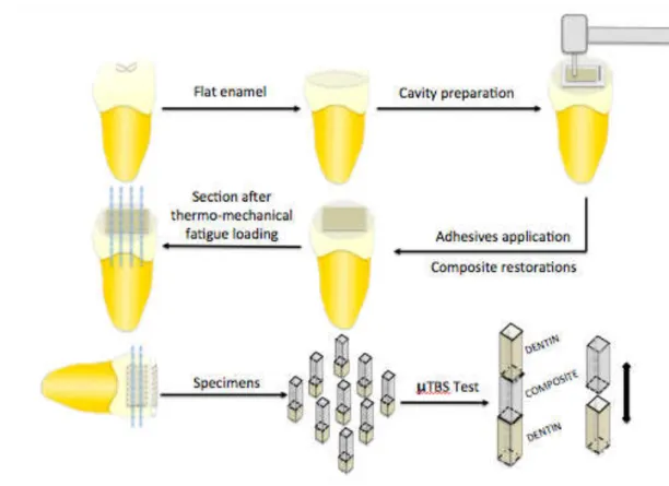
Bonded specimens were serially sectioned, parallel and perpendicular to the long axis of the tooth, using a slow-speed diamond saw (Isomet, Buehler Ltd., Lake Bluff, USA) under water cooling to obtain 1mm-thick beams, in accordance with microtensile test non-trimming technique (Fig. 1).
29
glue (SuperBonder flex gel, Henkel Ltda, Düsseldorf, Germany). Beams were stressed to failure under tension using a microtensile testing machine (Emic, DL2000, São José dos Pinhais, Brazil) at a crosshead speed of 0.5 mm/min. The number of prematurely debonded sticks per group during specimen preparation was also recorded.
Dentin side of the failed bonds was analyzed using a stereoscopic light microscopy (Stemi 2000–C, Carl Zeiss Jena, Jena, Germany) with 50 x magnification and classified according to the failure mode as adhesive (A), cohesive in dentin (CD), cohesive in composite (CC) or mixed (M).
Bond strength data were collected and analyzed using SigmaStat 3.5 (Systat, Chicago, USA). Premature failures from µTBS were included in the statistical analysis as 0 MPa. As the normality (Kolmogorov-Smirnov) test passed (p>0.05), a two-way ANOVA and Tukey’s test were employed to examine the effects of adhesive and dentin surface preparation methods (α=5%).
Scanning electron microscopy (SEM) analysis
for 5 min and placed in distilled water for futher 5 min.20 The specimes were fixed in 2.5% glutaraldehyde in 0.1 M sodium cacodylate buffer at pH 7.4 for 12 h at 4°C. After fixation, samples were rinsed with 20 ml of 0.2 M sodium cacodylate buffer at pH 7.4 for 1 h, with three changes, followed by deionized water for 1 min. All especimes were dehydrated in an ascending series of ethanol to 100% followed by immersion in hexamethyl disilazane (HMDS) for 10 min and allowed to air-dry.22 The specimens were gold sputter-coated and observed using SEM.
RESULTS
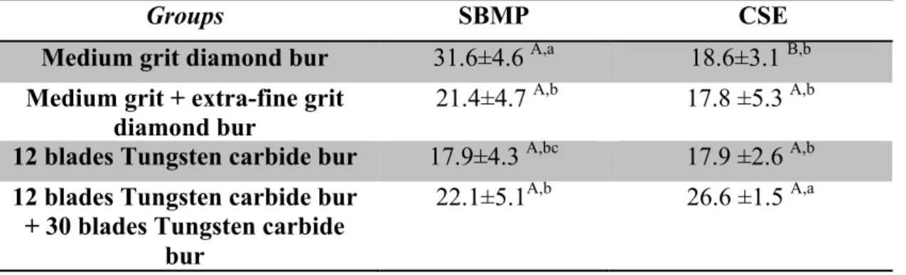
Microtensile bond strength
All information regarding microtensile bond strength are shown in table 2. The number of prematurely debonded sticks per test group affected all groups in a similar extent (Table 2).
Two-way ANOVA revealead that significant differences were observed for the main factors “dentin surface preparation method“ (p < 0.001) and “adhesive systems” (p < 0.001). The interaction between these two factors was also significant (p < 0.001), indicating that the effect of adhesive on bond strength results was dependent upon the dentin surface preparation method.
31
For the CSE, the highest bond strength was found using 12 blades carbide bur followed by 30 blades carbide bur group (26.6 ± 1.5 MPa), but no difference was shown among the remaining groups. However, CSE exhibited significantly lower mean bond strength (18.6 ± 3.1 MPa) than SBMP (31.6 ± 4.6 MPa) in medium grit diamond bur group.
Examination of failure mode by stereoscopic light microscopy indicated that the most of surfaces failed at adhesive interface (adhesive failure) and lower bond strengths were associated with higher percentages of adhesive failures (Table 3). Scanning electron microscopy (SEM) evaluation
Evaluation of prepared dentin surface
Under SEM analysis, medium grit diamond bur (100 µm grain size), produced the thickest SL (2.5 µm) with irregular and more corrugated surface, traversed by deep grooves (Fig. 2a and 2b). Extra-fine grit diamond bur (15 µm grain size) created a moderately thick SL (2.0 µm) with less irregular surface as compared to medium grit diamond bur (Fig. 2 c and 2d).
The group prepared with 12 blades Tungsten carbide bur produced a thin (1.5 µm) and relatively regular SL, showing uniform scratches on the cut surface (Fig. 3a and 3b). Specimes prepared with 30 blades Tungsten carbide bur showed the smoothest surface with narrow grooves on the dentin (Fig. 3c and 3d).
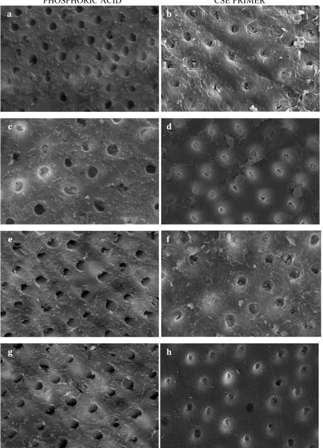
Evaluation of treated dentin surface
was also etched and the porosity of the intertubular dentin was similar than medium grit diamond bur prepared dentin. Dentin prepared with Tungsten carbide burs after CSE primer application exhibited most removal of SL and partial dissolution of smear plugs. Slight etching of peritubular dentin was observed (5f and 5h).
For phosphoric acid groups, there were no differences in terms of removal of the SL between diamond and carbide groups. The SL and smear plugs were completely removed (Fig. 5a, 5c, 5e, 5g). The only distinct differences were dissolution of smear plugs which seemed to be more resistant to the acid remained in some tubules (Fig. 5c) when extra-fine grit diamond bur was used.
DISCUSSION
The presence of the SL on ground dentin has been considered as a barrier for resin infiltration during bonding. This zone of debris is a mixture of partly denatured collagen fibrils, other organic materials and several compounds, according to the underlying dentin surface.7 Moreover, differences in surface preparation methods can be produce a variety of SL characteristics that have been reported to affect the bond strengths of resin to dentin.5,9,18,20,31 Furthermore, it has been related that the effect of dentin preparation on bond strength also depends on the adhesive used.9,18,19
Results of the current study showed that µTBS of SBMP or CSE were significantly different when bonded to dentin with distincts dentin surface preparation methods. Thus, since there was a difference, for each adhesive, when distincts dentin preparation methods were used, the null hypothesis must be rejected.
33
compact SL was observed with extra-fine diamond bur, created by tiny abrasive particles present on this cutting instrument (Fig. 2 c and 2d). It is speculated, that this variation in SL density may have contributed to partial removal of SL and smear plugs in some areas by phosphoric acid4 (Fig. 5c).
However, this observation is in contrast with previous studies that affirm phosphoric acid completely removes the SL and it is not sensivite to the method used to create the SL.20 Although, once the 37% phosporic acid removes the whole SL, the striated topography created by dentin surface preparation remained intact beneath. It is reasonable to assume that by increasing the roughness and surface area, the bond strenght would be higher, since it would increase the true area of surface bonded by resin.3,11 Thus, the lower roughness surface resulting from the extra-fine diamond bur would probably give lower bond strengths for SBMP. Indeed, on may suppose that the dentin surface roughness have a more prominent role than SL thickness on the bond strength of the etch-and-rinse adhesive to dentin.
Carbide burs generated the smoothest surface with apparently homogeneously distributed and thin SL and smear plug34 (Fig. 3). This might be due to the fact that carbide burs uses bladed cutting rather than the abrasive cutting of diamond burs.26 Although, extra-fine diamond bur produces a rougher surface and thicker SL than carbide burs,5 in this study, bond strength of these dentin surface preparation methods were similar, when SMPB was used. This outcome was expect once phosphoric acid enterily removes the SL23 and partially dissolve the surrounding peritubular dentin, allowing more resin to infiltrate into the dentin tubule.14
produced by 30 blades carbide bur had a higher permeability and allowed infiltration of the primer acidic monomers through the SL towards the underlying dentin to create a hybrid layer, resulting in higher resin-dentin bond strengths. Moreover, it was hypothesized that smooth dentin surface may improve adhesive wettability by reduction of the contact angle.3 Based on these findings, this might be the cutting instrument of choice for obtaining higher bond strenghts when self-etching systems are used in agreement with a previous investigation.5
Clearfil SE bond is a “mild” self-etching adhesive with ph 1.9 and incorporate the SL and smear plugs into the hybrized complex. As a result of its weak acidity, it may not be able to penetrate completely through the thick SL produced by diamond burs to demineralize the underlying dentin. Bonding of CSE to dentin surface prepared by Tungsten carbide burs was improved 33%, when compared to diamond burs.
35
For CSE groups, no difference in bond strength was observed when single medium grit diamond bur combined or not with a extra-fine grit was used. This finding is in agreement with results reported by others studies.20,24,30 Conversely, previous reports5,9,13,19 show that the thick of SL affected dentin bond strengths of self-etching systems. This results may be attributed to the similar removal pattern of the SL by the acidic primer in dentin surfaces prepared with diamond burs combined or not with extra-fine ones. Inou et al (2001)10 compared the bond strenghts of two self-etching adhesives using a medium grit and a extra-fine grit diamond bur prepared dentin. Lower bond strenghts were assumed to be related to remaining SL (after primer application) produced by a medium-grit diamond bur. This finding is not in agreement with our results, however, in that study, authors used all-in-one adhesives, which did not seem to remove the SL well enough due to their higher pH. Reported values of bond strenght of single-step adhesives have been lower than those of the two-step self-etching adhesive systems16, like CSE.
AKNOWLEDGMENTS
The authors would like to thank the Central Analítica of Federal University of Ceará for SEM images support. The authors declared no potential conflicts of interest with respect to the authorship and/or publication of this article.
Clinical relevance: Dentin surface preparation method affects surface topography and SL characteristics and it could influence the interaction between adhesive and substrate.
REFERENCES
1. Al-Omari WM, Mitchell CA, Cunningham, Jl. Surface roughness and
wettability of enamel and dentine surfaces prepared with different dental burs. J Oral Rehabil. 2001;28(7):645-50.
2. Ayad MF, Rosenstiel SF, Hassan MM. Surface roughness of dentin after tooth preparation with different rotary instrumentation. J Prosthet Dent.
1996;75(2):122-8.
3. Ayad MF, Johnston WM, Rosenstiel SF. Influence of dental rotary instruments on the roughness and wettability of human dentin surfaces. J Prosthet Dent. 2009;102(2):81-8.
4. Barros JA, Myaki SI, Nor JE, Peters MC. Effect of bur type and conditioning on the surface and interface of dentine. J Oral Rehabil. 2005;32(11):849-56. 5. Dias WR, Pereira PN, Swift EJ, Jr. Effect of bur type on microtensile bond
strengths of self-etching systems to human dentin. J Adhes Dent. 2004;6(3):195-203.
6. Dias WR, Pereira PN, Swift EJ, Jr. Effect of Surface Preparation on Microtensile Bond Strength of Three Adhesive Systems to Bovine Enamel. J Adhes Dent. 2004;6(4):279-85.
37
8. Eick JD, Johnson LN, Former JR, Good RJ, Neumann AW. Surface topography: its influence on wetting and adhesion in a dental adhesive system. J Dent Res. 1972;51(3):780-8.
9. Hosoya Y, Shinkawa H, Suefiji C, Nozaka K, Garcia-Godoy F. Effects of diamond bur particle size on dentin bond strength. Am J Dent. 2004;17(5):359-64.
10. Inoue H, Inoue S, Uno S, Takahashi A, Koase K, Sano H. Microtensile bond strength of two single-step adhesive systems to bur-prepared dentin. J Adhes Dent. 2001;3(2):129-36.
11. Jung M, Wehlen LO, Klimek J. Surface roughness and bond strength of enamel to composite. Dent Mater. 1999;15(4):250-6.
12. Kenshima S, Reis A, Uceda-Gomez N, Tancredo Lde L, Filho LE, Nogueira FN, et al. Effect of smear layer thickness and pH of self-etching adhesive systems on the bond strength and gap formation to dentin. J Adhes Dent. 2005;7(2)
13. Koibuchi H, Yasuda N, Nakabayashi N. Bonding to dentin with a self-etching primer: the effect of smear layers. Dent Mater. 2001;17(2):122-6.
14. Kwong SM, Cheung GS, Kei LH, Itthagarun A, Smales RJ, Tay FR, et al. Micro-tensile bond strengths to sclerotic dentin using a self-etching and a total-etching technique. Dent Mater. 2002;18(5):359-69.
15. Mine A, De Munck J, Vivan CM, Van Landuyt KL, Poitevin A, Kuboki T, Yoshida Y, Suzuki K, Van Meerbeek B. Enamel-smear compromises
bonding by mild self-etch adhesives. J Den Res. 2010;89(12):1505-9.
16. Nikaido T, Nakajima M, Higashi T, Kanemura N, Pereira PN, Tagami J. Shear bond strengths of a single-step bonding system to enamel and dentin. Dent Mater J. 1997;16(1):40-7.
17. Ogata M, Nakajima M, Sano H, Tagami J. Effect of dentin primer application on regional bond strength to cervical wedge-shaped cavity walls. Oper Dent. 1999;24(2):81-8.
19. Ogata M, Harada N, Yamaguchi S, Nakajima M, Tagami J. Effect of self-etching primer vs phosphoric acid etchant on bonding to bur-prepared dentin. Oper Dent. 2002;27(5):447-54.
20. Oliveira SS, Pugach MK, Hilton JF, Watanabe LG, Marshall SJ, Marshall GW, Jr. The influence of the dentin smear layer on adhesion: a self-etching primer vs. a total-etch system. Dent Mater. 2003;19(8):758-67.
21. Pashley DH, Carvalho RM. Dentine permeability and dentine adhesion. J Dent. 1997;25(5):355-72
22. Perdigao J, Lambrechts P, Van Meerbeek B, Vanherle G, Lopes AL. Field emission SEM comparison of four postfixation drying techniques for human dentin. J Biomed Mater Res. 1995;29(9):1111-20.
23. Perdigao J, Lopes M. Dentin bonding--state of the art 1999. Compend Contin Educ Dent. 1999;20(12):1151-8, 60-2; quiz 64.
24. Reis A, Grandi V, Carlotto L, Bortoli G, Patzlaff R, Rodrigues Accorinte Mde L, et al. Effect of smear layer thickness and acidity of self-etching solutions on early and long-term bond strength to dentin. J Dent. 2005;33(7):549-59.
25. Santini A, Mitchell S. A scanning electron microscopic study of the effect of Gluma CPS bonding system on dentinal smear layers produced by different bur types and rotational speeds and on the resin-dentin interface. Quintessence Int. 1998;29(11):737-47.
26. Sattabanasuk V, Vachiramon V, Qian F, Armstrong SR. Resin-dentin bond strength as related to different surface preparation methods. J Dent. 2007;35(6):467-75.
27. Sekimoto T, Derkson GD, Richardson AS. Effect of cutting instruments on permeability and morphology of the dentin surface. Oper Dent. 1999;24(3):130-
28. Stangel I, Ellis TH, Sacher E. Adhesion to tooth structure mediated by contemporary bonding systems. Dent Clin North Am. 2007;51(3): p.677-94. 29. Swift EJ, Jr., Perdigao J, Heymann HO. Bonding to enamel and dentin: a brief
history and state of the art, 1995. Quintessence Int. 1995;26(2):95-110.
39
31. Tay FR, Carvalho R, Sano H, Pashley DH. Effect of smear layers on the bonding of a self-etching primer to dentin. J Adhes Dent. 2000;2(2):99-116. 32. Van Meerbeek B, De Munck J, Yoshida Y, Inoue S, Vargas M, Vijay P, et al.
Buonocore memorial lecture. Adhesion to enamel and dentin: current status and future challenges. Oper Dent. 2003;28(3):215-35.
33. Watanabe I, Nakabayashi N, Pashley DH. Bonding to ground dentin by a phenyl-P self-etching primer. J Dent Res. 1994;73(6):1212-20.
34. Yiu CK, Hiraishi N, King NM, Tay FR. Effect of dentinal surface preparation on bond strength of self-etching adhesives. J Adhes Dent. 2008;10(3):173-82.
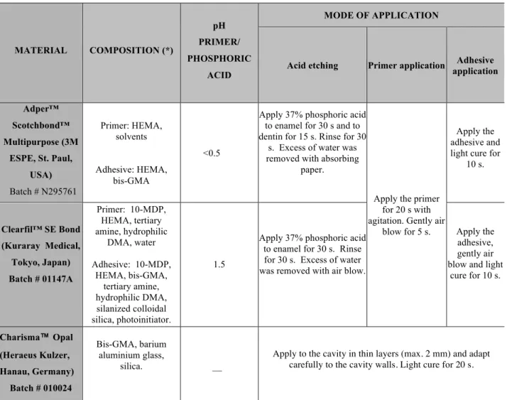
Table 1 - Materials compositions, pH and mode of application.
Abbreviations: HEMA= 2-hydroxyethylmethacrylate; BisGMA= bisphenol glycidyl methacrylate; MDP= 10-methacryloyloxydecyl dihydrogen phosphate; DMA= dimethacrylate.
MATERIAL COMPOSITION (*)
pH
PRIMER/
PHOSPHORIC
ACID
MODE OF APPLICATION
Acid etching Primer application Adhesive application
Adper™
Scotchbond™
Multipurpose (3M
ESPE, St. Paul,
USA)
Batch # N295761
Primer: HEMA, solvents Adhesive: HEMA, bis-GMA <0.5
Apply 37% phosphoric acid to enamel for 30 s and to dentin for 15 s. Rinse for 30
s. Excess of water was removed with absorbing
paper.
Apply the primer for 20 s with agitation. Gently air
blow for 5 s.
Apply the adhesive and light cure for
10 s.
Clearfil™ SE Bond
(Kuraray Medical,
Tokyo, Japan)
Batch # 01147A
Primer: 10-MDP, HEMA, tertiary amine, hydrophilic
DMA, water Adhesive: 10-MDP,
HEMA, bis-GMA, tertiary amine, hydrophilic DMA, silanized colloidal silica, photoinitiator. 1.5
Apply 37% phosphoric acid to enamel for 30 s. Rinse for 30 s. Excess of water was removed with air blow.
Apply the adhesive, gently air blow and light
cure for 10 s.
Charisma™ Opal
(Heraeus Kulzer,
Hanau, Germany)
Batch # 010024
Bis-GMA, barium aluminium glass,
silica.
__
41
Table 2 – Mean and standard deviation (MPa) for microtensile bond strength values according to each experimental group, as well as, statistical analysis (*)
Mean ± standard deviation (number of premature failed sticks/number of cohesive
failures/number of intact sticks tested) of µTBS results (expressed in MPa). (*) Different
lower case letters in column and capital letters in row indicate statistical difference (p<0.05).
Groups SBMP CSE
Medium grit diamond bur 31.6±4.6 A,a 18.6±3.1 B,b Medium grit + extra-fine grit
diamond bur
21.4±4.7 A,b 17.8 ±5.3 A,b 12 blades Tungsten carbide bur 17.9±4.3 A,bc 17.9 ±2.6 A,b 12 blades Tungsten carbide bur
+ 30 blades Tungsten carbide bur
43
Table 3 – Number of specimens (%) and failure mode according to each experimental group
A = adhesive failure; M = mixed failure; CD = cohesive failure in dentin and; CR =
cohesive failure in resin composite.
ADHESIVE SYSTEM GROUPS FAILURE MODE (%)
A M CD CR
Adper™
Scotchbond™
Multipurpose
Medium grit diamond bur 85.1 5.2 9.7 0.0
Medium grit + extra-fine grit
diamond bur 90.0 4.0 3.3 3.7
12 blades Tungsten carbide bur 96.0 4.0 0.0 0.0
12 blades Tungsten carbide bur +
30 blades Tungsten carbide bur 87.3 9.7 0.0 3.0
Clearfil™SE Bond
Medium grit diamond bur 93.0 3.5 3.5 0.0
Medium grit + extra-fine grit
diamond bur 93.7 3.3 3.0 0.0
12 blades Tungsten carbide bur 95.0 3.0 2.0 0.0
12 blades Tungsten carbide bur +
Fig. 2 – (a) and (b) SEM micrograph of dentin surface prepared with medium grit diamond bur. (a) An irregular smear layer with deep grooves was observed (500 X); (b) SEM longitudinal image of dentin tubules show a thick smear layer and dentinal tubules occluded by smear plugs (arrow) (2500 X); (c) and (d) SEM micrograph of dentin surface prepared with a medium grit followed by extra-fine grit diamond bur. (c) An irregular smear layer with more uniform scratches was observed (500 X); (d) SEM longitudinal image of dentin tubules show a thick and compact smear layer, and dentinal tubules closed by smear plugs (arrow) (2500 X).
a b
45
Fig. 3 – (a) and (b) SEM micrograph of dentin surface prepared with a Tungsten carbide
bur. (a) A smoother smear layer with regular scratches was observed (500 X); (b) SEM
longitudinal image of dentin tubules show a thin smear layer covered dentinal tubules
(2500 X). (c) and (d) SEM micrograph of dentin surface prepared with a Tungsten
carbide bur followed by a 30 blades carbide bur. (c) A relatively flat and groove-free
smear layer was observed (500 X). (d) SEM longitudinal image of dentin tubules show a
thin smear layer covered dentinal tubules (2500 X).
a b
Fig. 4 – SEM micrograph of cut dentin surface after treatment with 37% phosphoric
acid or CSE primer. Dentin surface cut with medium grit diamond bur after treament
with (a) phosphoric acid and (b) CSE primer. Dentin surface cut with medium grit
diamond bur followed by extra-fine diamond bur after treament with (c) phosphoric acid
and (d) CSE primer. Dentin surface prepared with Tungsten carbide bur after treament
with (e) phosphoric acid and (f) CSE primer. Dentin surface prepared with Tungsten
carbide bur followed by 30 blades carbide bur after treament with (g) phosphoric acid
and (h) CSE primer (2000 X). a
c d
e f
g h
b
48
3.2 CAPÍTULO 2
Effect of cavity preparation methods on marginal integrity and sealing ability of composite restorations
L.O. Barros1, L.A.M.S. Paulillo2, V.P.A. Saboia1 1
Department of Restorative Dentistry, Federal University of Ceará, Fortaleza, Brazil; 2
Department of Restorative Dentistry, Piracicaba Dental School, State University of Campinas, Piracicaba, Brazil.
Correspondence: Prof. Vicente Saboia, Department of Restorative Dentistry, Federal University of Ceará, Fortaleza, Brazil - Gilberto Studart street, 770 Aptº 901 – Cocó, CEP: 60.190-750, Fortaleza, Ceará, Brazil, Tel: +558588074623 Fax:
+558533668232; e-mail: vpsaboia@yahoo.com
ABSTRACT
Purpose: To evaluate the effect of cavity preparation method on marginal integrity and sealing ability of composite restorations bonded to human dentin using two adhesive systems. Materials and Methods: Standardized class I cavities (5 x 4 x 3 mm), with margins in enamel, were prepared in sound extracted human molars, according to the cavity preparation method (n=20): (1) Medium grit diamond bur; (2) Medium grit diamond bur followed by extra-fine grit diamond bur; (3) 12 blades Tungsten carbide bur and (4) 12 blades Tungsten carbide bur followed by 30 blades Tungsten carbide bur. Composite restorations were inserted in the cavities after Adper™ Scotchbond™ Multipurpose (SBMP) or Clearfil™ SE Bond (CSE) application. Specimens were submitted to 20,000 thermo-mechanical cycles. To evaluate the marginal integrity of the restorations after thermo-mechanical cycling, epoxy replicas were observed under scanning electron microscope (SEM) and the percentage of gap-free margins was analyzed. Data were statistically analyzed with non-parametric Kruskal-Wallis analysis (α=5%). Extra teeth, for each experimental group, were prepared, restored and immersed in 2% methylene blue solution for dye penetration analysis. Results: Preparation methods did not affect the marginal integrity and dye penetration of restorations bonded with SBMP (p>0.05). CSE exhibited the highest percentage of gap-free margins and lowest dye penetration when medium grit diamond bur was used (p<0.05). Conclusion: The cavity preparation method affected more significantly the marginal integrity and sealing ability of the self-etching adhesive. Diamond burs seemed to be the instrument of choice for obtaining better marginal quality and sealing ability for CSE.
50
INTRODUCTION
Microleakage around resin composite restorations results from the formation of gaps at the joint of restoration and cavosurface margin.14 Gap formation may be related to shrinkage of resin during polymerization1 and/or poor adhesion of bonding agents between tooth structure and composite material.29 Adhesive restorations with improved marginal integrity are less inclined to marginal staning, pulpal irritation, post operative sensitivity, tooth fracture and recurrent caries which may affect the longevity of restoration and ultimately the vitality of the dental pulp.29
One of the factors that can influence marginal integrity is the removal of carious dentin and cavity preparation method.21 To obtain an adequate bond to underlying dental substrate, smear layer (SL) must be treated prior to the application of adhesive resin8,26 using an acidic conditioner.
Moreover, cavity walls roughness may influence the wettability and bonding quality of adhesive agents.7 The development of an adhesive bonding requires estabilishing intimate contact between the adhesive and tooth surface. 2 Thus, the use of instruments for finishing of cavity walls may influence the adhesion. Finishing procedure brings to a uniformity and regularity of the margins that eliminates the asperities and the unsubstained prisms. A smooth surface makes easier the flowing of the adhesive resin, reducing the risk to hold air bubbles that could inhibit the resin polymerization.25
This in vitro study was designed to evaluate the effect of cavity preparation method on marginal integrity and sealing ability of composite restorations bonded to human dentin using an etch-and–rinse and a self-etching adhesives. The tested null hypothesis was that there are no differences in marginal gap formation and dye penetration at enamel/composite resin interface after different cavity preparation methods.
MATERIALS AND METHODS Tooth Preparation
52
Specimens were equally and randomly assigned to four groups (n= 20) and Class I standard cavities (4 mm buccolingually, 5 mm mesiodistally and 3 mm in depth) were prepared, using cavity preparation machine, according to the method of cavity preparation:
Group 1: Cavities were prepared using a cylindrical medium grit (100 µm grain size) diamond bur (#3098, KG Sorensen, Barueri, Brazil);
Group 2: The teeth were prepared as in Group 1, followed by a cylindrical extra-fine grit (15 µm grain size) diamond bur (#3098 FF, KG Sorensen, Barueri, Brazil);
Group 3: Cavities were prepared using a cylindrical 12 blades Tungsten carbide bur (#56, Beavers Dental, ON, Canada);
Group 4: The teeth were prepared as in Group 3, followed by a cylindrical 30 blades Tungsten carbide bur (#9572 FF, Beavers Dental, ON, Canada).
The occlusal margins of the cavities were located in the enamel. Each bur was mounted in air turbine using air and water spray coolant and used to cut five teeth. Bonding procedures
After application of adhesive on the cavity, the samples were restored with a resin composite (Charisma Opal, Shade A2, Heraeus Kulzer, Hanau, Germany) using the incremental technique (2 mm-thick oblique increments). Each layer was individually polymerized for 20 s at 1200 mW/cm2 (Radii-cal, SDI Limited, Victoria, Australia).
Specimens were stored in water at 37°C for 24 h and restorations were sequentially finished using a series of graded flexible discs (SofLex PopOn, 3M ESPE, St. Paul, MN, USA) under low speed. Restorations were then polished with 1200 and 2000-grit abrasive paper mounted in a polisher machine under running water (1 min, light pressure for each cycle) and fine polished using sequential wad discs and diamond paste (3, 0.5 and 0.25 µm; Buehler Ltd., Lake Bluff, USA) in order to generate a highly polished surface. The samples were submerged in an ultrasonic bath for 10 min to remove the debris over the margins.
Thermo-mechanical fatigue loading
Resin-bonded samples were then subjected to thermo-mechanical cycling (MSFT, Elquip, São Carlos, SP, Brazil) for 20,000 mechanical cycles under a load of 80 N, at a rate of 2 Hz, and thermocycling at a temperature of 5ºC and 50ºC with dwell time of 30 s.
After thermo-mechanical fatigue loading, impressions of the oclusal surface of the restorations were taken using individual moulds fabricated with vinyl polysiloxane putty (XT Express, 3M ESPE, St. Paul, MN, USA) and then filled with light body material (XT Express, 3M ESPE, St. Paul, MN, US). Epoxy replicas were obtained by pouring epoxy resin (Epoxy Cure Resin, Buehler Ltd., Lake Bluff, USA) into each impression of restorations.
54
Epoxy replicas were mounted on aluminium stubs with carbon tape (Koch Instrum Cient, São Paulo, Brazil) and were sputter-coated with gold. The enamel-composite margins of the replicas were examined under SEM (Quanta FEG 450, FEI, Amsterdan, Netherlands) at 180 x magnification. Marginal integrity was assessed according to the criteria “gap-free margin” versus “margin with gap”.6 The gap mensurements for each specimen was recorded and percentage of “gap-free margin” after thermo-mechanical fatigue loading was calculated in relation to the total marginal interface.
Dye penetration test
Additional teeth (n= 48) were prepared and restored using same procedures for each subgroups tested according describes previously. Restored teeth were divided equally in two subgroups: immediately tested and after 20,000 thermo-mechanical cycles.
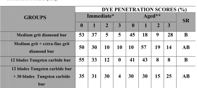
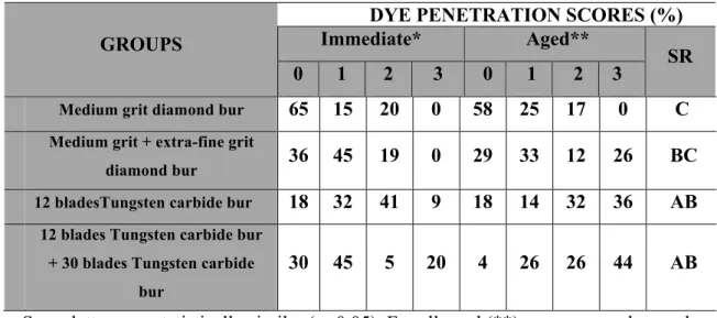
Each tooth was covered with nail varnish (Colorama, São Paulo, Brazil) except within 1 mm around margins of the restoration. The root apices sealed using sticky wax. Specimens were immersed in 2% methylene blue solution for 4 hours24, removed and rinsed under running water until all the dye was removed from the surface.
penetration less than half the length of lateral wall; 2- Dye penetration up to the full length of the lateral wall; and 3- Dye penetration reaching the pulpal wall.
Statistical analysis
Considering the marginal integrity assessment, the non-parametric data were analyzed using the Kruskal-Wallis one-way analysis of variance test by ranks at a significance level of p<0.05. The Dunn's test was used to determine significant differences among the groups.
The scores of dye penetration were tabulated, interpreted and the results were statistically analyzed using a nonparametric Kruskal-Wallis test and Tukey’s test for comparison (α= 5%). Regarding the dye penetration, there was a significant effect of thermomechanical loading for all groups (p<0.05). The statistical analysis was performed using SigmaStat 3.5 software (Systat, Chicago, IL, USA).
RESULT
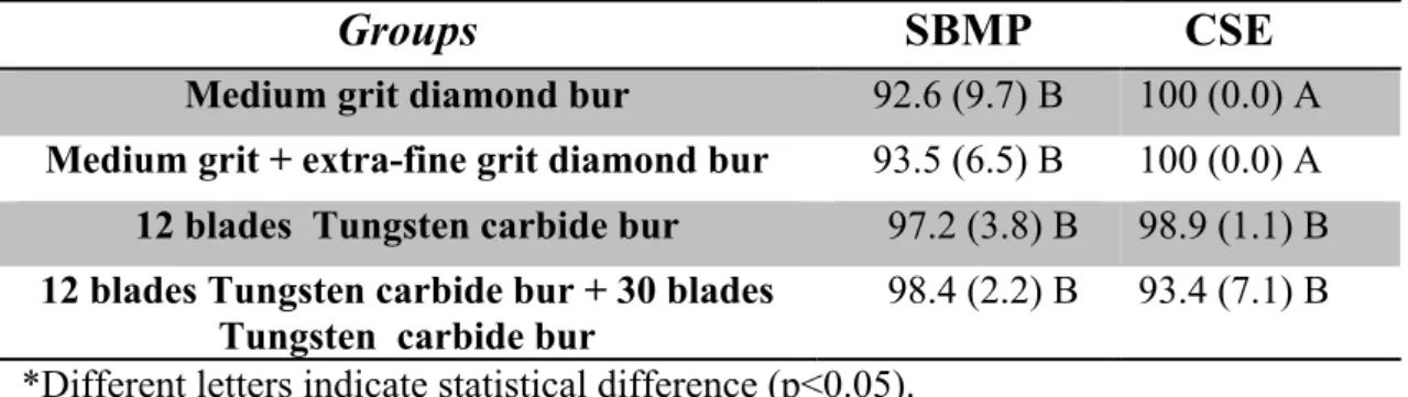
The percentages and standard deviations of gap-free margins for each group observed under SEM are shown in Table 2. These results shows that, for SBMP, there was no significant difference in all groups (p>0.05), regardless the cavity preparation method used. For CSE, the highest percentage of gap-free margins was observed in the diamond burs groups (gap-free margins = 100%). There were no statistical differences (p>0.05) between carbide burs. Representative SEM images are shown in Figure 1.
56
dye penetration) when preparation was performed using medium grit diamond bur, which resulted in a statistically significant difference (p < 0.05) when compared with cabide burs groups. All the groups showed more dye penetration after thermo-mechanical loading.
DISCUSSION
The present results demonstrated that cavity preparation method and SL caracteristics had no influence on the percentage of gap-free enamel margins, after termo-mechanical loading, for SBMP, agreeing with reported literature.11,13 In etch-and-rinse adhesive, phosphoric acid completely removes the SL and is also responsible for enamel and dentin demineralization,16 allowing resin infiltration through the nanometer spaces created on these substrates. This permits a strong mechanical interlocking between adhesives and tooth substrates, leading to higher bond strength.23 For the enamel, etching might have produced similar bonding substrate in spite of distincts preparation methods. This fact may explain the similar bonding performance, regardless of preparation method, when SBMP was used.
Regarding sealing ability of SBMP, no significant differences were observed between rotary preparation groups. However, a numeric reduction in dye penetration can be observed when medium grit diamond bur and carbide bur were used. It has been reported that the roughness of dentin and enamel surfaces is increased by coarseness of the rotary instrument2,10 and the surface topography caused by the cutting instrument continued onto the etched surface.10 An increase in roughness results in a greater area available for bonding4,9 and could, therefore, increased sealing ability, thus, justifying the results obtained in the current study.
were seem after thermo-mechanical loading (Fig. 1A) and less dye penetration was observed, although no statistical significant difference was observed in comparison to Tungsten carbide burs.
Diamond bur produces a thick, dense and rough SL20,27 and some researchers have reported reduced µTBS and higher mean gap widths for self-etching adhesives when applied on thick SL substrates.11,12,17,18 The higher the roughness of a substrate, the more difficult it is for an self-etching adhesive solution to spread over it.11
However, other studies11,15,19,22 related no correlation between µTBS and marginal integrity. It has been suggested that the lack of correlation between the bond strength and the gap measurements is due to the fact that these two properties are taken from different specimens, where other variables such as C-factor and polymerization shrinkage may alter the results.9 Besides, in the present work, all the restorations had margins in enamel which are less affected by the method of preparation,14 at least in terms of SL production. Therefore, a reduction in µTBS to dentin does not necessarily correlates with worse marginal adaptation. This may explain no influence of the SL thickness on gap formation13 or dye penetration observed in this study for CSE.
Retention, marginal and clinical dye penetration are usually the key parameters recorded to assess clinical effectiveness of adhesives.22 The self-etching adhesive evaluated in this study seemed to show a promising clinical prognosis compared to the etch-and-rinse adhesive, once it exhibited a better marginal integrity when diamond bur was used (Fig 1A and 1B), agreeing with reported literature.22
58
This chemical bonding promoted by 10-MDP is more effective and also more stable in water than that provided by other functional monomers like 4-MET and phenyl-P.30
The influence of cavity preparation method on the marginal integrity and sealing ability were dependent of the adhesive used once it affected more significantly the self-etching adhesive. Within the limits imposed in the experimental design, diamond burs seemed to be the instrument of choice for obtaining higher marginal integrity and lower dye penetration of resin-tooth interface produced by resin composite and CSE adhesive.
AKNOWLEDGMENTS
The authors would like to thank the Central Analítica of Federal University of Ceará for SEM images support. The authors declared no potential conflicts of interest with respect to the authorship and/or publication of this article.
Clinical relevance: Care should be taken in selecting proper instrument for each adhesive, as the best marginal integrity and sealing ability was observed for self-etching adhesive bonded to cavity prepared by diamond bur.
REFERENCES
1. Al-Boni R, Raja OM. Microleakage evaluation of silorane based composite
versus methacrylate based composite. J Conserv Dent. 2010;13(3):152-5.
2. Al-Omari WM, Mitchell CA, Cunningham JL. Surface roughness and
wettability of enamel and dentine surfaces prepared with different dental burs.
J Oral Rehabil. 2001;28(7):645-50.
3. Ayad MF, Rosenstiel SF, Hassan MM. Surface roughness of dentin after tooth
preparation with different rotary instrumentation. J Prosthet Dent.
4. Ayad MF, Johnston WM, Rosenstiel SF. Influence of dental rotary
instruments on the roughness and wettability of human dentin surfaces. J
Prosthet Dent. 2009;102(2):81-8.
5. Barros JA, Myaki SI, Nor JE, Peters MC. Effect of bur type and conditioning
on the surface and interface of dentine. J Oral Rehabil. 2005;32(11):849-56.
6. Benetti AR, Peutzfeldt A, Lussi A, Flurry S. Resin composites: Modulus of
elasticity and marginal quality. J Dent. 2014;42(9):1185-92.
7. Eick JD, Johnson LN, Former JR, Good RJ, Neumann AW. Surface
topography: its influence on wetting and adhesion in a dental adhesive system.
J Dent Res. 1972;51(3):780-8.
8. Eick JD, Gwinnett AJ, Pashley DH, Robinson SJ. Current concepts on
adhesion to dentin. Crit Rev Oral Biol Med. 1997;8(3):306-35.
9. Hilton TJ. Can modern restorative procedures and materials reliably seal
cavities? In vitro investigations. Part 2. Am J Dent 2002;15:279-289.
10.Jung M, Wehlen LO, Klimek J. Surface roughness and bond strength of
enamel to composite. Dent Mater. 1999;15(4):250-6.
11.Kenshima S, Reis A, Uceda-Gomez N, Tancredo Lde L, Filho LE, Nogueira
FN, et al. Effect of smear layer thickness and pH of self-etching adhesive
systems on the bond strength and gap formation to dentin. J Adhes Dent.
2005;7(2):117-26.
12.Koibuchi H, Yasuda N, Nakabayashi N. Bonding to dentin with a self-etching
primer: the effect of smear layers. Dent Mater. 2001;17(2):122-6.
13.Loguercio AD, Reis A, Bortoli G, Patzlaft R, Kenshima S, Rodrigues Filho
LE, et al. Influence of adhesive systems on interfacial dentin gap formation in
vitro. Oper Dent. 2006;31(4):431-41.
14.Malekipour MR, Shirani F, Tahmourespour S. The effect of cutting efficacy of
diamond burs on microleakage of class v resin composite restorations using
total etch and self etch adhesive systems. J Dent (Tehran). 2010;7(4):218-25.
15.Nakabayashi N, Saimi Y. Bonding to intact dentin. J Dent Res.
1996;75(9):1706-15.
16.Nakabayashi N, Pashley DH. Hybridization of dental hard tissues. Tokyo:
60
17.Ogata M, Harada N, Yamaguchi S, Nakajima M, Pereira PN, Tagami J.
Effects of different burs on dentin bond strengths of self-etching primer
bonding systems. Oper Dent. 2001;26(4):375-82.
18.Ogata M, Harada N, Yamaguchi S, Nakajima M, Tagami J. Effect of
self-etching primer vs phosphoric acid etchant on bonding to bur-prepared dentin.
Oper Dent. 2002;27(5):447-54.
19.Okuda M, Pereira PN, Nakajima M, Tagami J. Relationship between
nanoleakage and long-term durability of dentin bonds. Oper Dent
2001;26:482-490.
20.Oliveira SS, Pugach MK, Hilton JF, Watanabe LG, Marshall SJ, Marshall GW,
Jr. The influence of the dentin smear layer on adhesion: a self-etching primer
vs. a total-etch system. Dent Mater. 2003;19(8):758-67.
21.Pavuluri C, Nuvvula S, Kamatham RL, Nirmala S. Comparative Evaluation of
Microleakage in Conventional and RMGIC Restorations following
Conventional and Chemomechanical Caries Removal: An in vitro Study. Int J
Clin Pediatr Dent. 2014;7(3):172-5.
22.Peumans M, De Munck J, Mine A, Van Meerbeek B. Clinical effectiveness of
contemporary adhesives for the restoration of non-carious cervical lesions. A
systematic review. Dent Mater. 2014;30(10):1089-103.
23.Reis A, Loguercio AD, Azevedo CL, de Carvalho RM, da Julio Singer M,
Grande RH. Moisture spectrum of demineralized dentin for adhesive systems
with different solvent bases. J Adhes Dent. 2003;5(3):183-92.
24.Saboia VP, Pimenta LA, Ambrosano GM. Effect of collagen removal on
microleakage of resin composite restorations. Oper Dent. 2002;27(1):38-43.
25.Stangel I, Ellis TH, Sacher E. Adhesion to tooth structure mediated by
contemporary bonding systems. Dent Clin North Am. 2007;51(3): p.677-94.
26.Swift EJ, Jr., Perdigao J, Heymann HO. Bonding to enamel and dentin: a brief
history and state of the art, 1995. Quintessence Int. 1995;26(2):95-110.
27.Tani C, Finger WJ. Effect of smear layer thickness on bond strength mediated
by three all-in-one self-etching priming adhesives. J Adhes Dent.
28.Trivedi P, Dube M, Pandya M, Sonigra H, Vachhani K, Attur K. Effect of
different burs on the topography of smear layer formation on the dentinal
surface: a scanning electron microscope study. J Contemp Dent Pract.
2014;15(2):161-4.
29.Vinay S, Shivanna V. Comparative evaluation of microleakage fifth, sixth,
and seventh generation dentin bonding agents: An in vitro study. J Conserv
Dent. 2010;13(3):136-40.
30.Yoshida Y, Nagakane K, Fukuda R, Nakayama Y, Okazaki M, Shintani H, et
al. Comparative study on adhesive performance of functional monomers. J
Dent Res. 2004;83(6):454-8.
31.Yoshihara K, Yoshida Y, Nagaoka N, Fukegawa D, Hayakawa S, Mine A, et
al. Nano-controlled molecular interaction at adhesive interfaces for hard tissue
62
Table 1 - Materials compositions, pH and mode of application
Abbreviations: HEMA= 2-hydroxyethylmethacrylate; BisGMA= bisphenol glycidyl methacrylate; MDP= 10-methacryloyloxydecyl dihydrogen phosphate; DMA= Dimethacrylate.
MATERIAL COMPOSITION (*)
pH
PRIMER/ PHOSPHORIC
ACID
MODE OF APPLICATION
Acid etching Primer application Adhesive application Adper™ Scotchbond™ Multipurpose (3M
ESPE, St. Paul,
USA)
Batch # N295761
Primer: HEMA, solvents
Adhesive: HEMA, bis-GMA
<0.5
Apply 37% phosphoric acid to enamel for 30 s and to dentin for 15 s. Rinse for 30
s. Excess of water was removed with absorbing
paper.
Apply the primer for 20 s with agitation. Gently
air blow for 5 s.
Apply the adhesive and light cure for
10 s.
Clearfil™ SE
Bond (Kuraray
Medical, Tokyo,
Japan)
Batch # 01147A
Primer: 10-MDP, HEMA, tertiary amine,
hydrophilic DMA, water Adhesive: 10-MDP,
HEMA, bis-GMA, tertiary amine, hydrophilic DMA, silanized colloidal silica, photoinitiator. 1.5
Apply 37% phosphoric acid to enamel for 30 s. Rinse for 30 s. Excess of water was removed with air blow.
Apply the adhesive, gently air blow and light cure for
10 s.
Charisma™ Opal
(Heraeus Kulzer,
Hanau, Germany)
Batch # 010024
Bis-GMA, barium aluminium glass, silica.
__