~ 20 ~
Journal of Medicinal Plants Studies 2016; 4(1): 20-29
ISSN 2320-3862 JMPS 2016; 4(1): 20-29 © 2016 JMPS Received: 13-11-2015 Accepted: 14-12-2015
Elaine Cristina Pereira Lucetti Faculty of Medicine of the Federal University of Ceará (UFC), Brazil.
Daniel Luna Lucetti Faculty of Medicine of the Federal University of Ceará (UFC), Brazil.
Ana Elisa da Silva Ribeiro Faculty of Medicine Estácio of Juazeiro do Norte (FMJ), Ceará, Brazil.
Rafael Barbosa de Moura Faculty of Medicine Estácio of Juazeiro do Norte (FMJ), Ceará, Brazil.
Thayga Maria Araújo Sampaio Faculty of Medicine Estácio of Juazeiro do Norte (FMJ), Ceará, Brazil.
Viviane Leandro de Almeida Faculty of Medicine Estácio of Juazeiro do Norte (FMJ), Ceará, Brazil.
Andreza Sérvula Pereira da Silva Faculty of Medicine Estácio of Juazeiro do Norte (FMJ), Ceará, Brazil.
Louise Rayna Alves Bezerra Faculty of Medicine Estácio of Juazeiro do Norte (FMJ), Ceará, Brazil.
Kelly Rose Tavares Neves Faculty of Medicine of the Federal University of Ceará (UFC), Brazil.
Glauce Socorro de Barros Viana a) Faculty of Medicine of the Federal University of Ceará (UFC), Brazil.
b) Faculty of Medicine Estácio of Juazeiro do Norte (FMJ), Ceará, Brazil.
Correspondence Dr. Glauce Viana Rua Barbosa de Freitas, 130/1100
Fortaleza 60170-020 Brazil.
Curcumin reversion of neurochemical and
immunohistochemical alterations in brain ischemia is
related to its antioxidant and anti-inflammatory properties
Elaine Cristina Pereira Lucetti, Daniel Luna Lucetti, Ana Elisa da Silva
Ribeiro,
Rafael Barbosa de Moura, Thayga Maria Araújo Sampaio,
Viviane Leandro de
Almeida, Andreza Sérvula Pereira da Silva, Louise
Rayna Alves Bezerra, Kelly Rose
Tavares Neves, Glauce Socorro de
Barros Viana
Abstract
Stroke is one leading cause of mortality and morbidity worldwide where free radicals production and inflammation play important roles. Curcumin, a curcuminoid from Curcuma longa presents several biological properties and is a potent anti-inflammatory and antioxidant agent. The objectives were to study the neuroprotective effects of curcumin on ischemic pups from mothers treated with this drug (25, 50 or 100 mg/kg) during lactation. Twenty-one-day old pups were anesthetized with ketamine and xylazine, and submitted to global ischemia by clamping the carotid arteries, followed by 15 min reperfusion of the right artery and cut of the left artery. At the next day, the pups were treated with curcumin (25, 50 or 100 mg/kg) or vehicle, for 3 days. The sham operated group (SO) was subjected to the same procedure, except for carotid clamping. After 3 days, the animals were euthanized and brain areas isolated for neurochemical (monoamines, lipid peroxidation and nitrite determinations), histological (fluoro-jade staining) and immunohistochemical (TNF-alpha, iNOS and COX-2) assays. The results show that ischemia decreased DA and DOPAC as well as NE contents in the pups’ striatum, as related to the SO group, and curcumin treatments reversed these alterations. Brain ischemia decreased hippocampal neuronal viability also reversed by curcumin. Furthermore, ischemia drastically increased TNF-alpha, iNOS and COX-2 immunore activities in the hippocampus, and these effects were completely reversed after curcumin treatments. In conclusion, curcumin treatments reversed brain alterations presented after ischemia, representing a potential drug to be considered for prevention or treatment of pathological conditions as stroke.
Keywords:brain ischemia, curcumin, inflammation, oxidative stress
1. Introduction
Brain ischemia is a very important and leading cause of morbidity and mortality worldwide, producing brain injury through a variety of cellular and molecular mechanisms that impair the
energy requirements to maintain ionic-gradients [1]. Aging is the most important risk for
ischemic stroke and, although being negatively correlated to infarct volumes, is associated
with worsened functional recovery after stroke [2]. Although the immature brain be considered
resistant to the damaging effects of hypoxia and hypoxia-ischemia [3], ischemic brain injury is
an important cause of disability in infants and children, and a major cause of acute mortality
and chronic neurologic morbidity [4]. In addition, clinical evidences demonstrate that the
incidence of perinatal stroke is high, similar to that in the elderly, leading to significant
morbidityand severe long-term neurological and cognitive deficits, including cerebral palsy [5].
~ 21 ~
However, reperfusion after thrombolytic therapy often leads to cellular, biochemical and metabolic manifestations of cerebral ischemia, including ROS(s) generation, calcium overload, excitotoxicity and inflammation that ultimately lead to irreversible brain injury. Many natural compounds are designed to protect the brain from irreversible injury after ischemia-reperfusion or to retard the pathological process. Furthermore, by presenting antioxidant, anti-inflammatory and anti-apoptotic effects and antagonizing calcium some natural products exhibit protective effects on brain injury and are then potential candidates for prevention and treatment of brain
ischemia [6, 7].
Curcumin isolated from the rhizomes of Curcuma longa L.
(Zingiberaceae) is a polyphenol compound and a major component among the turmeric curcuminoids. Evidences indicate that curcumin presents not only anti-inflammatory, antioxidant and anti-tumor properties, but also neuroprotective
effects against a wide variety of neurologic disorders [8].
Recently, we showed [9] that curcumin administered prenatally
protects rat pups from ischemic brain injury. Thus, the objectives of the present study were to extend further those findings, attempting to clarify the early mechanisms involved in brain ischemia in young rats. The focus was mainly on neurochemical (striatal monoamines determinations and oxidative stress in cerebral cortex and hippocampus), histological (fluoro-jade staining) and immunohistochemical assays for TNF-alpha, iNOS and COX-2.
2. Materials and Methods Drugs and reagents
Curcumin was purchased from Sigma-Aldrich (St. Louis, MO, USA); ketamine and xylazine were from Konig (Santana de Parnaíba, São Paulo, Brazil). Antibodies for immunohistochemistry assays were from Santa Cruz Biotechnology (Dallas, TX, USA) or Merck-Millipore (Darmstadt, Germany). All other reagents were of analytical grade.
Animals
Pregnant female Wistar rats (200 g), at its 3rd week of
pregnancy were placed in separated cages until giving birth.
Mothers and litters were maintained at a 24±2 oC temperature,
in a 12 h dark/12 h light cycle, with standard food and water
ad libitum. The study was submitted to the Ethical Committee for Animal Experimentation of the Faculty of Medicine of the Federal University of Ceará (Brazil) and was approved under the number 23/2010. All experiments followed the ethical principles established in the Guide for the Care and Use of Laboratory Animals, USA, 1986.
Experimental protocol
Pregnant female Wistar rats were placed in cages individually and, 1 day after pups birth, they were treated for 21 days with curcumin (25, 50 or 100 mg/kg, p.o.) or vehicle (1% Cremophor EL, 10 mL/kg) up to the end of lactation. At this time, pups from both sexes were separated from their mothers and placed in different cages. Then, they were anesthetized with ketamine (75 mg/kg, i.p.) and xylazine (10 mg/kg, i.p.) and subjected to global cerebral ischemia by clamping both common carotid arteries. After a 15 min reperfusion, the right carotid was released and the left carotid was tied at both sides and cut. The sham-operated (SO) group was subjected to all the procedure, except for the carotid clamping. In order to prevent hypothermia, the pups were exposed to an incandescent light. At the next day, they were treated daily by gavage with curcumin (25, 50 or 100 mg/kg, p.o.) or vehicle
(SO group), for 3 days, and euthanized for dissection of striatum, cerebral cortex and hippocampus that were reserved for neurochemical studies. Besides, hippocampal slices were used for histological and immunohistochemical assays.
Neurochemical determinations of NE, DA and DOPAC by HPLC
The striatal contents of NE, DA and DOPAC were determined
by HPLC. Homogenates were prepared in 10% HClO4 and
centrifuged at 4ºC (15,000 rpm, 15 min). The supernatants were filtered and 20 µL injected into the HPLC column. For that, an electrochemical detector (model L-ECD-6A from Shimadzu, Japan) coupled to a column (Shim-Pak CLC-ODS, 25 cm) with a flux of 0.6 mL/min were employed. A mobile phase was prepared with monohydrated citric acid (150 mM), sodium octil sulfate (67 mM), 2% tetrahydrofuran and 4% acetonitrile, in deionized water. The mobile phase pH was adjusted to 3.0 with NAOH (10 mM). Monoamines were quantified by comparison with standards, processed the same manner as the samples. The results are expressed as ng/g tissue.
TBARS determination
Thiobarbituric acid reactive substances (TBARS) are formed as a byproduct of lipid peroxidation which can be detected by the TBARS assay, using thiobarbituric acid as a reagent. Because reactive oxygen species (ROS) have extremely short half-lives, they are difficult to measure directly and then can be measured as several products of the damage produced by oxidative stress, such as TBARS. Assays of TBARS measure malondialdehyde (MDA) present in the sample. MDA is one of several low-molecular-weight end products, formed by decomposition of lipid peroxidation products. These assays were carried out in rat cerebral cortices and hippocampi. Lipid peroxidation is an important indication of oxidative stress, induced by reactivity of oxygen free radicals. Homogenates at 10% were prepared in 1.15% KCl solution. Samples (250 µL) were then mixed with 1mL 10% trichloroacetic acid solution, and added to 1 mL 0.6% thiobarbituric acid. After shaking, the
mixture was maintained in a water-bath at 100 oC for 15 min,
and then cooled in an ice-bath and centrifuged (4000 rpm/5 min). The TBARS content was determined in a plate reader, at 540 nm, and the results expressed as µmol MDA/g tissue. The standard curve was obtained by using several MDA concentrations (0.6; 1.2; 2.4; 4.08; 8.16; 16.32 µmol).
Determination of nitrite contents
The Griess test is a chemical analysis test which detects the presence of organic nitrite compounds. It is based on the chemical diazotization reaction which uses sulfanilamide and
N-1-napthylethylenediamine dihydrochloride under acidic
(phosphoric acid) conditions. A typical Griess reagent may contain 0.2% N-1-naphthylethylenediamine dihydrochloride (NEED), and 2% sulphanilamide in 5% phosphoric acid. Under this acid condition, nitrites react with sulphanilamide by means of a diazotization reaction, forming a diazonium salt. This salt reacts with the NEED reagent forming a pink colored azo-compound which absorbs at 560 nm. Homogenates (10%) from brain cortex or hippocampus were prepared in KCl buffer
and centrifuged (12,000 rpm for 10 min). Then, 100 μL
supernatants were added to 100 μL Griess reagent and left at
room temperature for 10 min. The standard curve was obtained
at several NaNO2 decreasing concentrations (from 100
~ 22 ~
Fluoro-jade staining
Fluoro-jade is an anionic fluorescein derivative, useful for the histological staining of neurons undergoing degeneration. After paraffin removal (by immersion in xylol), hippocampal sections (5 µm) were mounted on slides surrounded by gelatin. The tissue was rehydrated by immersion in ethanol for 3 min, followed by immersions in 70 and 50% ethanol solutions and distilled water. The slices were placed into a 0.06% potassium permanganate solution, for 15 min, washed in distilled water and transferred to a fluoro-jade solution where they stayed for 30 min (with gentle stirring). After staining, the slices were washed in distilled water (3 times, 2 min each time). The excess water was discarded and the dry slices mounted in
Fluoromount® media and examined with a fluorescence
microscope.
Immunohistochemistry assays for TNF-alpha, iNOS and COX-2
Brain hippocampal sections (5 µm) were fixed in 10% buffered formol, for 24 h, followed by a 70% ethanol solution. The sections were embedded into paraffin wax for slices processing on appropriate glass slides. These were placed in
the oven at 58oC, for 10 min, followed by deparaffinization in
xylol, rehydration in alcohol at decreasing concentrations, washing in distilled water and PBS (0.1 M sodium phosphate buffer, pH 7.2), for 10 min. The endogenous peroxidase was blocked with a 3% hydrogen peroxide solution, followed by incubation with the appropriate primary anti-antibody for TNF-alpha, iNOS and COX-2 and diluted according to the manufacturers’ instructions (Santa Cruz or Millipore, USA), for 2 h, at room temperature in a moist chamber. The glass slides were then washed with PBS (3 times, 5 min each) and incubated with the biotinylated secondary antibody, for 1 h, at room temperature. Then, they were washed again in PBS and incubated with streptavidin-peroxidase, for 30 min, at room temperature. After another wash in PBS, they were incubated in 0.1% DAB solution (in 3% hydrogen peroxide). Finally, the
glass slides were washed in distilled water and counterstained with Mayers hematoxylin, washed in tap water, dehydrated in alcohol (at increasing concentrations), diaphonized in xylol and mounted on Entelan® for optic microscopy examination. The data were quantified by the Image J software (National Institute of Health, USA).
Statistical Analyses
For statistical analyses, One-way ANOVA, followed by the
Newman-Keuls or Tukey as post hoc tests, were used for
multiple comparisons. Whenever needed, the paired or unpaired Student’s t-test was used for comparisons between
two means. Differences were considered significant at p<0.05.
3. Results
Monoamines determinations in the rat striatum by HPLC
We showed that ischemia decreased by 80% dopamine (DA) levels in the striatum of young rats, 3 days after ischemia, as related to the SO group. This effect was completely reversed in a dose-dependent manner in the ischemic groups, after curcumin treatments with the doses of 25, 50 and 100 mg/kg. Interestingly, DA levels in the ISC+C100 group tended to be even higher than those seen in the SO group (Fig. 1A). Although to less extent, ischemia also decreased DOPAC levels (32%), as related to the SO group. Also, similar decreases were observed in the ischemic groups, after curcumin treatments with the doses of 25 and 50 mg/kg (58 and 42%, respectively). However, no significant changes were demonstrated in the ISC+C100 group, as related to the SO group (Fig. 1B). Decreases of 60 and 54% were demonstrated in NE contents in the ISC and ISC+C25 groups, as related to the SO group. These alterations were completely reversed in the ischemic groups, after curcumin treatments with the doses of 50 and 100 mg/kg, respectively. NE values in the ISC+C100 group were even higher than those of the SO group (Fig. 1C).
Fig 1: Effects of curcumin on monoamine contents in striata from rat pups, at the 3rd post-ischemia day. DA: a. vs. SO, q=5.287; b. vs. ISC+C50, q=3.486; c. vs. ISC+C100, q=6.010; d. vs. ISC+C100, q=3.836. DOPAC: a. vs. SO, q=5.561; b. vs. ISC+C100, q=4.805; c. vs. SO, q=4.604; d. vs. ISC+C100, q=3.758; e. vs. SO, t=2.301. df=18, p=0.0336; f. vs. ISC+C25, t=3.214, df=12, p=0.074. NE: a. vs. SO, q=4.063; b. vs. ISC+C50, q=4.220; c. vs. ISC+C100, q=6.551; d. vs. SO, q=3.847; e. vs. ISC+C50,
q=4.041; f. vs. ISC+C100, q=6.509 (One-way ANOVA and Newman-Keuls as the post hoc test).
TBARS determinations in the rat cerebral cortex and hippocampus
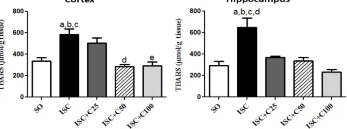
Our results showed a 1.7-fold increase in TBARS contents in the cerebral cortex of ischemic groups, as related to those of the SO group. This increase was similar to that observed in the ischemic group treated with the lower curcumin dose (ISC+C25). On the other hand, TBARS values even lower than those of the SO group were observed in the ischemic groups, treated with curcumin at the doses of 50 and 100
~ 23 ~
Fig 2: Determination of lipid peroxidation (TBARS assay) in brain cortex and hippocampus from ischemic rat pups, at the 3rd post-ischemia day.
Cortex: a. vs. SO, q=9.114; b. vs. ISC+C50, q=10.20; c. vs. ISC+C100, q=9.335; d. vs. ISC+C50, q=4.424. Hippocampus: a. vs. SO, q=8.058; b. vs. ISC+C25, q=5.265; c. vs. ISC+C50, q=7.143; d. vs. ISC+C100, q=7.863 (One-way ANOVA and Tukey as the post hoc test).
Nitrite determination in rat cerebral cortex and hippocampus
A 2.4-fold increase were demonstrated in nitrite contents, in
the rat cortex after ischemia, as related to the SO group, also maintained in the ISC+C25 and ISC+C50 groups (3.0- and 1.6-fold increases, respectively). On the other hand, ischemic groups treated with the higher curcumin dose (ISC+C100)
showed a 50% increase in nitrite levels, as related to the SO group (Fig. 3A). In the hippocampus, the ischemic group presented a 6.5-fold increase in nitrite contents, while a much lower increase was observed in the ISC+C25 group (2.9-fold), relatively to the SO group. Values close or even lower than those shown in the SO group were demonstrated in the ISC+C50 and ISC+C100 groups (Fig. 3B).
Fig 3: Determination of nitrite contents in brain cortex and hippocampus from ischemic rat pups, at the 3rd post-ischemia day. Cortex: a. vs. SO,
q=7.373; b. vs. ISC+C50, q=6.704; c. vs. ISC+C100, q=8.241. Hippocampus: a. vs. SO, q=7.811; b. vs. ISC+C25, q=4.458; c. vs. ISC+C50, q=8.110; d. vs. ISC+C100, q=8.612 (One-way ANOVA and Tukey as the post hoc test).
Histological studies (Fluoro-jade staining)
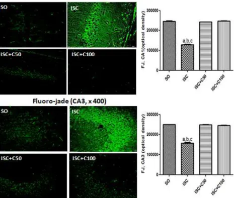
We showed a 48% decrease in cell viability after ischemia in the CA1 area, as related to the SO group. Interestingly, the decrease in cell viability was somewhat less (38% decrease) in the CA3. On the other hand, even higher percentage decreases in cell viability were observed in the temporal cortex (55%
~ 24 ~
Fig 4: Representative photomicrographs (magnification x400, scale bars=200 µm) of fluoro-jade staining in hippocampal CA1 and CA3 areas. The data were analyzed by the Image J software (NIH, USA). CA1: a. vs. SO, q=50.91; b. vs. ISC+C50, q=49.20; c. vs. ISC+C100, q=51.57; CA3: a. vs. SO, q=56.56; b. vs. ISC+C50, q=59.10; c. vs. ISC+C100, q=57.36. (One-way ANOVA and Tukey as the post hoc test).
Immunohistochemistry assays for TNF-alpha
While ischemia increased by 6.5-fold TNF-alpha immunostaining in the CA1 area, as related to the SO group, treatments with curcumin at the doses of 25 and 50 mg/kg decreased TNF-alpha immunoreactivity by 68 and 49%,
respectively, and thus to values even lower than those of the SO group. Similar increases (6.8-fold) were observed in the temporal cortex (TC) in the ischemic group, in relation to the SO group (Fig. 5).
~ 25 ~
Immunohistochemistry assays for iNOS
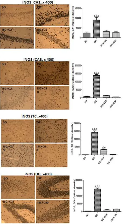
A 4.9-fold increase in the immunostaining for TNF-alpha was seen in the CA1 area after ischemia, as related to the SO group. An even higher increase (19-fold) was observed in the CA3 area. These alterations were reversed in the ischemic pups after curcumin treatments, mainly with the higher dose (50 mg/kg), and sometimes the values were lower than those of the SO group. The highest increases (111-fold) were
detected in the temporal cortex, after ischemia, suggesting that this area is very sensitive to brain injury and this alteration was partially reversed after curcumin treatments. A similar profile, although a less intense increase (42-fold) was seen in the dentate gyrus, in the ischemic group, and also a partial recovery towards the SO group values, after curcumin treatments. These last results are not shown (Fig. 6).
~ 26 ~
Immunohistochemistry assays for COX-2.
We showed that ischemia increased by 9-fold the immunoreactivity for COX-2 in the CA1, relatively to the SO group. These alterations were reversed by curcumin treatments. A similar profile was observed in the CA3 area and, at the higher dose, the values were even lower than those
of the SO group. While smaller increases in the immunoreactivity for COX-2 were seen after ischemia, in the dentate gyrus (DG), as related to the SO group, higher increases (33-fold) were demonstrated in the pups’ cerebral cortex (TC). Again, in both areas these increases were reversed after curcumin treatments, mainly at its higher dose (Fig. 7).
~ 27 ~
4. Discussion
Stroke is one of the most significant health problems in the world. Considering that the majority of strokes occur in the elderly, its prevalence is expected to increase dramatically with the increased life expectancy and advancing age of the
population. Evidences [10] indicate that the incidence of stroke
more than doubles in each successive decade, for people over the age of 55. A few therapeutic options are available in the clinic, as the thrombolytic tissue plasminogen activator (tPA) which has been proven effective for the stroke treatment, when given within 3 h after the onset of neurological symptoms. Furthermore, the high costs associated with stroke point out to the increased need for preventive therapy, early critical care
and rehabilitation [11].
Furthermore, the hypoxic-ischemic brain injury is one of the main causes of disabilities in term-born infants, with a
complex and multifactorial etiology [5, 12-14]. In addition,
evidences indicate that the incidence of perinatal stroke is high
and similar to that in the elderly [5]. It is considered the single
most important cause of acute mortality and chronic disability
in newborns worldwide [15-17]). However, there are differences
between the adult and neonatal brain injuries after hypoxia-ischemia. Thus, the immature brain may be more vulnerable to oxidative damage than the adult brain, due to the high concentration of unsaturated fatty acids and the availability of
redox-active iron in the developing brain [14, 18, 19].
It is widely accepted that inflammation plays an important role in the pathogenesis of ischemic stroke, and evidences from experimental and clinical studies show that the brain respond to ischemic injury with an inflammatory process characterized by rapid activation of resident cells, production of pro-inflammatory mediators and infiltration of various types of
inflammatory cells [20]. In addition, the involvement of
oxidative stress in ischemic reperfusion injury is well
established [21].
Curcumin, a main curcuminoid from Curcuma longa, besides
being used since ancient times as food spice in Asian countries, is known to possess several biological properties, including antioxidant, anti-inflammatory and anti-tumor ones
[22, 23]. Curcumin has been shown to present a neuroprotective
effect in experimental models of ischemic brain injury in
rodents [24-27].
In the present study, we showed curcumin neuroprotective actions in a global ischemia model, in young rats treated with this drug for a short period of time. While ischemia significantly decreased monoamines (DA, DOPAC and NE) contents in the striatum, this effect was reversed by curcumin
treatments. Earlier studies [28] demonstrated that carotid
ligations and 2 h of 8%-oxygen environment in 7-day-old rat pups led to a 70% decrease in endogenous striatal dopamine levels. According to these authors, their data suggest that dopamine release from striatal nerve terminals is associated with events causing brain injury during perinatal hypoxia-ischemia. Although in our study we used a slight different model, our results are similar to the above ones.
The levels of DA, DOPAC and HVA were shown to increase sharply after 1 h, in a model of transient brain ischemia in adult rats. They remained high after 24 h and normalized after
72 h of recirculation [29]. According to these authors, the
increased DA metabolism in striatal nerve terminals, in response to ischemic injury, may reflect an early degenerative
change in DA terminals. Recently [30], others have also
observed that brain ischemia triggers excessive release of neurotransmitters that mediate neuronal damage, following ischemic injury. The release of DA from ischemic neurons directly contributes to cell death in compromised areas. Our
group has already demonstrated significant decreases in striatal DA contents, 3 and 7 days after global ischemia, in adults as well as in rat pups. The reason for this discrepancy is probably due to the time windows for monoamine measurements. We also showed a high percentage of non-viable neurontin the hippocampus after brain ischemia, visualized as an intense fluorescence, as evaluated by fluoro-jade staining. The neuronal degeneration was observed not only in the CA1 and CA3 areas, but also in the dentate gyrus and brain cortex, as related to the SO group. These effects were completely reversed in the ischemic group after curcumin treatments. The immunoreactivity for the pro-inflammatory cytokine TNF-alpha increased by almost 7-foldin the CA1 and CA3 areas, after ischemia, and went to values even lower than those of the SO group, in ischemic groups after curcumin treatments.
Previously [31], curcumin was shown to inhibit expression of
pro-inflammatory cytokines, including TNF-alpha in HaCaT cells, and blocked the activation of NF-kappaB by TNF-alpha,
in TNF-alpha-stimulated human endothelial cells [32].
Evidences [31] indicated that TNF-alpha induces
pro-inflammatory cytokines, as IL-1beta, IL-6, IL-8, and itself by activation of NF-kappaB or MAPKs. According to these authors, curcumin exerts anti-inflammatory and growth inhibitory effects in TNF-alpha-treated HaCaT cells through inhibition of those pathways.
In addition, in the present study we showed that immunoreactivities for iNOS and COX-2 were drastically increased in several hippocampal areas, after ischemia, and these changes were reversed by curcumin treatments. Interestingly, in both cases the greatest effects were observed in the temporal cortices, followed by CA1 and CA3 areas. Similar data were recently observed by us, after prenatal curcumin administration in a model of global ischemia in rat
pups [9].
Evidences [33-35] have already indicated curcumin to be a potent
scavenger of free radicals and nitric oxide as well. Curcumin also reduces oxidative damage, in a model of Alzheimer
disease in transgenic mice [36], and suppressed iNOS activity
by promoting the ubiquitination and degradation of iNOS,
after LPS stimulation in RAW 264.7 cells [37]. Besides,
curcumin also inhibits COX-2 expression in HL-60 cells
stimulated by LPS [38]. Furthermore, the anti-inflammatory
effect of curcumin is probably mediated by its ability to inhibitCOX-2, LOX and iNOS, important enzymes that
mediate inflammatory processes [39]. Curcumin has also been
shown to regulate a number of inflammation signaling pathways, including the eicosanoid pathway involving COX
and LOX enzymes [40].
Much of our understanding on the pharmacological effects of
Curcuma longa derives from researches on curcumin. It can protect DNA against single strand breaks induced by a single
oxygen, and is a potent anti-inflammatory agent [41]. Orally, it
inhibits neutrophil function, platelet aggregation and
lymphocyte activity [42]. Curcumin also promotes fibrinolysis,
stabilizes lysosomal membranes and inhibits NF-kappaB activation (resulting in downregulation of multiple
inflammatory genes) [43].
~ 28 ~
derivative presenting a better bioavailability.
5. Acknowledgments
The authors are grateful to Ms. Auryclennedy Callou de Araújo, Ms. Maria Janice Lopes and Ms. Maria Vilani Rodrigues Bastos for technical assistance and to Prof. M.O.L. Viana for the orthographic revision of the manuscript. The work had the financial support from the Foundation for Research and Development of the State of Ceará (FUNCAP) and the Brazilian National Research Council (CNPq). Conflict of Interest the authors declare there are no conflicts of interest.
Conflict of Interest
The authors declare there are no conflicts of interest.
6. References
1. Traystman RJ. Animal models of focal and global cerebral
ischemia. ILAR Journal. 2003; 44:85-95.
2. Manwani B, Friedler B, Verma R, Venna VR,
McCullough LD, Liu F. Perfusion of ischemic brain in young and aged animals: a laser speckle flowmetry study. Stroke, 2014; 45:571-578.
3. Vannucci SJ, Hagberg H. Hypoxia-ischemia in the
immature brain. J Exp Biol. 2004; 207:3149-3154.
4. Comi AM, Trescher WH, Abi-Raad R, Johnston MV,
Wilson MA. Impact of age and strain on ischemic brain injury and seizures after carotid ligation in immature mice. Int J Dev Neurosci. 2009; 27:271-277.
5. Kratzer I, Chip S, Vexler Z. Barrier mechanisms in
neonatal stroke. Frontiers in Neuroscience Doi: 10.3389 fnins, 2014; 8:359.
6. Wu PF, Zhang Z, Wang F, Chen JG. Natural compounds
from traditional medicinal herbs in the treatment of cerebral is chemia/reperfusion injury. Acta Pharmacologica Sinica. 2010; 31:1523-1531.
7. Gupta YK, Briyal S, Gulati A. Therapeutic potential of
herbal drugs in cerebral ischemia. Indian J Physiol Pharmacol. 2010; 54:99-122.
8. Hatcher H, Planalp R, Cho J, Torti FM, Torti SV.
Curcumin: from ancient medicine to current clinical trials. Cell Mol Life Sci. 2008; 65:1631-1652.
9. Telles MVL, Nobre MEP, Alencar LP, Siqueira KP,
Borges AMFS, Tavares MWL et al. Prenatal curcumin
administration reverses behavioral and neurochemical effects and decreases iNOS and COX-2 expressions in ischemic rat pups. Int J Brain Sci. 2014, Article ID 907581. http://dx.doi.org/10.1155/2014/907581.
10. Bramlett HM, Dietrich WD. Pathophysiology of cerebral
ischemia and brain trauma: similarities and differences. J Cereb Blood Flow & Metabolism. 2004; 24:133-150.
11. Demaerschalk BM, Huang HM, Leung G. US cost burden
of ischemic stroke: a systematic literature review. Am J Manag Care. 2010; 16:525-533.
12. Cerio FG, Lara-Celador I, Alvarez A, Hilario E.
Neuroprotective therapies after perinatal hypoxic-ischemic brain injury. Brain Sci. 2013; 3:191-214.
13. Thornton C, Rousset CI, Kichev A, Miyakuni Y, Vontell
R, Baburamani AA et al. Molecular mechanisnms of
neonatal brain injury. Neurology Res Int. 2012, Article ID 506320,
doi: 10.1155/2012/506320.
14. Kleman NW, Sun D, Cengiz P. Mechanisms underlying
neonatal hypoxia ischemia. The Open Discovery Journal. 2010; 2:129-137.
15. Arteaga O, Revuelta M, Montalvo H, Canãvate ML,
Alonso-Alconada D, Martinez-Ibargüen A et al.
Neuroprotective effect of antioxidants in neonatal rat brain after hypoxia-ischemia. In: Microscopy: advances in scientific research and education (A. Méndez-Vilas, Ed.), Formatex Research Center. 2014; 1:335-343.
16. Du Plessis AJ, Volpi JJ. Perinatal brain injury in the
preterm and term newborn. Curr Opin Neurol. 2002; 2:151-157.
17. AzraHaider B, Bhutta ZA. Birth asphyxia in developing
countries: current status and public health implications. Curr Probl Pediatr Adolesc Health Care. 2006; 5:178-188.
18. Rice JE 3rd,Vannucci RC, Brierley JB. The influence of
immaturity on hypoxic-ischemic brain damage in the rat. Ann Neurol. 1981; 2:131-141.
19. Towfighi J, Zec N, Yager J, Housman C, Vannucci RC.
Temporal evolution of neuropathologic changes in an immature rat model of cerebral hypoxia: a light microscopic study. Acta Neuropathol. 1995; 4:375-386.
20. Jin R, Yang G, Li G. Inflammatory mechanisms in
ischemic stroke: role of inflammatory cells. J Leukoc Biol. 2010; 87:779-789.
21. Cruzzocrea S, Riley D, P<Caputi AP, Salvemini D.
Antioxidant therapy: a new pharmacological approach in shock, inflammation and ischemic/reperfusion injury. Cardiovasc Res. 2008; 47:537-548.
22. Basnet P, Skalko-Basnet N. Curcumin: an
anti-inflammatory molecule from a curry spice on the path to cancer treatment. Molecules. 2011; 16:4567-4598.
23. Kulkarni SK, Dhir A. An overview of curcumin in
neurological disorders. Indian J Pharm Sci. 2010; 72:149-154.
24. Zhao J, Zhao Y, Zhang W. Iu Y, Gang F, Yu S.
Neuroprotective effect of curcumin on transient focal cerebral ischemia in rats. Brain Res. 2008; 1229:224-232.
25. Yang C, Zhang X, Fan H, Liu Y. Curcumin upregulates
transcription factor Nrf2, HO-1 expression and protects rat brains against focal is chemia. Brain Res. 2009; 1282:133-141.
26. Wu J, Li Q, Wang X, Yu S, Li L, Wu X et al.
Neroprotection by curcumin in ischemic brain injury involves the Akt/Nrf2 pathway. PLoS One 8:359843. Doi: 10.137/journal.pone.0059843, 2013.
27. Liu ZJ, Liu W, Liu L, Xiao C, Wang Y, Jiao JS. Curcumin
protects neuron against cerebral ischemia-induced inflammation through improving PPAR-gamma function. Evid Based Complement Alternat Med, 2013. 470975. Doi: 10.1155/2013/470975.
28. Silverstein F, Johnston MV. Effects of hypoxia-ischemia
on monoamine metabolism in the immature brain. Ann Neurol. 1984; 15:342-347.
29. Nemeth G, Cintra A, Herb JM, Ding A, Goldstein M,
Agnati LF et al. Changes in striatal dopamine
neurochemistry and biochemistry after incomplete transient cerebral ischemia in the rat. Exp Brain Res. 1991; 86:545-554.
30. Oliva I, Fernandez M, Martin ED. Dopamine release
regulation by astrocytes during cerebral ischemia. Neurobiol Dis. 2001; 58:231-241.
31. Cho JW, Lee KS, Kim CW. Curcumin attenuates the
expression of IL-1B, IL-6, and TNF-alpha as well as cyclin E in TNF-alpha-treated HaCaT cells; NF-kB and MAPKs as potential upstream targets. Int J Mol Med. 2007; 19:469-474.
32. Kim YS, Ahn Y, Hong MH, Joo SY, Kim KH, Sohn IS et
al. Curcumin attenuates inflammatory responses of
~ 29 ~
33. Sreejayan N, Rao MNA. Nitric oxide scavenging by
curcuminoids. J Pharm Pharmacol. 1997; 49:105-107.
34. Sumanont Y, Murakami Y, Tohda M, Vajragupta O,
Matsumoto K, Watanabe H. Evaluation of the nitric oxide radical scavenging activity of manganese complexes of curcumin and its derivatives. Biol Pharm Bull. 2004; 27:170-173.
35. Zheng M, Ekmekcioglu S, Walch ET, Tang CH, Grimm
EA. Inibition of nuclear factor-kappa B and nitric oxide by curcumin induces G2/M cell cycle arrest and apoptosis in human melanoma cells. Melanoma Res. 2004; 14:165-171.
36. Lim GP, Chu T, Yang F, Beech W, Frautschy SA, Cole
GM. The curry spice curcumin reduces oxidative damage and amyloid pathology in an Alzheimer transgenic mouse. J Neurosci. 2001; 21:8370-8377.
37. Ben P, Liu J, Lu C, Xu Y, Xin Y, Fu J et al. Curcumin
promotes degradation of inducible nitric oxide synthase and suppresses its enzyme activity in RAW 264.7 cells. Int Immuno pharmacol, 2011; 11:179-186.
38. Lantz RC, Chen GJ, Solyom AM, Jolad SD, Timmermann
BN. The effect of turmeric extracts on inflammatory mediator production. Phytomedicine, 2005; 12:445-452.
39. Menon VP, Sudheer AR. Antioxidant and
anti-inflammatory properties of curcumin. Adv Exp Med Biol. 2007; 595:105-125.
40. Rao CV. Regulation of COX and LOX by curcumin. Adv
Exp Med Biol. 2007; 595:213-226.
41. Jurenka JS. Anti-inflammatory properties of curcumin, a
major constituent of Curcuma longa: a review of
pre-clinical and pre-clinical research. Altern Med Rev. 2009; 14:141-153.
42. Srivastava KC, Bordia A, Verma SK. Curcumin, a major
component of food spice turmeric (Curcuma longa)
inhibits aggregation and alters eicosanoid metabolism in human blood platelets. Prostaglandins Leukot Essent Fattt Acids. 1995; 52:223-227.
43. Ammon HP, Wahl MA. Pharmacology of Curcuma longa.