Abstract
Background: Haemophilia A is a hereditary haemorrhagic disorder that can cause bleeding in the intestinal loops and, on rare occasions, simulate an acute surgical abdomen. Careful assessment of coagula-tion must be performed in these patients, followed by an attempt to correct the dysfunctions. Often, the administration of the deficient factor is sufficient to resolve the problem, avoiding unnecessary sur-geries.
Case report: We present a male patient, 15-years-old, of indige-nous descent, who was a diagnosed with haemophiliaA. The young manwas admitted with abdominal pain in the right iliac fossa; ultraso-nography suggested acute appendicitis. He underwent an exploratory laparotomy that revealed a normal appendix and the presence of a caecal wall haematoma, without other abnormalities.
Conclusion: This case describes an unusual instance of decompen-sation of a patient with haemophilia A that simulated an acute surgi-cal abdomen. The case suggests the need for further evaluation of ca-rriers of coagulopathies, whether acquired or congenital, when they suffer abdominal pain. Otherwise, clinically treatable dysfunctions are prone to surgical treatment, with a potential for increased morbidity
Acute abdomen in a patient
with haemophilia A:
A case study
CASE REPORT
Hermes Melo Teixeira Batista1, Gylmara Bezerra de Menezes Silveira2,
Ivo Cavalvante Pita Neto1, Woneska Rodrigues Pinheiro1, Italla Maria Pinheiro Bezerra1, Vitor Engracia Valenti3,
Demostênia Rodrigues Coelho2, Sérgio de Araújo2,
Luiz Carlos de Abreu1
1 Laboratory of Study Design and Scientific Writing, ABC Faculty of Medicine GBdeMS: nurse,Cariri Regional Hospital.
2 Cariri Regional Hospital.
3 Department of Speech and Hearing Pathology at FFC-UNESP/ Marília. São Paulo
Contact information:
Juc Luiz Carlos de Abreu.
Background
Haemophilia A is a hereditary disease linked to the X chromosome and occurs in 1 out of 10,000 men [1]. This coagulopathy is caused by a fac-tor VIII deficiency that manifests as spontaneous bleeding of the joints and tissues, including those forming the digestive and nervous systems [2]. These patients may develop conditions that re-quire surgical treatment, whether they are related to the underlying pathology or to another disor-der [3].Haemorrhagic complications in the small or large intestine may simulate an acute surgical abdomen, causing a potential increase in morbi-dity and mortality [4]. The case described involves the surgical treatment of an abdominal problem, suggestive of appendicitis, in a patient with hae-mophilia A [5].
Case presentation
A 15-year-old male of indigenous descent was admitted to hospital with an 8-day history of ab-dominal pain. The patient had been previously diagnosed with haemophilia A, and was being monitored by the haematology service of another department. His pain began in the epigastric re-gion and remained localized to the right iliac fos-sa. Over time, he developed vomiting, asthenia,
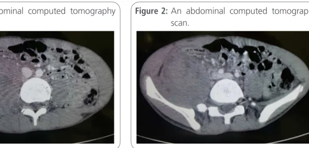
anorexia, and abdominal guarding. As a result, he underwent abdominal ultrasonography, which ggested appendicitis. Abdominal tomography su-ggested a lesion to the right of the psoas muscle, but the appendix could not be visualized (figure 1 and figure 2). The results of a laboratory exam revealed a haemoglobin level of 9.4 g/dL, a leuco-cyte count of 16,000/µL, a left-shifted leukogram; an international normalized ratio of 1.21; an activa-ted partial thromboplastin time (APTT/INR) of 2.86; and a platelet count of 326,000/µL. The patient’s renal and liver function tests werewithin normal limits [6].
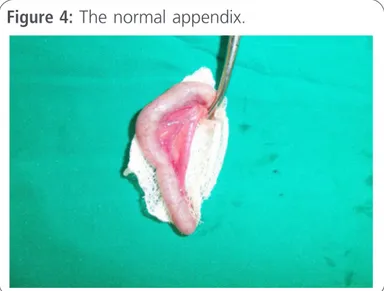
The patient received3000 IU of factor VIII, preoperatively, and was anaesthetised with pro-pofol, fentanyl, cisatracurium, and lidocaine; anaesthesia was maintained with 2.5% sevoflura-ne. Standard monitoring, using pulse oximetry, ele-trocardiography, non-invasive blood pressure mea-surement, and capnographic analysis of inhaled and exhaled gases, was continuously performed. A midline incision was performed and a normal caecal appendix was visualized. After inspection of the loops, extensive haematoma was found in the caecum, near the base of the appendix (figure 3). An appendectomy was performed, with laye-red suturing, and a drain was placed (figure 4). At the end of the procedure,a bilateral transverse
ab-Figure 1: An abdominal computed tomography scan.
dominis planeblock was performed with 20 mLof 0.5% ropivacaine. The patient awoke well, without pain or signs of bleeding. Post-surgically, his APTT was again determined and another dose of factor VIII was administered.
Discussion
Hemophilia A and B are hereditary disease that re-sult from functional deficiencies in specific circula-ting blood clotcircula-ting factors termed factor VIII and factor IX [7]. The gene for factor VIII is located on the X chromosome and is relatively large. Changes resulting from deletion or inversion portions of the genome or changing wrong direction, result in fac-tor VIII activity below 1% of normal, causing severe forms of the disease. Point mutations and smaller depletions result in blood levels of Factor VIII larger than 1% and milder disease [8].
truction and necrosis of bowel segments [10]. In addition, the carrier of hemophilia A is subject to abdominal surgical pathologies common as sus-pected appendicitis in our case, the clinical symp-toms (nausea, vomiting and abdominal pain well located in the right iliac fossa with the presence of positive Blumberg sign). In addition, features high valeucograma, suggestive of an infectious process (16,000 leukocytes per mcL) which led to a strong suspicion of appendicitis.
It was contemplated the possibility of intramu-ral hematoma handles, however, there was no tomographic confirmation and held Factor VIII replacement at a dose of 30 IU/kg 8/8 h without worsening of symptoms [11].
For any surgical intervention it is important to determine the factor VIII level and make appro-priate preoperative replacement to ensure good surgical hemostasis [12]. In our case, the APTT
Figure 3: Intraoperative appearance of the appen-dix and caecum (with the haematoma).
reason we chose general anesthesia, followed by TAP Block guided by ultrasound to postoperative analgesia, which was adequate.
The case report suggests that, in frames of acute abdomen, even with clinical and laboratory eviden-ce of mimicking appendicitis, suspected intramural hematoma handles, which, in most cases resolves with appropriate replacement of Factor VIII (defi-cient) [14]. Clinical treatment avoids surgery, high morbidity and mortality factor in these patients
Conclusion
In patients with coagulopathies, particularly the more severe coagulopathies, who present with abdominal pain, complications of the underlying disease must always be assumed [6, 12]. The com-plications may include retroperitoneal hematoma, hematoma of the intestinal loops or hematoma of the abdominal musculature [7, 8, 14]. These com-plications may arise spontaneously or after minor trauma, simulating an acute abdomen [9]. Often these complications can be reversed with appro-priate clinical treatment [10, 11, 15].
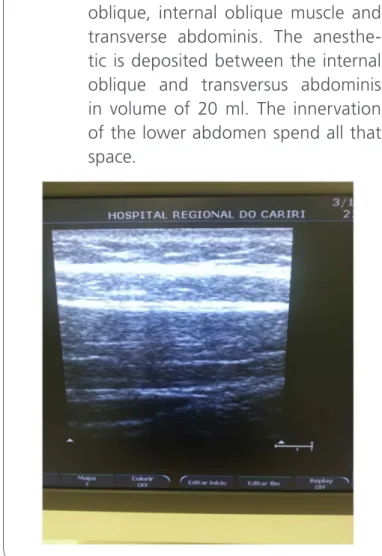
General anaesthesia is indicated in these pa-tients. Central blocks as spinal anesthesia or epi-dural anesthesia are not indicated in patients with coagulopathy patients at high risk of progressing to spinal hematoma. In exploratory laparotomy, always related to the possibility of holding the TAP Block guided by ultrasound with local anesthetics long term as bupivacaine or ropivacaine for postoperati-ve analgesia (Figure 5).
Consent
Written informed consent was obtained from the patient's mother for publication of this Case report and any accompanying images. A copy of the writ-ten consent is available for review by the Editor-in-Chief of this journal.
Ethics questions
The current study was approved by the ethical com-mittee of the Regional Hospital of Cariri, for case presentation.
List of abbreviations
APTT=activated partial thromboplastin time TAP= transversusabdominis plane
Competing interests
The authors declare no conflicts of interest. All re-search was conducted with their own resources.
Financial resources
The authors state that they used their own resou-rces and did not receive financial aid from any ins-titution.
The authors state that they did not receive fi-nancial aid from any institution (political, personal, religious, ideological, academic,intellectual, com-mercial, or any other).
Authors' contributions
HMTB conceived the study. ICPN, MPLN, GBM DE S, WRP, IMPB, JFC, LTA, RDR, LCA analysed the results and helped to draft the document. All authors read and approved the final manuscript.
Authors' information
HMTB: anaesthesiologist, Cariri Regional Hospital, Laboratory of Study Design and Scientific Writing, ABCFaculty of Medicine ICPN: dentist, Master of Public Health, Hospital Regional ofCariri. Laboratory of Study Design and Scientific Writing, Faculty of Medicine at ABC.
GBdeMS: nurse,Cariri Regional Hospital.
IMPB: Master of Public Health. Laboratory of Study Design and Scientific Writing, ABCFaculty of Medicine. Cariri Regional Hospital. Laboratory of Study Design and Scientific Writing, ABC Faculty of Medicine.
WRP: nurse, Master of Public Health, Laboratory of Study Design and Scientific Writing, ABC Faculty of Medicine
References
1. Flores RPG, Bagatini A, Santos ATL, Gomes CR, Fernandes MS, Molon RP. Hemophilia and anesthesia. Rev Bras Anestesiol
2004, 54: 6: 865-871.
2. Redwine J, Bold RJ:Perioperative care of the patient with hemophilia undergoing urgent appendectomy. Arch Surg 2007, 142(9): 894-897.
3. Rabie ME, Al-Tayeb AR, Zafer MH, Sheikha AK.: Hemorrhagic episodes in hemophiliacs simulating abdominal surgical emergencies. Saudi Med J 2001, 25(1): 95-8.
4. Roy V, Tillyer ML, Colvin BT: Acute abdominal pain due to an acquired disorder of coagulation. BMJ 1998, 296: 1460.
5. Carkman S, Ozben V, SaribeyogluK, Somuncu E, Ergüney S, Korman U, Pekmezci S: Spontaneous intramural hematoma of the small intestine. Turkish J of Trauma & Emergsurg; 2010, 16(2): 165-169.
6. Ramadan KM, Lowry JP, Wilkinson A, McNulty O, McMullin MF, Jones FG. Acute intestinal obstruction due to intramural haemorrhage in small intestine in a patient with severe haemophilia A and inhibitor. Eur J Haematol; 2005, 75(2): 164-166.
7. Girolami A, Luzzatto G, Varvarikis C, Pellati D, Sartori R, Girolami B.: Main clinical manifestations of a bleeding diathesis: an often disregarded aspect of medical and surgical history taking.
Haemophilia 2005, 11(3): 193-202.
8. Gyanesh P, Dhiraaj S: Anesthetic management of a patient with hemophilia A with spontaneous acute subdural hematoma. J Anaesthesiol Clin Pharmacol 2013, 29(1): 117-120.
9. Harry e, macCoy III, Craig S, Kitchens MD: Small bowel hematoma in a hemophiliac as a cause of pseudoappendicitis: Diagnosis by ct imaging. Americ J of Hematol 1991, 38(2): 138-139.
10.Pieper GR, Perry S, Burroughs: Surgical intervention in hemophilia:review of the literature and report of two cases.
11. Gamba G, Maffé GC, Mosconi E, Tibaldi A, Di Domenico G, Frego R.: Ultrasonographic images of spontaneus intramural hematomas of the intestinal wall in two patients with congenital bleeding tendency. Haematologica 1995, 80: 388-389.
12. Rabie ME, Al-Tayeb AR, Zafer MH, Sheikha AK. Hemorrhagic episodes in hemophiliacs simulating abdominal surgical emergencies. Saudi Med J. 2004; 25(1): 95-8.
13. Ellison N, Diagnosis and management of bleeding disorders. Anesthesiology. 1977; 47: 17-80.
14. Carkman S, Ozben V, Saribeyoğlu K, Somuncu E, Ergüney S, Korman U, Pekmezci S.Spontaneous intramural hematoma of the small intestine. Ulus Travma Acil Cerrahi Derg. 2010; 16(2): 165-9.
15. Ramadan KM, Lowry JP, Wilkinson A, McNulty O, McMullin MF, Jones FG. Acute intestinal obstruction due to intramural haemorrhage in small intestine in a patient with severe haemophilia A and inhibitor. Eur J Haematol. 2005; 75(2): 164-6.
Where Doctors exchange clinical experiences, review their cases and share clinical knowledge. You can also access lots of medical publications for free. Join Now!
http://medicalia.org/ Comment on this article:
International Archives of Medicine is an open access journal publishing articles encompassing all aspects of medical scien-ce and clinical practiscien-ce. IAM is considered a megajournal with independent sections on all areas of medicine. IAM is a really international journal with authors and board members from all around the world. The journal is widely indexed and classified Q1 in category Medicine.
Publish with iMedPub