J of Evidence Based Med & Hlthcare, pISSN- 2349-2562, eISSN- 2349-2570/ Vol. 2/Issue 53/Dec. 03, 2015 Page 8746
HISTOPATHOLOGICAL STUDY OF GASTRIC TUMORS - 4 YEARS ANALYSIS IN A TERTIARY
CARE HOSPITAL
Archana V1, Venkatraman J2, Dhananjay S. Kotasthane3, Kannan R4
1Assistant Professor, Department of Pathology, Sri Venkateswara Medical College, Ariyur, Pondicherry.
2Assistant Professor, Department of Pathology, Sri Manakula Vinayagar Medical College, Madagadipet, Pondicherry. 3Professor and Head, Department of Pathology, Mahatma Gandhi Medical College, Pondicherry.
4Assistant Professor, Department of Surgery, Mahatma Gandhi Medical College, Pondicherry.
ABSTRACT
BACKGROUND
Gastric tumors is one of the leading cause of death in developing countries like India. Gastric carcinoma is fourth most common tumor in the world accounting for 11%.
AIM
To determine the occurrence of gastric tumors and its correlation with variables like age, gender distribution and anatomical site in a tertiary care hospital in Puducherry.
MATERIALS AND METHODS
This was a descriptive study of gastric tumors which included all gastric Biopsies/specimens diagnosed on histopathology from May 2009 to May 2013.
RESULTS
Total 65 gastric tumours were observed, representing 6.22% of all tumours during study period. Out of these, 60 cases were malignant and 5 cases were benign. Benign cases constitute around 1.01% of all benign tumours and commonest being hyperplastic polyps. In malignant tumours all cases were adenocarcinoma except one case of gastric Carcinoid and lymphoma. Gastric tumours showed a peak incidence in the 7th decade of life. Mean age at presentation was 62.2 years in males and 46.6
years in females. The male to female was 1.6:1. Gastric antrum was the commonest site of presentation of the lesion. Tubular variant of adenocarcinoma is the commonest with 76.7% followed by mucinous adenocarcinoma. Moderately differentiated adenocarcinoma and stage II carcinomas was commonly observed in our study.
CONCLUSION
Malignant gastric tumors were more common than benign tumors. Adenocarcinoma–tubular type was the most common malignancy, with pyloric antrum being the commonest site. Gastric carcinomas were seen in earlier age in females than in males.
KEYWORDS
Gastric Tumors, Histopathology.
HOW TO CITE THIS ARTICLE: Archana V, Venkatraman J, Dhananjay S. Kotasthane, Kannan R. “Histopathological Study of Gastric Tumors - 4 Years Analysis in a Tertiary Care Hospital”. Journal of Evidence based Medicine and Healthcare; Volume 2, Issue 53, December 03, 2015; Page: 8746-8750, DOI: 10.18410/jebmh/2015/1218
INTRODUCTION: Gastric tumours comprises of benign tumours, malignant tumours and polyps. Gastric tumours have environmental and genetic predisposition. Polyps accounts for 0.9% to 1.9% of overall patients undergoing gastroscopy.1 Gastric carcinoma is fourth most common
tumour in the world accounting for 11% of all cancer,one of the leading causes of cancer death (2th in male and female) in India.2
The Percentage of newly diagnosed cancer cases has been estimated in the year 2010 based on the National Cancer Registry Program which showed 556,400 cancer deaths in India. 395,400(71%) cancer deaths occurred in people aged 30-69 years (200,100 men and 195,300 women). At 30-69 years, most common fatal cancers were oral (45,800 [22·9%]), stomach (25,200 [12·6%]), and lung (including trachea and larynx, 22,900 [11·4%]) in men, and cervical (33,400 [17·1%]), stomach (27,500
[14·1%]), and breast (19,900 [10·2%]) in women.3
Different parts of the world show varying pattern and incidence of gastric tumours. Costa Rica and Japan were leading having occupied the first and second highest rate in the world with a death rate of 77.7 and 50.5 per 100000, respectively.4, 5 Incidence is 20 times higher in Japan, Chile,
Costa Rica and Eastern Europe than in North America, Africa and Southeast Asia.6 Incidence and prognosis is Submission 21-11-2015, Peer Review 23-11-2015,
Acceptance 27-11-2015, Published 03-12-2015. Corresponding Author:
Dr. Venkatraman J, Assistant Professor, Department of Pathology,
Sri Manakula Vinayagar Medical College, Madagadipet, Pondicherry.
J of Evidence Based Med & Hlthcare, pISSN- 2349-2562, eISSN- 2349-2570/ Vol. 2/Issue 53/Dec. 03, 2015 Page 8747 different for various histological types of gastric tumours.
Adenocarcinoma constitutes around 90% of all gastric carcinomas.5, 6 The present study was aimed to determine
the occurrence of gastric tumours and its correlation with variables like age & gender distribution, anatomical site in tertiary care hospital in Puducherry.
METHODOLOGY: This was a descriptive study which was conducted in a Tertiary care Hospital, Puducherry. The present study comprise of 65 cases of gastric biopsies/specimens received From May 2009 to May 2013 in the Department of Pathology. Detailed history of the patient such as age, sex, clinical presentation, site, nature of specimen, pathological findings including gross features like size and type of the growth and number of lymph node identified were noted. Multiple bits were taken from large specimens from representative sites, processed and paraffin blocks were made. Small biopsies were processed as a whole after noting the gross features. The blocks were sectioned and stained with haematoxylin and eosin. The biopsies were analysed with reference to clinical features, gross and light microscopic findings with special emphasis on the histological type and grade of tumours in case of malignant tumours and it was classified according to WHO classification. Data generated was analysed using statistical methods like chi square test and unpaired t test.
RESULTS: Sixty five of Gastric tumours over a period of four years from May 2009 to May 2013 were studied. A total of 1045 neoplastic lesions were diagnosed during this period. Out of these, 65 were gastric tumours accounting for 6.2% of all tumours. Five cases of benign lesions were found, which constitutes 1.01% of all benign tumours. Sixty cases of malignant gastric tumours were found accounting for 10.9% of all malignant tumours.
Age and Gender Distribution of Gastric Tumours: Of all ages ranged from 31 to 80 years, the peak incidence was in the 7th decade of life. Mean age of presentation of
gastric tumour was 56.8 years (Table 1). The percentage of gastric tumours were more in males (63.1%) than in females (36.9%).
Age No. of Cases Percent
31-40 10 15.4
41-50 11 16.9
51-60 16 24.6
61-70 24 36.9
71-80 4 6.2
Total 65 100.0
Table 1: Age Distribution of Gastric Tumors
Age and Gender Distribution of Benign Gastric tumours: Hyperplastic polyps were found to be the commonest benign tumour accounting for 60% of all benign lesions of stomach which occurred more commonly in the 5th decade (Table 2). The percentage of female
patients having benign lesion is around 60% which is more than the males.
Benign lesions 41-50 Age group
51-60 age group %
Inflammatory polyp 1 0 20
Hyperplastic polyp 1 2 60
Benign gastrointestinal
Stromal tumor 1 0 20
Total 3 2 100
Table 2: Age Distribution of Benign Gastric Tumors
Age and Gender Distribution of Malignant Tumours: Males showed the peak age presentation in the 6th and 7th
decade of life however females showed a peak presentation at 4th decade of life. Malignant tumours
showed a mean age of 62.2 years in males and 46.6 years in females in the ratio of 1.6:1.
Site Distribution: The commonest site of presentation of gastric tumours was pyloric antrum accounting to 44.6% followed by body of the stomach (Table 3).
Site No. of Cases Percent
Fundus 14 21.5
Body 22 33.8
Pyloric antrum 29 44.6
Total 65 100.0
Table 3: Site Distribution of Gastric Tumors
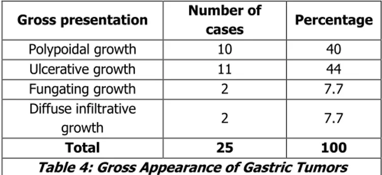
Gross Appearance: The most common gross appearance was ulcerative growth which constituted about 44% of all gross presentations. Followed by Polypoidal growth which constituted the second most common type (Table 4).
Gross presentation Number of
cases Percentage
Polypoidal growth 10 40
Ulcerative growth 11 44
Fungating growth 2 7.7
Diffuse infiltrative
growth 2 7.7
Total 25 100
Table 4: Gross Appearance of Gastric Tumors
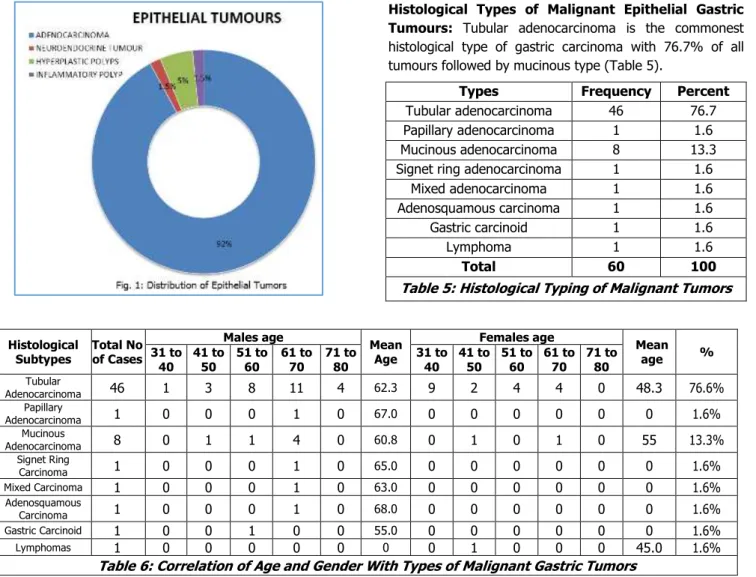
J of Evidence Based Med & Hlthcare, pISSN- 2349-2562, eISSN- 2349-2570/ Vol. 2/Issue 53/Dec. 03, 2015 Page 8748 Histological Types of Malignant Epithelial Gastric Tumours: Tubular adenocarcinoma is the commonest histological type of gastric carcinoma with 76.7% of all tumours followed by mucinous type (Table 5).
Types Frequency Percent
Tubular adenocarcinoma 46 76.7
Papillary adenocarcinoma 1 1.6
Mucinous adenocarcinoma 8 13.3
Signet ring adenocarcinoma 1 1.6
Mixed adenocarcinoma 1 1.6
Adenosquamous carcinoma 1 1.6
Gastric carcinoid 1 1.6
Lymphoma 1 1.6
Total 60 100
Table 5: Histological Typing of Malignant Tumors
Histological Subtypes
Total No of Cases
Males age
Mean Age
Females age
Mean
age %
31 to 40
41 to 50
51 to 60
61 to 70
71 to 80
31 to 40
41 to 50
51 to 60
61 to 70
71 to 80 Tubular
Adenocarcinoma 46 1 3 8 11 4 62.3 9 2 4 4 0 48.3 76.6%
Papillary
Adenocarcinoma 1 0 0 0 1 0 67.0 0 0 0 0 0 0 1.6%
Mucinous
Adenocarcinoma 8 0 1 1 4 0 60.8 0 1 0 1 0 55 13.3%
Signet Ring
Carcinoma 1 0 0 0 1 0 65.0 0 0 0 0 0 0 1.6%
Mixed Carcinoma 1 0 0 0 1 0 63.0 0 0 0 0 0 0 1.6%
Adenosquamous
Carcinoma 1 0 0 0 1 0 68.0 0 0 0 0 0 0 1.6%
Gastric Carcinoid 1 0 0 1 0 0 55.0 0 0 0 0 0 0 1.6%
Lymphomas 1 0 0 0 0 0 0 0 1 0 0 0 45.0 1.6%
Table 6: Correlation of Age and Gender With Types of Malignant Gastric Tumors
Correlation of Age and Gender with Types of Malignant Gastric Tumours: Tubular adenocarcinoma was found to be the most common carcinoma accounting for 75.5% of carcinoma with mean age of presentation of 62.3 years (SD±10.5) in males and 48.3 years (SD±14.0) in females. The difference in mean age presentation in males and females was found to be statistically significant (p value=0.0003 using un-paired t test). Mucinous adenocarcinoma was found to be the second most common with mean age of presentation of 60.8 years (SD±7.2) in males and 52.5 years (SD±14.1) in females.
DISCUSSION: Gastric carcinoma is the fourth commonest cancer in the world with second position in India.2 Gastric
cancer accounts for 11% of all cancers. In our country gastric cancer remains in second position in both male and female after oral cancer in males and cervical cancer in
females.3 Benign tumours such as polyps accounts
for1.01% of all benign lesions. Early detection of the disease due to various modalities has increased early
treatment and good survival rates. In advanced cancer prognosis still remains poor. On reviewing literature, it was found that different studies had many varying factors like number of cases, duration of study and geographic region of study which might be the cause for the differences in frequency when compared with studies of other authors.
Occurrence of Gastric Carcinoma: Occurrence of gastric carcinoma in our study when compared with the study of David et al 2002(10%) was slightly higher.7
Age and Gender Distribution: In this study, the peak incidence was found in the 7th decade of life. This was
consistent with the study conducted by Mohammad et al whereas all other studies showed a peak incidence in 6th
decade of life (Table 7).8,9,10 Also the percentage of gastric
tumours in males were higher than in females which was similar with all the other authors.8,11,12
Authors 00-10 11-20 21-30 31-40 41-50 51-60 61-70 71-80
Present study 0 0 0 10 11 16 24 4
Mohammad et al (2006) 0 0 0 1 3 1 7 3
Prabhakar et al (1981) 0 3 23 40 71 53 37 0
Leena Devi et al (1980) 1 4 20 72 133 149 91 30
J of Evidence Based Med & Hlthcare, pISSN- 2349-2562, eISSN- 2349-2570/ Vol. 2/Issue 53/Dec. 03, 2015 Page 8749 Location of the Tumour: The studies conducted by Rao
et al, 11 Chanda et al13 and Jijo et al.14 Showed similarities
with our study where antrum is the commonest site for tumours of stomach. In contrast, Ming et al15 proved that
the body of the stomach was the commonest site of presentation.
Gross Appearance of the Lesion: In our study gross appearance of the tumour was classified as polypoidal, fungating, ulcerative and infiltrative. Our study is similar to the study of Usha et al where the commonest presentation of the lesion was ulcerative.12
Malignant Gastric Tumours: In our study the commonest malignant tumour was adenocarcinoma of stomach (92%) which is similar with the study conducted by Leena Devi et al and Mohammed et al.8,9
Tubular Adenocarcinoma: Microscopically, tumour showed tubules formation, differentiation depending upon percentage of tubule formation shown by tumour cells. Most adenocarcinoma were moderately differentiated (53.2), followed by poorly differentiated (37.5%) in our study. These finding was comparable and showed a variation with study conducted by Shivaraj et al.16
Papillary Adenocarcinoma: A single case of papillary adenocarcinoma was seen in stomach in a 67 years old male patient. Similar observation were quoted by Leena et al.8
Mucinous Adenocarcinoma: This type of adenocarcinoma was found to be the second most common variant comprising of 13.3% in our study. Mucinous adenocarcinoma was found to be the second most common with mean age of presentation of 60.8 years (SD±7.2) in males and 52.5 years (SD±14.1) in females. This is different from the other study conducted in Kerala.8
SignetRingCarcinoma: In this study one case of signet ring carcinoma was reported which comprises of 1.6% of all adenocarcinoma. This observation was different from other study conducted by Leena Devi et al which showed higher percentage of signet ring carcinoma than the present study.8
Adenosquamous Carcinoma: A single case of this rare variant accounting to 1.6% of all adenocarcinoma was seen in a 68 year old male patient. Similar observation was seen in Bengal in the study conducted by Indranil et al.17
MixedCarcinoma: In the present study, a single case of mixed carcinoma was reported accounting for 1.6% of all tumours. This a new variant included in the new WHO (2010) classification. These carcinomas shows a mixture of discrete glandular and signet ring components.
Other Tumors: Neuroendocrine tumours and Lymphoma accounts for 2-3% of malignant tumours. Our study
showed one case of neuroendocrine and one case of lymphoma which was similar with the study conducted by Leena Devi et al.8
CONCLUSION: Malignant gastric tumours were more
common than benign tumours. Adenocarcinoma–tubular
type was the most common malignancy, with pyloric antrum being the commonest site. Gastric carcinomas were seen in early age group in females than in males. Gastric carcinomas showed male preponderance with M: F ratio 1.6:1. However, mean age of presentation of gastric carcinomas in females (46.6 years) was earlier as compared to males (62.2 years), which was statistically significant (p value=0.0025). Tubular adenocarcinoma was the commonest tumour found amongst all malignant gastric tumour, accounting for 76.7% with mean age of presentation of 62.3 years (SD±10.5) in males and 48.3 years (SD±14.0) in females. The difference in mean age presentation in males and females was found to be statistically significant (p value=0.0003 using un-paired t test). Endoscopy is the most useful modality for screening gastric carcinoma especially in females with positive family history and age <40 years.
REFERENCES:
1. Burt RW. Gastric fundic gland polyps.
Gastroenterology. 2003; 125: 1462–69. [pubmed] 2. Kumar V, Abbas K, Fausto N, Aster C. Robbins and
Cotran Pathological Basis of Disease. 2010; 8: 763-831.
3. Dikshit R, Gupta PC, Ramasundarahettige C,
Gajalakshmi V, Aleksandrowicz L, Badwe R, Kumar R, Roy S, Suraweera W, Bray F, Mallath M, Singh PK, Sinha DN, Shet AS, Gelband H, Jha P; Cancer Mortality in India: A Nationally representative survey 2010.
4. Rao DN, Ganesh B, Dinshaw KA, Mohandas KM. A
case-control study of stomach cancer in Mumbai, India. Int J Cancer. 2002; 99: 727–31.
5. Parkin DM, Bray F, Ferlay J, Pisani P. Global cancer statistics, 2002. CA Cancer J Clin. 2005; 55: 74–108. 6. Wingo PA et al.: Long–term trends in cancer mortality
in the United states, 1930-1998. Cancer 97: 3133, 2003.
7. David M. Roder. The epidemiology of gastric cancer.
International and Japanese Gastric Cancer
Associations 2002; 5(1): 5-11.
8. Leena Devi KK, Suvarna N. Patterns of
Gastrointestinal tumors in North Kerala. Indian Journal of Cancer 1980; 17: 159-163.
9. Mohammad A, Makaju R. Retrospective
J of Evidence Based Med & Hlthcare, pISSN- 2349-2562, eISSN- 2349-2570/ Vol. 2/Issue 53/Dec. 03, 2015 Page 8750
10.Prabhakar BR, Prabhakar H, Tung BS.
Gastro-intestinal Malignant tumors in Amristsar (Punjab). Indian Journal of Surgery 1981; 343-345.
11.Koteshwar K Rao. Profile of Gastric carcinoma as seen in a rural institute. Indian Journal of Cancer 1983; 20: 1-4.
12.Usha SD, Shukla HS, Gupta S, Aryya NC, Khanna S et
al. A clinicopathological study of carcinoma stomach. Indian J pathol Microbiol 1988; 31: 266-71.
13.Nassima Chanda, Khan AR. Histopathology of Gastric Cancer in Kashmir. A five year Retrospective Analysis. JK Science 2007; 9: 21-24.
14.Jijo Velliyapillil cherian, Ramalingam Sivaraman et al. Stomach carcinoma in Indian subcontinent. The Saudi Journal of Gastroenterology 2007; 13(3): 114-7.
15.Ming SC, Hirota T. malignant epithelial tumor of the stomach. Pathology of gastrointestinal tract. 2nd ed. Williams & Wilkins 1998; 607-47.
16.Shiva Raj KC, GL Amatya, A Lakhey, S Basnet, G Aryal. Incidence of gastric cancer, its subtypes, and correlation with Helicobacter Pylori. Journal of Pathology of Nepal 2013; Vol. 3, No. 1, Issue 5, 403-407.
17.Indranil Chakrabarti, Anuradha De A, Kaushik