of Pandemic (H1N1) 2009 Influenza Virus in Japan
between 2009 and 2010
Mayo Yasugi1,2, Shota Nakamura3,4, Tomo Daidoji1,8, Norihito Kawashita3,5, Ririn Ramadhany1,2, Cheng-Song Yang1,2, Teruo Yasunaga3, Tetsuya Iida1,4, Toshihiro Horii1,4,6, Kazuyoshi Ikuta2, Kazuo Takahashi7, Takaaki Nakaya1,4,8*
1International Research Center for Infectious Diseases, Research Institute for Microbial Diseases (RIMD), Osaka University, Suita, Osaka, Japan,2Department of Virology, RIMD, Osaka University, Suita, Osaka, Japan,3Department of Genome Informatics, RIMD, Osaka University, Suita, Osaka, Japan,4Department of Infection Metagenomics, RIMD, Osaka University, Suita, Osaka, Japan,5Department of Environmental Pharmacometrics, Graduate School of Pharmaceutical Sciences, Osaka University, Suita, Osaka, Japan,6Department of Molecular Protozoology, RIMD, Osaka University, Suita, Osaka, Japan,7Osaka Prefectural Institute of Public Health, Higashinari-ku, Osaka, Japan,8Department of Infectious Diseases, Kyoto Prefectural University of Medicine, Kamigyo-ku, Kyoto, Japan
Abstract
Background:In April 2009, a novel swine-derived influenza A virus (H1N1pdm) emerged and rapidly spread around the world, including Japan. It has been suggested that the virus can bind to both 2,3- and 2,6-linked sialic acid receptors in infected mammals, in contrast to contemporary seasonal H1N1 viruses, which have a predilection for 2,6-linked sialic acid.
Methods/Results:To elucidate the existence and transmissibility ofa2,3 sialic acid-specific viruses in H1N1pdm, amino acid
substitutions within viral hemagglutinin molecules were investigated, especially D187E, D222G, and Q223R, which are related to a shift from human to avian receptor specificity. Samples from individuals infected during the first and second waves of the outbreak in Japan were examined using a high-throughput sequencing approach. In May 2009, three specimens from mild cases showed D222G and/or Q223R substitutions in a minor subpopulation of viruses infecting these individuals. However, the substitutions almost disappeared in the samples from five mild cases in December 2010. The D187E substitution was not widespread in specimens, even in May 2009.
Conclusions:These results suggest thata2,3 sialic acid-specific viruses, including G222 and R223, existed in humans as a
minor population in the early phase of the pandemic, and that D222 and Q223 became more dominant through human-to-human transmission during the first and second waves of the epidemic. These results are consistent with the low substitution rates identified in seasonal H1N1 viruses in 2008.
Citation:Yasugi M, Nakamura S, Daidoji T, Kawashita N, Ramadhany R, et al. (2012) Frequency of D222G and Q223R Hemagglutinin Mutants of Pandemic (H1N1) 2009 Influenza Virus in Japan between 2009 and 2010. PLoS ONE 7(2): e30946. doi:10.1371/journal.pone.0030946
Editor:Patrick C. Y. Woo, The University of Hong Kong, China
ReceivedSeptember 2, 2011;AcceptedDecember 27, 2011;PublishedFebruary 17, 2012
Copyright:ß2012 Yasugi et al. This is an open-access article distributed under the terms of the Creative Commons Attribution License, which permits unrestricted use, distribution, and reproduction in any medium, provided the original author and source are credited.
Funding:This work was supported in part by a Grant-in-Aid for Scientific Research from the Ministry of Education, Science, Sports, Culture, and Technology (MEXT) to TN; and a Grant-in-Aid for Young Scientists from the Japan Society for the Promotion of Science (JSPS) to TD and MY. No additional external funding received for this study. The funders had no role in study design, data collection and analysis, decision to publish, or preparation of the manuscript.
Competing Interests:The authors have declared that no competing interests exist.
* E-mail: tnakaya@biken.osaka-u.ac.jp
Introduction
In April 2009, a novel swine-derived influenza A virus (H1N1pdm) emerged and spread rapidly around the world [1,2], causing the World Health Organization to declare a pandemic in June. To date, most countries have confirmed infections by the new virus.
Since the first appearance of H1N1pdm, one particular amino acid substitution {aspartic acid to glycine at position 222 (D222G)} within the hemagglutinin (HA) molecule has appeared sporadically [1,3,4,5]. The D222G substitution is known to cause a shift from
a2,6-sialic acid receptor specificity to mixeda2,3/a2,6-sialic acid
receptor specificity [6,7]. It is noteworthy that G222 is highly conserved among avian viruses [8]. Previously,a2,3-specific avian
viruses have been isolated from patients during the initial phases of the pandemics of 1957 and 1968, and avian HA in humans has been
shown to be selected for increased affinity for thea2,6 receptor [8].
Also, the G222 substitution was present in the Spanish Flu out-break of 1918 [9]; however, the existence and transmissibility of H1N1pdma2,3 sialic acid-specific viruses remain unclear.
To identify whethera2,3 sialic acid-specific viruses, which
re-plicate well in swine, were spread during the early phase of the H1N1pdm pandemic and whethera2,3 sialic acid-specific viruses
are easily transmitted, the nucleotide sequences of the HA receptor binding site of H1N1pdm in clinical specimens were determined in this study.
for receptor binding, which cause a shift froma2,6 toa2,3 sialic
acid receptor specificity [8,10]. Changes in HA amino acid diver-sity were identified in individual cases using a high-throughput sequencer.
Results
Sequencing analysis of H1N1pdm HA receptor binding site (RBS) from nasal specimens in May 2009
Three nasal swabs (#1–#3) from mild H1N1pdm cases were obtained in May 2009 in Osaka, Japan. Total RNA was extracted from each sample and the receptor binding site (RBS) within HA was successfully amplified using One Step RT-PCR. The PCR products (369 bp) were cloned into pGEM-T easy vector and analyzed by conventional Sanger technology. More than 100 clones in each specimen were sequenced, and the deduced amino acid sequences were analyzed (Figures 1, S1, S2, and S3). The substitution rate of the deduced amino acids was 0.29–0.41% (average: 0.34%) in the three samples, and mutations commonly observed in more than two patients are listed in Table 1. Four substitutions, K119N, N125D, D222G, and Q223R, were detected in more than 2.0% of the samples (Table 1). Among these substitutions, D222G and Q223R had already been shown to be critical for receptor binding and to cause a shift froma2,6 to a2,3 sialic acid receptor specificity [10]. In addition, the D187E
substitution could be critical for the human-to-avian–type receptor switch [9]. A homology modeling and docking study also showed that D222G and Q223R mutants had reduced affinity for thea2,6
sialic acid-linked receptor (Figure S4). Thus, we focused on these five mutations and tried to measure the mutation ratio of un-isolated viruses in upper respiratory airways. For a larger scale analysis, the PCR products were examined via a high-throughput sequencing approach using a 454/Roche GS-Junior sequencer.
Deep sequencing of more than 3,000 clones (reads) per specimen showed high rates of amino acid diversity, with the K119N, N125D, D222G, and Q223R substitutions in specimens #1–#3 (Table 2). The results obtained by deep sequencing were consistent with those obtained using Sanger sequencing (Table 1). The D187E substitution was also identified in two specimens through deep sequencing analysis; however, the substitution rate was only slightly increased in one of these (by 0.72% in specimen #2) (Table 2). These results suggest thata2,3 sialic acid-specific
viruses, including G222 and/or R223, were present as a minor population in the upper respiratory tract even in mild cases.
Multiple passages of H1N1pdm viruses in embryonated chicken eggs
To elucidate how the genomic diversity (and population) of the RBS, including the D222G and/or Q223R substitutions, might change, we inoculated these specimens into embryonated chicken eggs and serially passaged them five or six times in eggs. Hemagglutination was not detected in any samples of the first passage (P1) but was detected in the third or fourth passage and showed 25 to 26 hemagglutination titers on the fifth or sixth passage (P5 or P6) (Figure S5). PCR products amplifying the same region as in Figure 1 were prepared from early- and late-passaged viruses (P1 and P5/P6), as well as MDCK-passaged viruses (except #3). The amplicons, approxi-mately 200–500 reads, were examined using deep sequencing.
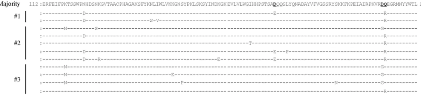
Figure 1. Alignment of the amino acid sequences including D187E, D222G, and Q223R mutants within the receptor binding site of H1N1pdm hemagglutinin.E187, G222, and R223 variants obtained from three clinical specimens (#1,#2, and#3) from the first wave of the outbreak. Three clinical nasal swabs were each subjected to RNA extraction, RT-PCR, and TA cloning. More than one hundred clones per specimen were sequenced using conventional Sanger technology. Positions 187, 222, and 223 are shown in bold and are underlined.
doi:10.1371/journal.pone.0030946.g001
Table 1.Deduced amino acid mutations commonly observed in more than two patients in May 2009.
HA position Wild type Mutant Mutant Numbera Ratio (%)
119 K N 8 2.6
125 N D 9 2.9
129 N S 4 1.3
145 Y H 2 0.6
147 N D 2 0.6
154 K E 4 1.3
157 S P 3 1.0
160 K E 2 0.6
179 I T 3 1.0
182 P Q 5 1.6
187 D E 5 1.6
191 L P 4 1.3
204 S P 3 1.0
210 F L 2 0.6
214 I V 2 0.6
222 D G 8 2.6
223 Q R 10 3.2
aNumber of each mutant clone out of a total of 313 sequenced clones (#1–#3
specimens).
D222G and/or Q223R substitutions were detected at even higher rates in the early passaged (P1) viruses (Table 3) than in the parental viruses (Table 1). More than half (98.6%, 95.0%, and 59.7% in #1, #2, and #3, respectively) possessed either D222G or Q223R substitutions (Table 3). K119N, N125D and D187E mutations were also detected at higher ratios in the P5 or P6 passages than in the P1 passage, suggesting that these mutations were being selected for effective growth in avian cells. We also detected these substitutions at a lower level in the MDCK-passaged viruses compared with the egg-MDCK-passaged viruses (Table 3). In contrast to the HA gene, no amino acid substitutions were de-tected in the RNA polymerase B2 (PB2), RNA polymerase B1 (PB1 and PB1-F2), RNA polymerase A (PA), neuraminidase (NA), matrix proteins (M1 and M2) and non-structural proteins (NS1 and NS2) of the P5/P6 viruses compared with the nucleotide sequence of the original H1N1pdm derived from the#2 clinical specimen (data not shown).
P5/P6 in#1,#2, and#3 specimens reduced hemagglutination titers when RBCs were treated with a2,3 sialidase whereas the
titers did not change in MDCK-passaged viruses (Table 4). These results suggested that egg-passaged viruses, including G222 and/or R223, were a2,3 sialic acid receptor tropic and that
MDCK-passaged viruses including D222 and Q223 had a2,6 sialic acid
receptor specificity. From these results, we can extrapolate that the major populations in the original specimens #1–#3 had a2,6
sialic acid receptor tropism. These results are coincident with the homology modeling and docking of D222G and Q223R mutants (Figure S4).
Deep sequencing analysis of H1N1pdm HA D222G or Q223R mutants from the second wave of the outbreak
To elucidate the transmissibility of the G222 and/or R223 viruses in humans, five nasal swabs (#4–#8) obtained from individuals with a mild case of H1N1pdm in December 2010 were examined by deep sequencing analysis. The same region of HA RBS (369 bp) as shown in Figure 1 was amplified by One Step RT-PCR. The results showed
that, by December 2010, both the G222 and R223 variants had almost disappeared (0.01–0.07% and 0.47–0.63%, respectively) (Table 2). The K119N mutation frequency also shifted to an un-detectable level except for one specimen (#8). By contrast, the N125D mutation could still be detected (0.66–2.15%; average, 1.42%) (Table 2). Taken together, these results suggest that the H1N1pdm D222 and Q223 variants are competent for human-to-human transmission but that thea2,3 sialic acid-specific G222 and
R223 mutations resulted in low transmissibility. We also examined the substitution rates in seasonal H1N1 viruses using five nasal swabs (#9–#13) from patients diagnosed in 2008 (Table 2). The results showed low substitution rates for D222G and Q223R (0.01–0.11% and 0.1–0.41%, respectively), consistent with the results of the second epidemic, in H1N1pdm.
Discussion
This study used a high-throughput sequencing approach to analyze more than 2,000 clones from each of eight H1N1pdm samples obtained in 2009–2010. The results showed thata2,3 sialic
acid-specific viruses containing D222G and/or Q223R substitu-tions within the HA molecule were present in the upper respiratory tract as a minor population in patients with mild H1N1pdm infections in the early phase (May 2009) of the pandemic in Japan. However, these substitutions nearly disappeared in the samples from five mild cases in Dec 2010, suggesting that the D222G and/or Table 2.Frequency distribution of N119, D125, E187, G222,
and R223 variants within the HA receptor binding site of viruses obtained from clinical specimens infected with influenza A viruses.
Speci-men
Total read
K119N (%)
N125D (%)
D187E (%)
D222G (%)
Q223R (%)
H1N1pdm #1 3,308 8.59 4.99 0.15 0.74 4.64
1st wave #2 29,607 4.61 1.01 0.72 21.49 2.39
(2009) #3 15,514 3.45 4.88 0 3.16 3.62
H1N1pdm #4 15,485 0.00 1.07 0 0.04 0.57
2nd wave #5 25,429 0.00 1.33 0 0.02 0.53
(2010) #6 14,183 0.02 2.15 0 0.07 0.47
#7 18,046 0.01 1.87 0 0.01 0.63
#8 11,179 3.10 0.66 0 0.05 0.48
H1N1 #9 2,014 0.00 0.00 0 0.1 0.1
Seasonal #10 7,170 0.00 0.00 0 0.04 0.15
(2008) #11 3,652 0.00 0.00 0 0.08 0.16
#12 3,733 0.00 0.00 0 0.11 0.13
#13 7,268 0.00 0.00 0 0.01 0.41
Plasmid (control) 12,428 0.00 0.00 0 0 0.38
doi:10.1371/journal.pone.0030946.t002
Table 3.Frequency of amino acid mutations in the hemagglutinin receptor binding site in egg-passaged or MDCK-passaged samples.
Specimen Isolation Total
Read Mutant Ratio (%)
K119N N125D D187E D222G Q223R
#1 P1 495 0.6 0.2 0 3 95.6
P6 330 0 96.7 12.1 7.9 95.8
MDCK 481 0.2 0.8 1.5 1.9 2.7
#2 P1 340 0.6 12.1 0.3 24.4 70.6
P6 202 60.4 33.7 25.7 60.4 33.2
MDCK 515 0 0 0 0.4 0
#3 P1 300 0.7 0 0 59 0.7
P5 193 49.2 36.3 0 85 0.5
doi:10.1371/journal.pone.0030946.t003
Table 4.Hemagglutination assay using chicken, guinea pig, and horse erythrocytes with or withouta2,3 sialidase
treatment.
Chicken Guinea pig Horse
a2,3 sialidase 2 + 2 + 2 +
#1-P6 512 8 1,024 32 128 16
#2-P6 512 256 512 1,024 64 4.
#3-P5 256 128 512 512 64 4.
#1-MDCK 256 256 512 1,024 128 128
#2-MDCK 32 16 256 256 8 8
Q223R mutants showed low rates of human-to-human transmis-sion. Thus, the newly emergent influenza A viruses may have been dual specific but not exclusivelya2,6-sialic acid-specific during the
early phase of the pandemic and adapted during multiple cycles of human-to-human transmission. These results are consistent with previous reports showing that, during previous pandemics, the receptor binding properties of avian influenza viruses changed after introduction into mammals [8]. In particular, positions 187 and 222 within the HA of H1 viruses and position 223 in H2 and H3 strains are critical for receptor binding specificity [8]. In the present study, the D187E substitution was not striking, suggesting that E187 had already disappeared in humans, or the original swine lineages, by May 2009. The N125D mutation, however, was detected (0.66– 2.15%), even in the second wave of the outbreak (Table 2). However, as far as we know, there is no report indicating that this mutation affects receptor-binding affinity and specificity. The K119N mutation was also significantly detected in one specimen (3.1%) from a patient who had similar clinical symptoms as those of other patients during the second wave of the outbreak (Table 2). Neither mutation was detected in specimens of seasonal H1N1 in 2008, suggesting that further prospective study, as well as virological study, is required to understand the functions of these mutations.
HA residue 222 plays a critical role in the binding affinity of the galactose moiety of sialosaccharides for the RBS. The D222G mutation would result in a loss of interaction between galactose and K219 (Figure S4) because of the loss of charge and the side-chain. In turn, this would open up a cavity on the side of the RBS to accommodate the a2,3-linked receptor [11,12]. Residue 223
is also important for the binding affinity of galactose and the ter-minal sialic acid of the receptor. The emergence of a positively charged and bulky Arginine(R) instead of Glutamine(Q) at this position would disturb the interaction between K219 and ga-lactose, leading to decreased virus affinity for thea2,6-linked
re-ceptor (Figure S4) [13,14]. Also, the homology modeling results showed that the double mutations, D187E and Q223R, would decrease virus affinity for thea2,6-linked receptor because the
salt-bridge between E187 and R223 would lead to narrowing of the receptor binding pocket (data not shown).
In this study, we performed deep amplicon sequencing us-ing Roche/454 GS-Junior technology. We compared the results obtained by using deep sequencer with those obtained by the con-ventional Sanger method to evaluate this newly developed te-chnology. Sequencing analysis using next-generation technology is necessary to reveal H1N1pdm genetic diversity in detail; our preliminary sequencing analysis using a conventional cloning ap-proach failed to detect the D222G mutation in specimen #1 (Figure 1 and Table 2). The frequency of mutations such as D187E in specimens#1 and#2 and D222G in specimen #2 was not the same between GS-Junior technology and conventional Sanger sequencing (Figures 1, S1, S2, and S3, Tables 1 and 2). This discrepancy might be due to the different number of clones sequenced in each method. The proportion of HA sequences observed with D222G and Q223R in the#1 and#2 P6 and in the #3 P5 PCR products (Table 3) was consistent with results obtained by direct sequencing analysis using the ABI Sanger sequencer (Figure S6), suggesting that high-throughput amplicon sequencing analysis was highly quantitative.
The present study did not confirm whether viruses with the D222G and/or Q223R substitutions actually replicated in hu-mans; however, the D222G and Q223R mutant viruses formed the dominant population when specimens were inoculated into embryonic chicken eggs (Table 3), indicating that the G222 and R223 mutants may be able to replicate in humans.
Several reports have shown that the D222G substitution was associated with severe, and sometimes fatal, cases of H1N1pdm [10,15,16,17,18]. It has also been reported that two different amino acids, D and E, may be polymorphic variants at position 222 [19], and that amino acids, G or N, would be present in potentially more pathogenic mutants or circulating variants [20]. However, the re-levance of the mutation at position 222 to H1N1pdm pathogenesis remains unclear. In this study, D222E, D222N, and D222V variants were not detected, even when deep sequencing was per-formed (Table S1). D222G mutants were detected as a minor population even in mild cases; thus, every patient may have been exposed to D222G mutant viruses. Minoritya2,3 sialic acid-tropic
G222 mutants could be amplified ina2,3 sialic acid expressing cells
in the lower respiratory tract of certain populations, which would result in severe to fatal respiratory illness in these populations. No conclusions can be drawn regarding the relationship between the D222G substitution and pathogenesis from the present study. Therefore, further study is required to determine whether G222 viruses were ever present as a major population in the lower respiratory tract of patients with severe or fatal pneumonia due to H1N1pdm infection.
Materials and Methods
Clinical samples
Three and five individual H1N1pdm-positive nasal swabs were provided in May 2009 and December 2010, respectively, in Osaka, Japan. All of the patients examined in 2009 had fever, sore throat, and cough, but were not hospitalized. They were administered oseltamivir after nasal swab-sampling. The patients examined in 2010 had upper respiratory symptoms but were not hospitalized. As a control, five specimens diagnosed with seasonal H1N1 in 2008 in Osaka were investigated. The analyzed samples were unlinked and anonymous in the Osaka Prefectural Institute of Public Health. This study was approved by the ethical review committees of the Osaka Prefectural Institute of Public Health and the Research Institute for Microbial Diseases (RIMD), Osaka University. The Both ethical committees specifically waived the need for consent.
RNA extraction
Each nasal swab was collected in Hanks solution and was centrifuged at 20,000 g for 10 min. The supernatants were suspended in TRIzol LS reagent (Life Technologies/Invitrogen, Carlsbad, USA) for 60 min. Total RNA was extracted using a PureLinkTM RNA Mini Kit (Life Technologies/Invitrogen) according to the manufacturer’s instructions. Contaminating DNA was eliminated with DNAase I (Life Technologies/Invitrogen).
One-step reverse transcription-PCR (RT-PCR)
Total RNA was subjected to One-step RT-PCR (SuperScript III/Platinum Taq HiFi One-step RT-PCR Kit; (Life Technolo-gies/Invitrogen) to detect the receptor binding site in the HA molecule. The primers used were as follows: forward primer, 59-416TTGAAAGGTTTGAGATATTC435-39; reverse primer, 59-784CTAGTGTCCAGTAATAGTTC765-39.
Plasmid cloning
Virus isolation and serial passage in embryonated eggs and Madin-Darby canine kidney (MDCK) cells
Clinical specimens were injected into 9-day-old embryonated chicken eggs for virus isolation [21]. After incubation at 37uC for 72 h, the allantoic fluids were collected and filtered (Passage 0: P0). The samples were then diluted 1–100 fold in phosphate buffered saline (PBS) and passaged either six (samples derived from#1 and #2) or five (sample derived from#3) times in embryonated chi-cken eggs. Identical specimens derived from#1 and#2 were also inoculated into MDCK cells for virus isolation [22] and the amplified viruses were serially passaged two or three times in MDCK cells.
Hemagglutination titration
Viral samples were serially diluted with PBS and added at a concentration of 0.5% to chicken red blood cells. After incubation at room temperature for 30 min, hemagglutination was observed.
High-throughput pyrosequencing and data analysis PCR products were purified using the QIAquickHPCR purifica-tion kit (Qiagen GmbH, Hilden, Germany) and were ligated with RL MID adaptors (Roche Diagnostics GmbH, Mannheim, Germany). The PCR products were further purified with Agencourt AMPure XP beads (Beckman Coulter, Brea, USA). The quality and quantity of the library was assessed using an Agilent 2100 Bioanalyzer (Agilent Technologies, Santa Clara, USA) and TBS380 mini fluorometer (A. Daigger & Company Inc., Vernon Hills, USA), respectively. After amplifying the samples by emulsion PCR, high-throughput pyro-sequencing was performed using a GS Junior (Roche Diagnostics GmbH). Data analysis was performed on each sequence read using computational tools as described previously [23,24,25].
a2,3 sialidase treatment
100ml of 10% chicken, guinea pig, or horse red blood cells were treated with 10 U ofa2,3 sialidase (New England Biolabs,
Ipswich, MA) at 37uC for 18 h and were washed with PBS. After the erythrocyte suspension was diluted to 0.5% (chicken) or 0.75% (guinea pig and horse), the hemagglutination titer was measured as described above.
Homology modeling and docking
The crystal structure of HA of influenza virus, A/Brevig Mission/1/1918 (H1N1) (Protein Data Bank ID code 2WRG), was used as a template for homology modeling by using a Mo-lecular Operating Environment. SAa2,6-linked structural analogs
were used as inputs for docking with the model HA structure.
Supporting Information
Figure S1 Alignment of the amino acid sequences within the receptor binding site of variants other than E187, G222, and R223. Variants other than E187, G222, and R223 (Figure 1) obtained from clinical specimen#1 from the first wave of the outbreak.
(TIFF)
Figure S2 Alignment of the amino acid sequences within the receptor binding site of variants other than E187, G222, and R223.Variants other than E187, G222, and R223 (Figure 1) obtained from clinical specimen#2 from the first wave of the outbreak.
(TIFF)
Figure S3 Alignment of the amino acid sequences within the receptor binding site of variants other than E187, G222, and R223.Variants other than E187, G222, and R223 (Figure 1) obtained from clinical specimen#3 from the first wave of the outbreak.
(TIFF)
Figure S4 Stereo views of the homology-based structur-al model for binding of sistructur-alosaccharides to the RBS of H1N1pdm hemagglutinin.The structural complex of the RBS of H1N1pdm HA with human receptor (orange) was constructed using the co-crystal structure of A/Brevig Mission/1/1918 (PDB ID: 2WRG) as a template. (A) HA of wild type (pink) overlapped with the HA of D222G mutants (green). Position 222 is shown in deep colors. Putative interactions salt-bridge between the sugar and the RBS are shown as black dashed lines. (B) HA of wild type (pink) overlapped with the HA of Q223R mutants (blue). Q223 and R223 are shown in deep pink and yellow, respectively. (TIFF)
Figure S5 Kinetics of hemagglutination in viruses isolated from egg-passaged samples (#1,#2, and#3). Clinical specimens were injected into 9-day-old embryonated chicken eggs and incubated for 72 h. Allantoic fluid was collected (passage 0) and serially passaged in chicken eggs (passage 5–6). The viral growth of each sample was measured by hemaggluti-nation assay using (0.5%) chicken red blood cells.
(TIFF)
Figure S6 Direct sequencing analysis of PCR products (positions 221–227 aa in HA-RBS) amplified from egg-passaged P6 (#1 and#2) and P5 (#3) viruses using a conventional ABI sequencer.The arrows indicate nucleotide substitutions. WT indicates wild type.
(TIFF)
Table S1 Frequency distribution of D222G, D222N, D222V, and D222E variants within the hemagglutinin receptor binding site of viruses obtained from clinical specimens (#1–#3). (TIFF)
Acknowledgments
We are grateful to R. Koketsu, Y. Watanabe, N. Higashi, S. Morikawa, S. Hiroi, T. Kase, M. Tujikawa, M. Yunoki, S. Yoshiki, and A. Yoshioka for technical support and to Y. Okuno for helpful discussions.
Author Contributions
and designed the experiments: MY SN TN. Performed the experiments: MY C-SY TD RR TN. Analyzed the data: MY SN NK KI TN. Contributed reagents/materials/analysis tools: TY TI TH KT. Wrote the paper: MY TN.
References
1. Miller MA, Viboud C, Balinska M, Simonsen L (2009) The signature features of influenza pandemics–implications for policy. N Engl J Med 360: 2595–2598. 2. Neumann G, Noda T, Kawaoka Y (2009) Emergence and pandemic potential of
swine-origin H1N1 influenza virus. Nature 459: 931–939.
3. Ikonen N, Haanpaa M, Ronkko E, Lyytikainen O, Kuusi M, et al. (2010) Genetic diversity of the 2009 pandemic influenza A(H1N1) viruses in Finland. PLoS One 5: e13329.
4. Kuroda M, Katano H, Nakajima N, Tobiume M, Ainai A, et al. (2010) Characterization of quasispecies of pandemic 2009 influenza A virus (A/H1N1/ 2009) by de novo sequencing using a next-generation DNA sequencer. PLoS One 5: e10256.
6. Chutinimitkul S, Herfst S, Steel J, Lowen AC, Ye J, et al. (2010) Virulence-associated substitution D222G in the hemagglutinin of 2009 pandemic influenza A(H1N1) virus affects receptor binding. J Virol 84: 11802–11813.
7. Liu Y, Childs RA, Matrosovich T, Wharton S, Palma AS, et al. (2010) Altered receptor specificity and cell tropism of D222G hemagglutinin mutants isolated from fatal cases of pandemic A(H1N1) 2009 influenza virus. J Virol 84: 12069–12074.
8. Matrosovich M, Tuzikov A, Bovin N, Gambaryan A, Klimov A, et al. (2000) Early alterations of the receptor-binding properties of H1, H2, and H3 avian influenza virus hemagglutinins after their introduction into mammals. J Virol 74: 8502–8512.
9. Tumpey TM, Maines TR, Van Hoeven N, Glaser L, Solorzano A, et al. (2007) A two-amino acid change in the hemagglutinin of the 1918 influenza virus abolishes transmission. Science 315: 655–659.
10. Chen Z, Wang W, Zhou H, Suguitan AL, Jr., Shambaugh C, et al. (2010) Generation of live attenuated novel influenza virus A/California/7/09 (H1N1) vaccines with high yield in embryonated chicken eggs. J Virol 84: 44–51. 11. Nunthaboot N, Rungrotmongkol T, Malaisree M, Kaiyawet N, Decha P, et al.
(2010) Evolution of human receptor binding affinity of H1N1 hemagglutinins from 1918 to 2009 pandemic influenza A virus. J Chem Inf Model 50: 1410–1417.
12. Stevens J, Blixt O, Glaser L, Taubenberger JK, Palese P, et al. (2006) Glycan microarray analysis of the hemagglutinins from modern and pandemic influenza viruses reveals different receptor specificities. J Mol Biol 355: 1143–1155. 13. Mochalova L, Gambaryan A, Romanova J, Tuzikov A, Chinarev A, et al. (2003)
Receptor-binding properties of modern human influenza viruses primarily isolated in Vero and MDCK cells and chicken embryonated eggs. Virology 313: 473–480.
14. Viswanathan K, Chandrasekaran A, Srinivasan A, Raman R, Sasisekharan V, et al. (2010) Glycans as receptors for influenza pathogenesis. Glycoconj J 27: 561–570.
15. Chan PK, Lee N, Joynt GM, Choi KW, Cheung JL, et al. (2011) Clinical and virological course of infection with haemagglutinin D222G mutant strain of 2009 pandemic influenza A (H1N1) virus. J Clin Virol 50: 320–324. 16. Chen H, Wen X, To KK, Wang P, Tse H, et al. (2010) Quasispecies of the
D225G substitution in the hemagglutinin of pandemic influenza A(H1N1) 2009 virus from patients with severe disease in Hong Kong, China. J Infect Dis 201: 1517–1521.
17. Mak GC, Au KW, Tai LS, Chuang KC, Cheng KC, et al. (2010) Association of D222G substitution in haemagglutinin of 2009 pandemic influenza A (H1N1) with severe disease. Euro Surveill 15: pii19534.
18. Melidou A, Gioula G, Exindari M, Chatzidimitriou D, Diza E, et al. (2010) Molecular and phylogenetic analysis of the haemagglutinin gene of pandemic influenza H1N1 2009 viruses associated with severe and fatal infections. Virus Res 151: 192–199.
19. Ledesma J, Pozo F, Ruiz MP, Navarro JM, Pineiro L, et al. (2011) Substitutions in position 222 of haemagglutinin of pandemic influenza A (H1N1) 2009 viruses in Spain. J Clin Virol 51: 75–78.
20. Valli MB, Selleri M, Meschi S, Zaccaro P, Vincenti D, et al. (2011) Hemagglutinin 222 variants in pandemic (H1N1) 2009 virus. Emerg Infect Dis 17: 749–751. 21. Daidoji T, Koma T, Du A, Yang CS, Ueda M, et al. (2008) H5N1 avian
influenza virus induces apoptotic cell death in mammalian airway epithelial cells. J Virol 82: 11294–11307.
22. Ueda M, Daidoji T, Du A, Yang CS, Ibrahim MS, et al. (2010) Highly pathogenic H5N1 avian influenza virus induces extracellular Ca2+ influx, leading to apoptosis in avian cells. J Virol 84: 3068–3078.
23. Nakamura S, Maeda N, Miron IM, Yoh M, Izutsu K, et al. (2008) Metagenomic diagnosis of bacterial infections. Emerg Infect Dis 14: 1784–1786.
24. Nakamura S, Nakaya T, Iida T (2011) Metagenomic analysis of bacterial infections by means of high-throughput DNA sequencing. Exp Biol Med (Maywood) 236: 968–971.