Inflammatory Markers in Patients with Stable Coronary
Artery Disease
Jan-Willem E. M. Sels1,3*, Ellen H. A. M. Elsenberg2, Imo E. Hoefer2, Anton Jan van Zonneveld4, Johan Kuiper5, J. Wouter Jukema6, Nico H. J. Pijls1,3, Gerard Pasterkamp2
1Department of Cardiology, Catharina Hospital Eindhoven, Eindhoven, The Netherlands,2Laboratory of Experimental Cardiology, University Medical Center Utrecht, Utrecht, The Netherlands,3Department of Biomedical Engineering, Eindhoven University of Technology, Eindhoven, The Netherlands,4Department of Nephrology, Leiden University Medical Center, Leiden, The Netherlands,5Division of Biopharmaceuticals, Leiden University, Leiden, The Netherlands,6Department of Cardiology, Leiden University Medical Center, Leiden, The Netherlands
Abstract
Background:Atherosclerosis is an inflammatory condition and increased blood levels of inflammatory biomarkers have been observed in acute coronary syndromes. In addition, high expression of inflammatory markers is associated with worse prognosis of coronary artery disease. The presence and extent of inducible ischemia in patients with stable angina has previously been shown to have strong prognostic value. We hypothesized that evidence of inducible myocardial ischemia by local lesions, as measured by fractional flow reserve (FFR), is associated with increased levels of blood based inflammatory biomarkers.
Methods:Whole blood samples of 89 patients with stable angina pectoris and 16 healthy controls were analyzed. The patients with stable angina pectoris underwent coronary angiography and FFR of all coronary lesions. We analyzed plasma levels of cytokines IL-6, IL-8 and TNF-aand membrane expression of Toll-like receptor 2 and 4, CD11b, CD62L and CD14 on monocytes and granulocytes as markers of inflammation. Furthermore, we quantified the severity of hemodynamically significant coronary artery disease by calculating Functional Syntax Score (FSS), an extension of the Syntax Score.
Results:For the majority of biomarkers, we observed lower levels in the healthy control group compared with patients with stable angina who underwent coronary catheterization. We found no difference for any of the selected biomarkers between patients with a positive FFR (#0.75) and negative FFR (.0.80). We observed no relationship between the investigated biomarkers and FSS.
Conclusion:The presence of local atherosclerotic lesions that result in inducible myocardial ischemia as measured by FFR in patients with stable coronary artery disease is not associated with increased plasma levels of IL-6, IL-8 and TNF-a or increased expression of TLR2 and TLR4, CD11b, CD62L and CD14 on circulating leukocytes.
Citation:Sels J-WEM, Elsenberg EHAM, Hoefer IE, van Zonneveld AJ, Kuiper J, et al. (2012) Fractional Flow Reserve Is Not Associated with Inflammatory Markers in Patients with Stable Coronary Artery Disease. PLoS ONE 7(10): e46356. doi:10.1371/journal.pone.0046356
Editor:Andreas Zirlik, University of Freiburg, Germany
ReceivedJune 20, 2012;AcceptedAugust 29, 2012;PublishedOctober 16, 2012
Copyright:ß2012 Sels et al. This is an open-access article distributed under the terms of the Creative Commons Attribution License, which permits unrestricted use, distribution, and reproduction in any medium, provided the original author and source are credited.
Funding:This research was performed within the framework of CTMM, the Center for Translational Molecular Medicine (www.ctmm.nl), project CIRCULATING CELLS (grant 01C-102), and supported by the Dutch Heart Foundation, St Jude Medical Inc. and Stichting Vrienden van het Hart Zuid-Oost Brabant, The Netherlands. The funders had no role in study design, data collection and analysis, decision to publish, or preparation of this manuscript.
Competing Interests:The authors have read the journal’s policy and have the following potential conflicts: GP is founder of Cavadis, a biomarker company. There is no association with the content of this paper. JJ has received research grants from and was speaker on (CME accredited) meetings sponsored by Astellas, Astra-Zeneca, Bayer, Biotronik, Boston Scientific, Daiichi Sankyo, Lilly, Genzyme, Medtronic, Merck-Schering-Plough, Pfizer, Orbus Neich, Novartis, Roche, Servier, Sanofi Aventis, the Netherlands Heart Foundation, the Interuniversity Cardiology Institute of the Netherlands and the European Community Framework KP7 Programme. NHJP has received research grants from St Jude Medical, Abbott and Maquet and is a consultant for St Jude Medical. Other authors have no potential conflicts to disclose.
* E-mail: jan-willem.sels@cze.nl
Introduction
Atherosclerosis is the leading cause of mortality and morbidity in the Western world and coronary artery disease is its most prevalent manifestation. Atherosclerosis has been recognized as a chronic inflammatory disease. The influx of inflammatory cells into the vascular wall and release of pro-inflammatory substances drives plaque initiation and progression. In addition, local inflammation is the key factor in the biological events leading to
cytokines have been associated with impaired prognosis in patients with acute coronary syndromes [5,6].
The detrimental effect of increased inflammatory activity also applies to stable atherosclerotic disease and even asymptomatic individuals. In a healthy cohort, increased levels of interleukin (IL) -6 were associated with increased occurrence of myocardial infarction and coronary death [7]. In another study, IL-8 was predictive for the occurrence of cardiovascular events in patients with stable coronary artery disease [8]. The presence and extent of inducible ischemia is a strong prognostic factor in stable coronary artery disease. It has consistently been shown, both invasively and non-invasively, that coronary artery lesions that give rise to ischemia negatively affect prognosis. On the other hand, if coronary stenoses do not cause ischemia, prognosis is excellent with low event rates [9–12]. It is unknown if the inflammatory activity in patients at risk is explained by the local functional severity of a lesion or that it reflects the inflammatory status of the entire vasculature. Considering the reports that both increased inflammatory activity and the presence of inducible ischemia affect the clinical course of stable coronary disease, we hypothesized that functional parameters of locally inducible ischemia and systemic inflammatory parameters are related.
Fractional Flow Reserve (FFR) is an invasive, lesion-specific index of myocardial ischemia and is considered the gold standard for the assessment of ischemic potential of coronary lesions. FFR has a well defined cut-off value for ischemic lesions and has shown excellent reproducibility. FFR-guided treatment of coronary lesions has been shown to be superior to conventional angiogra-phy-guided treatment and FFR is of prognostic significance with respect to future acute myocardial infarction or death [11,12].
In the current study we assessed the relationship of functional coronary lesion significance, assessed by FFR, with both cell-based as well as secreted markers of inflammatory activity.
Methods
Patient selection
Our analysis included 89 patients with chronic stable angina that were presented at the catherization lab of the Catharina Hospital Eindhoven, the Netherlands. All patients were investi-gated for ischemia with FFR.
As a negative control group, 16 healthy individuals aged.40 years without known coronary artery disease or atherosclerosis were included. All participants provided written informed consent prior to participation. This study was approved by the local ethics committee. Exclusion criteria were active inflammatory condi-tions, autoimmune disease, malignancies, use of immunosuppres-sive drugs and known hematological disorders. Patients with ST-elevation myocardial infarction were also excluded. Blood of patients with suspected unstable angina or NSTEMI was studied but retrospectively excluded from this analysis.
Invasive assessment of ischemia by FFR
The technical aspects of FFR measurement have been extensively described previously [13]. In short, a stenosis to be investigated is crossed with a standard pressure-wire (Pressur-eWiretm Certus, St Jude Medical Inc,USA) through a coronary guiding catheter after which myocardial hyperemia is induced. This is achieved by continuous infusion of adenosine 140mg/kg/
min through a central venous catheter until steady-state maximum hyperemia is achieved after which pressure proximal (Pa) and distal to the stenosis (Pd) are measured simultaneously. FFR is calculated as the ratio of distal coronary pressure divided by proximal coronary pressure during steady-state maximum
hyper-emia. This procedure is the state-of-the-art operating procedure for performing FFR.
Patients with inducible myocardial ischemia were defined as having at least one coronary lesion with an FFR#0.75, while patients without inducible myocardial ischemia were defined as having no coronary lesion with an FFR#0.80. These cutoff values have been extensively validated [14–16]. To clearly demarcate ischemic status, patients with an intermediate FFR-value (0.76– 0.80) were excluded from the analysis. Chronic total occlusions were arbitrarily assigned a FFR value of 0.50. In the remainder of the text patients with at least one FFR-value#0.75 will be referred to as FFR-positive, while patients with all FFR values.0.80 will be referred to as FFR-negative.
Coronary angiography was not performed in the healthy control group. Control subjects were randomly selected from local laboratory staff and had to meet the following criteria: age above 40, no previous vascular history, no medication use and absence of inflammatory or autoimmune disease. These subjects were questioned for coronary risk factors and symptoms of coronary artery disease. A routine electrocardiogram and standard blood analysis was performed as described below.
Measurement of systemic markers of inflammation
After inclusion, blood was collected in lithium-heparin (LH) and ethylenediaminetetraacetic acid (EDTA) anticoagulated tubes. In the patient group blood samples were drawn immediately before angiography from the inserted arterial sheath while in healthy controls blood samples were obtained from a large antecubital vein.
Standard whole blood analysis, including complete blood cell count, renal function and lipid spectrum was performed in all participants.
Cytokine measurements
Portions of 100ml of whole blood LH – anticoagulated samples
were transferred to 96 wells plates. 100ml PBS was added and
incubated at 37uC and atmosphere containing 5% CO2 for 2 hours. This was executed to allow valid comparison with the measurements of cell based parameters obtained with flow cytometry.
Afterwards, the samples were centrifuged for 5 minutes at 4006g, the supernatant was carefully transferred to sterile tubes and frozen at 280uC until further analysis. Cytokine levels of interleukine (IL)-6, IL-8 and tumor necrosis factor (TNF)-a were measured in these supernatants by Luminex cytometric bead analysis, according to the manufacturer’s instructions. Undetect-able values were imputed as 0.1 pg/ml. All levels of cytokines were corrected for total white blood cell count.
Flow cytometry analysis
To assess expression of surface markers 50ml of
Severity of ischemic coronary disease and inflammation
To investigate a possible correlation between the severity of ischemic coronary disease and inflammatory markers we calcu-lated Functional Syntax Score (FSS) for FFR-positive patients. FSS is a functional extension of the Syntax Score (a scoring system to grade anatomical severity of coronary artery disease) which has been shown to have predictive value in patients with multivessel disease and left main coronary artery disease treated with either PCI or CABG [17]. In this scoring system, each lesion is individually scored on the basis of location and morphological characteristics. The total score directly reflects the angiographic extent and severity of coronary artery disease. In a recent study, FSS showed superior predictive value over SS in FFR-guided PCI treated cohort [18]. Since all patients in our cohort were treated with an FFR-guided strategy, this scoring system is appropriate for this purpose. We hypothesized that increasing severity of the ischemic coronary disease would be associated with increased levels of inflammatory markers. To obtain FSS, Syntax score (SS) was calculated according to the instructions of the web-based Syntax Score Calculator (www.syntaxscore.com). Next, FSS was calculated by subtracting the individual scores of lesions with an FFR.0.80 from the total score. In this way, only hemodynam-ically significant lesions participate in the total score and thus reflect the severity of ischemic coronary artery disease (‘‘ischemic burden’’). The FFS scores of the FFR-positive group were divided into tertiles. We then compared inflammatory markers of the individual tertiles of FSS to inflammatory markers measured in the FFR-negative group and to each other.
Statistical analysis
Results are presented as means6SD or medians (interquartile range; IQR) in case of a skewed distribution. Continuous variables were compared between groups with either student’s T, Mann Whitney U-test in case of comparison between two groups and ANOVA or Kruskal-Wallis test or Jonckheere-Terpstra for comparison between multiple groups, as appropriate. Discrete variables were compared using Chi-square testing. A p val-ue,0.05 was considered statistically significant. All analysis were performed using SPSS 17.0 (SPSS inc, Chicago, Ill, USA)
Results
Procedural results
In 108 patients with stable angina, FFR was measured and inflammatory markers were assessed. Twelve patients with an intermediate FFR-value (0.76–0.80) were not included in this analysis. Due to technical errors an additional 7 patients were excluded from further analysis. Consequently, 89 patients were analyzed in the current study. Demographics and history of patients according to ischemic status are presented in Table 1. Of the patients with an FFR#0.75 included in this analysis 45 patients (76.3%) were treated with PCI, 9 (15.3%) with CABG and 5 (8.5%) treated conservatively. There were no statistically significant differences in risk profiles between the FFR-positive and FFR-negative patients, neither in age, sex, risk factors, body mass index or relevant cardiovascular history. The majority of patients had a normal Left ventricular ejection fraction (LVEF) (Table 1). In the healthy control group, 12 (75%) were male with a mean age of 49.865.6. None of the healthy controls used any medication. Five healthy control subjects (31.2%) reported a positive family history and 1 had known hyperlipidemia for which no medication was taken. None of the control subjects had any other relevant medical history. BMI of the healthy control subjects was lower (23.662.0 versus 27.164.3 and 27.663.4, p,0.01).
Medication use in the patient group before undergoing catheter-ization and FFR-measurement is described in Table 2. Clopido-grel loading dose was administered depending on whether PCI was expected to be performed. Use of beta-blockers and administration of a loading dose clopidogrel was significantly higher in patients with lowest FFR#0.75 (both p,0.01). Chronic use of clopidogrel, however, was similar in both groups (p = 0.83).
White blood cell (WBC) count
In the group of patients with stable angina total WBC count, neutrophil and monocyte-counts did not differ between FFR-positive and FFR-negative patients, as shown in Table 1. Patients with stable angina had significantly higher total WBC, neutrophil and monocyte counts than healthy controls, respectively 6.7261.666109/l versus 5.3761.356109/l leukocytes, p,0.01, 4.3262.236109/l versus 2.9961.116109/l neutrophils, p,0.01 and 0.5660.186109/l versus 0.4360.126109/l monocytes,
p,0.01.
Cytokine concentrations
Concentrations of IL-6, IL-8 and TNF-awere measured in the supernatants of all patients and healthy controls and corrected for total white blood cell count. In 2 patients cytokine-concentration could not be measured, due to technical reasons. Concentrations
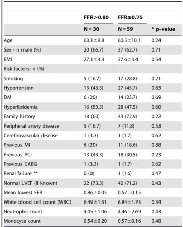
Table 1.Baseline Characteristics of patients with stable angina.
FFR.0.80 FFR#0.75
N = 30 N = 59 * p-value
Age 63.169.8 60.5610.1 0.24
Sex - n male (%) 20 (66.7) 37 (62.7) 0.71
BMI 27.164.3 27.663.4 0.54
Risk factors- n (%)
Smoking 5 (16.7) 17 (28.8) 0.21
Hypertension 13 (43.3) 27 (45.7) 0.83
DM 6 (20) 14 (23.7) 0.69
Hyperlipidemia 16 (53.3) 28 (47.5) 0.60
Family history 18 (60) 43 (72.9) 0.22
Peripheral artery disease 5 (16.7) 7 (11.8) 0.53
Cerebrovascular disease 1 (3.3) 1 (1.7) 0.62
Previous MI 6 (20) 11 (18.6) 0.88
Previous PCI 13 (43.3) 18 (30.5) 0.23
Previous CABG 1 (3.3) 1 (1.7) 0.62
Renal failure ** 0 (0) 1 (1.6) 0.47
Normal LVEF (if known) 22 (73.3) 42 (71.2) 0.43
Mean lowest FFR 0.8660.05 0.5760.15
White blood cell count (WBC) 6.4961.51 6.8461.73 0.34
Neutrophil count 4.0561.06 4.4662.69 0.43
Monocyte count 0.5460.20 0.5760.16 0.48
Continuous values are presented as means6standard deviation (SD). Categorical values are presented as number (percentages).
*Significance level 0.05. FFR = fractional flow reserve BMI = body mass index ;DM = diabetes mellitus; MI = myocardial infarction; PCI = percutaneous coronary intervention; CABG = coronary artery bypass grafting; LVEF = left ventricular ejection fraction.
of the above mentioned cytokines were compared among groups. Results are depicted in Figure 1. We did not observe differences in concentrations for any of the cytokines in supernatants of FFR-positive patients compared to those of FFR-negative patients. Median concentration (IQR) of IL-6 in FFR-positive patients compared to FFR-negative patients was 0.19 (0.04–0.45) versus 0.09 (0.02–0.54), p = 0.21, concentration of IL-8 was 3.4 (1.9–7.2) versus 2.7 (1.8–6.9), p = 0.56 and concentration of TNF-a 0.30 (0.10–0.75) versus 0.34 (0.14–0.66), p = 0.94. We did observe a significantly lower concentration of both IL-6 and TNF-ain the healthy controls compared to the patients with stable angina (p = 0.001 and p = 0.03, respectively).
Concentration of IL-8 in the supernatants did not significantly differ between all three groups (p = 0.21).
Expressions of TLR2, TLR4, CD14, CD11b and CD62L on peripheral blood cells
Expression of TLR2 and TLR4 quantified by mean fluores-cence intensity (MFI), was measured by flow cytometry on monocytes and granulocytes in blood samples of all 89 patients. We did not observe any significant differences between the expression levels for TLR2 and TLR4 on both monocytes (Figure 2A) and granulocytes (Figure 2B) between FFR-positive and FFR-negative patients. TLR2 and TLR4 expression on monocytes and TLR4 expression on granulocytes in healthy
control subjects was significantly lower compared to both FFR-positive and FFR-negative patients (p,0.01, p = 0.01 and p = 0.03).
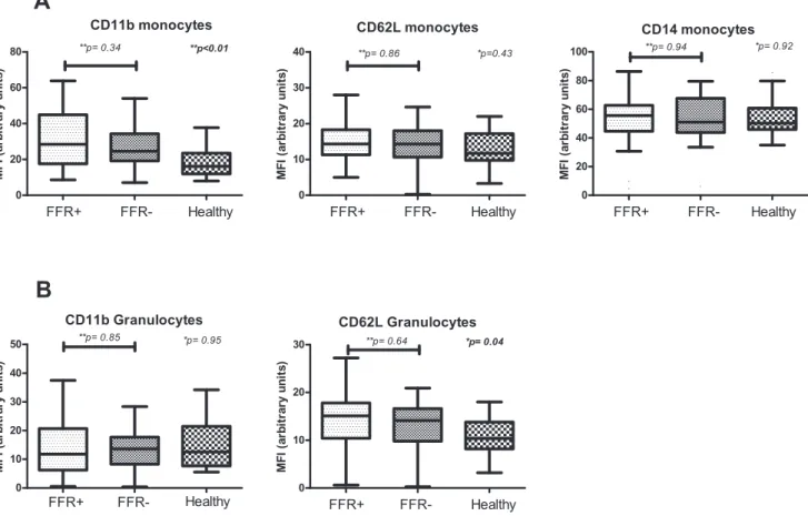
We then compared expression levels of CD11b and CD62L on both monocytes and granulocytes and CD14 on monocytes (Figure 3A and 3B). We observed no differences between the FFR-groups in expression of CD11b on monocytes (p = 0.34) or granulocytes (p = 0.85). Expression of CD62L on both monocytes and granulocytes also did not differ between FFR-positive and FFR-negative patients (p = 0.86 and p = 0.64, respectively). Healthy control subjects had significantly lower expression levels of CD11b on monocytes (p,0.01) and CD62L on granulocytes (p = 0.04). CD11b on granulocytes and CD62L on monocytes did not differ between groups (p = 0.95 and p = 0.43 respectively). Moreover, no difference in expression of CD14 on monocytes could be detected (p = 0.92).
Influence of severity of ischemic coronary disease on inflammatory markers
To assess the influence of theseverity and extent of ischemic coronary disease on the measured markers of inflammation we calculated FSS for each FFR-positive patient (as described above), divided FSS-scores into tertiles (range 1 to 28, intertertile range 7 to 14) and compared inflammatory markers of each tertile to each other and to FFR-negative patients. No between-subgroup differences were noted (p-values all.0.05). More importantly, there was no incremental relation between FSS tertiles and the measured markers (Figure 4A–C).
Discussion
The presence of inducible myocardial ischemia is of key importance for the prognosis of stable coronary artery disease. Numerous studies have shown increased rates of myocardial infarction and cardiovascular death when substantial myocardial ischemia is present as measured by FFR. Systemically measured inflammatory markers have also been associated with adverse outcome in patients suffering from cardiovascular disease. The origin of these inflammatory parameters can be the local inflammatory unstable plaque, can be a reflection of the atherosclerotic burden in the vascular system or an epi-phenom-enon. Since plasma levels of pro-inflammatory cytokines as well as cell-based markers on circulating cells have been found to be elevated in acute coronary syndromes, and the presence and
Table 2.Medication use of patients with stable angina at inclusion.
FFR.0.80 FFR#0.75
Medication – n(%) N = 30 N = 59 p-value*
ASA 24 (80) 55 (93) 0.08
b-blocker 18 (60) 51 (86) ,0.01
ACE-inhibitor 8 (26.7) 15 (25.4) 0.90
Statin 28 (93.3) 54 (91.5) 0.77
Clopidogrel (chronic use) 9 (30) 19 (32.2) 0.83
Clopidogrel (loading dose) 3 (10) 32 (54.2) ,0.01
Proportions were compared using Chi-square testing.
*Significance level 0.05. FFR = fractional flow reserve; ASA = Acetylsalicylic acid; ACE = angiotensin-converting enzyme.
doi:10.1371/journal.pone.0046356.t002
Figure 1. Comparison of levels of cytokines for FFR-positive and FFR-negative patients and healthy controls.*Comparison by Kruskal-Wallis test.** Between subgroup differences compared with Mann Whitney U test.
extent of inducible ischemia associated with increased occurrence of myocardial infarction and death, we hypothesized that inducible ischemia is associated with increased levels of blood derived inflammatory markers.
In this study, however, we found no relation between the investigated inflammatory markers and the presence of inducible myocardial ischemia as evidenced by FFR. In line with previous reports, we observed a difference in inflammatory markers between healthy subjects and patients with stable angina, pointing to a chronically elevated inflammatory status in patients with coronary artery disease [19]. Our observations suggest that this elevated inflammatory state is not altered by the presence of inducible ischemia by local atherosclerotic luminal narrowing, but probably a reflection of co-morbidities or systemic risk factors with concomitant generalized atherosclerotic disease, independently of its functional severity.
The latter is supported by the baseline characteristics that reveal that the FFR-positive and negative patient groups have compa-rable atherosclerotic risk profiles and history of previous MI or PCI as opposed to the healthy control group, which has virtually no risk factors and no history of coronary pathology. This study therefore shows that local lesions that may induce local ischemia do not explain the increased inflammatory blood profile in patients suffering from stable coronary artery disease. Even if the extent
and severity of ischemic coronary disease is taken into account according to FSS, no relationship between the inflammatory markers and inducible ischemia could be identified.
Fractional Flow Reserve (FFR) is the most reliable method for assessing the ischemic potential of coronary stenoses. Reducing myocardial ischemia is associated with improved prognosis [20] and by use of FFR it is possible to adequately pinpoint which lesions are responsible for ischemia. FFR-guided revascularization provides optimal relief of ischemia, preventing unnecessary stenting or bypass surgery of non-ischemia causing lesions while at the same time preventing undertreatment of lesions that appear moderate on angiography but do cause ischemia [21].
In this study, we investigated a specific subset of inflammation markers known to be associated with coronary artery disease. Toll-like receptors (TLRs) are a key part of the innate immune system and have been shown to play an important role in both the initiation and progression of coronary disease [22,23]. Increased expression of TLR4 on circulating monocytes and increased transcription of pro-inflammatory cytokines and co-stimulatory molecules, reflecting downstream activation of TLR stimulation, have been found in patients with unstable angina (UA) and acute myocardial infarction (AMI) compared to patients with stable angina and healthy controls [24,25].
Figure 2. Expression levels of TLR2 and TLR4 on monocytes (A) and granulocytes (B) FFR-positive and FFR-negative patients and healthy control subjects.*Comparison by Kruskal-Wallis test for all groups.** Between subgroups with Mann Whitney U test. TLR = toll-like receptor.
CD11b, also known as IntegrinaM, is involved in adhesion and transendothelial migration an early marker for cellular activation of leukocytes [26]. Furthermore, CD11b-expression has been shown to be upregulated immediately after stimulation of TLRs [27]. CD62L (L-selectin) is a member of the selectin family, expressed on both monocytes and granulocytes and is involved in cell adhesion [28]. Both granulocytes and monocytes readily shed CD62L upon stimulation.
It has been shown in both experimental and clinical settings that short lasting myocardial ischemia elicits an inflammatory response. In a porcine experiment 30 minutes of induced ischemia resulted in marked increase of plasma levels of IL-6 and TNF-a [29]. In patients with coronary artery disease, dobutamine-induced ische-mia resulted in an increase in plasma levels of IL-6 and tissue factor. Furthermore, IL-6 levels were related to left ventricular dysfunction at peak stress and rate of recovery of left ventricular function [30]. In patients with a reversible perfusion defect in myocardial perfusion imaging after stress, TLR mediated leuko-cyte activation was attenuated [31]. These findings provide both experimental as well as clinical evidence that an acute inflamma-tory response to ischemic episodes occurs which is detectable in peripheral blood samples. However, these results pertain to acutely induced ischemia and it is unknown whether inflammatory status is permanently altered by short repetitive ischemic episodes. In this study we found no evidence for this.
Limitations
Due to the fact that FFR measurements are obviously unknown prior to angiography, patient groups are inevitably unequal in size. Also, FFR-positive patients were more frequently pre-treated with a loading dose of clopidogrel. Clopidogrel has been shown to have anti-inflammatory properties [32], and thus it is possible that inflammatory status in the FFR-positive group could be underes-timated by pre-treatment. It must be noted however, that any anti-inflammatory properties of clopidogrel have mainly been observed during chronic treatment [32–35], and chronic clopidogrel treatment did not differ between the FFR positive and negative groups. Quinn et al specifically investigated effects of clopidogrel treatment (.24 hours) before PCI on inflammatory markers and observed that serum CD40 ligand and IL-6 were not affected by clopidogrel treatment [36]. It is therefore unlikely that pre-treatment with a loading dose of clopidogrel would have played a significant role in our study. In any case, we feel withholding clopidogrel pretreatment in patients if stenting is anticipated is unethical and potentially hazardous. Over 90% of the patients with stable angina in this study are treated with statins (HMG-CoA reductase inhibitors), which have been shown to have anti-inflammatory properties [37–39]. Despite the use of statins, inflammatory markers are increased in the patient groups as compared to the healthy controls. In the patient groups it is conceivable that use of statins may mask small differences related to the presence or absence of inducible ischemia. Blood samples were taken from an arterial sheath in the patient groups.
Figure 3. Expression levels of CD11b, CD62L and CD14 on monocytes (A) and CD11b and CD62L on granulocytes (B) in FFR-negative patients, FFR-positive and healthy control subjects.*Comparison by Kruskal-Wallis test for all groups.** Between subgroups with Mann Whitney U test.
Consequently, no statements can be made on possible differences in other locations of the vascular system (e.g. coronary artery or coronary sinus) in these patients. In the healthy control group, blood was obtained from an antecubital vein. Although differences in inflammatory markers between locally (coronary sinus) obtained blood and peripheral blood in coronary artery disease have been
observed [40,41], to our knowledge differences of these markers between peripheral venous and peripheral arterial blood have not been reported [42]. BMI of the healthy control subjects was significantly lower than that of the patient groups. As increased levels of inflammatory markers have been reported in obese subjects [43–45], we cannot exclude the possibility that this may
Figure 4. Concentrations of cytokines and surface markers on monocytes and granulocytes according to FSS tertiles and FFR-negative patients.Concentrations of cytokines (A) and surface markers on monocytes (B) and granulocytes (C) according to FSS tertiles (of FFR-positive patients) and FFR-negative patients. Comparison of all subgroups was performed with the Jonckheere-Terpstra test. FSS = functional syntax score.
have influenced biomarker levels in the patient groups, although BMI was only moderately increased in the FFR positive and FFR-negative patients. In this study we only analyzed a specific subset of inflammatory markers; IL-6, IL-8 and TNF-aas inflammatory cytokines and CD11b, CD62L, TLR2 and TLR4 as markers for cellular activation of circulating cells. Although we investigated cell based expression as well as secreted markers for inflammation, we cannot discount the possibility that repetitive ischemia exerts effects through different mechanisms and is reflected by different biomarkers than the ones investigated here.
Conclusions
Inducible myocardial ischemia is not associated with increased concentrations of IL-6, IL-8 and TNF-ain blood or expression of CD11b, CD62L, CD14, TLR2 or TLR4 on circulating leukocytes in patients with stable angina.
Author Contributions
Conceived and designed the experiments: JS EE GP IH JJ NP. Performed the experiments: JS EE. Analyzed the data: JS EE IH. Contributed reagents/materials/analysis tools: JK AvZ EE. Wrote the paper: JS EE GP NP.
References
1. Virmani R, Burke AP, Kolodgie FD, Farb A (2003) Pathology of the thin-cap fibroatheroma: a type of vulnerable plaque. J Interv Cardiol 16: 267–272. 2. Tziakas DN, Chalikias GK, Tentes IK, Stakos D, Chatzikyriakou SV, et al.
(2008) Interleukin-8 is increased in the membrane of circulating erythrocytes in patients with acute coronary syndrome. Eur Heart J 29: 2713–2722. 3. Ueda K, Takahashi M, Ozawa K, Kinoshita M (1999) Decreased soluble
interleukin-6 receptor in patients with acute myocardial infarction. Am Heart J 138: 908–915.
4. Mizia-Stec K, Gasior Z, Zahorska-Markiewicz B, Janowska J, Szulc A, et al. (2003) Serum tumour necrosis factor-alpha, interleukin-2 and interleukin-10 activation in stable angina and acute coronary syndromes. Coron Artery Dis 14: 431–438.
5. Anguera I, Miranda-Guardiola F, Bosch X, Filella X, Sitges M, et al. (2002) Elevation of serum levels of the anti-inflammatory cytokine interleukin-10 and decreased risk of coronary events in patients with unstable angina. Am Heart J 144: 811–817.
6. Ridker PM, Rifai N, Pfeffer M, Sacks F, Lepage S, et al. (2000) Elevation of tumor necrosis factor-alpha and increased risk of recurrent coronary events after myocardial infarction. Circulation 101: 2149–2153.
7. Luc G, Bard JM, Juhan-Vague I, Ferrieres J, Evans A, et al. (2003) C-reactive protein, interleukin-6, and fibrinogen as predictors of coronary heart disease: the PRIME Study. Arterioscler Thromb Vasc Biol 23: 1255–1261.
8. Inoue T, Komoda H, Nonaka M, Kameda M, Uchida T, et al. (2008) Interleukin-8 as an independent predictor of long-term clinical outcome in patients with coronary artery disease. Int J Cardiol 124: 319–325.
9. Hachamovitch R, Berman DS, Kiat H, Cohen I, Lewin H, et al. (1997) Incremental prognostic value of adenosine stress myocardial perfusion single-photon emission computed tomography and impact on subsequent management in patients with or suspected of having myocardial ischemia. Am J Cardiol 80: 426–433.
10. Iskandrian AS, Chae SC, Heo J, Stanberry CD, Wasserleben V, et al. (1993) Independent and incremental prognostic value of exercise single-photon emission computed tomographic (SPECT) thallium imaging in coronary artery disease. J Am Coll Cardiol 22: 665–670.
11. Pijls NH, van Schaardenburgh P, Manoharan G, Boersma E, Bech JW, et al. (2007) Percutaneous coronary intervention of functionally nonsignificant stenosis: 5-year follow-up of the DEFER Study. J Am Coll Cardiol 49: 2105– 2111.
12. Tonino PA, De Bruyne B, Pijls NH, Siebert U, Ikeno F, et al. (2009) Fractional flow reserve versus angiography for guiding percutaneous coronary intervention. N Engl J Med 360: 213–224.
13. Pijls NH, Sels JW (2012) Functional measurement of coronary stenosis. J Am Coll Cardiol 59: 1045–1057.
14. De Bruyne B, Pijls NH, Bartunek J, Kulecki K, Bech JW, et al. (2001) Fractional flow reserve in patients with prior myocardial infarction. Circulation 104: 157– 162.
15. Pijls NH, van Gelder B, Van der Voort P, Peels K, Bracke FA, et al. (1995) Fractional flow reserve. A useful index to evaluate the influence of an epicardial coronary stenosis on myocardial blood flow. Circulation 92: 3183–3193. 16. Pijls NH, De Bruyne B, Peels K, Van Der Voort PH, Bonnier HJ, et al. (1996)
Measurement of fractional flow reserve to assess the functional severity of coronary-artery stenoses. N Engl J Med 334: 1703–1708.
17. Serruys PW, Morice MC, Kappetein AP, Colombo A, Holmes DR, et al. (2009) Percutaneous coronary intervention versus coronary-artery bypass grafting for severe coronary artery disease. N Engl J Med 360: 961–972.
18. Nam CW, Mangiacapra F, Entjes R, Chung IS, Sels JW, et al. (2011) Functional SYNTAX score for risk assessment in multivessel coronary artery disease. J Am Coll Cardiol 58: 1211–1218.
19. Mizia-Stec K, Mandecki T, Zahorska-Markiewicz B, Janowska J, Szulc A, et al. (2002) Selected cytokines and soluble forms of cytokine receptors in coronary artery disease. Eur J Intern Med 13: 115–122.
20. Shaw LJ, Berman DS, Maron DJ, Mancini GB, Hayes SW, et al. (2008) Optimal medical therapy with or without percutaneous coronary intervention to reduce ischemic burden: results from the Clinical Outcomes Utilizing Revascularization and Aggressive Drug Evaluation (COURAGE) trial nuclear substudy. Circulation 117: 1283–1291.
21. Tonino PA, Fearon WF, De Bruyne B, Oldroyd KG, Leesar MA, et al. (2010) Angiographic versus functional severity of coronary artery stenoses in the FAME study fractional flow reserve versus angiography in multivessel evaluation. J Am Coll Cardiol 55: 2816–2821.
22. Li H, Sun B (2007) Toll-like receptor 4 in atherosclerosis. J Cell Mol Med 11: 88–95.
23. Vink A, Schoneveld AH, van der Meer JJ, Van Middelaar BJ, Sluijter JP, et al. (2002) In vivo evidence for a role of toll-like receptor 4 in the development of intimal lesions. Circulation 106: 1985–1990.
24. Liuzzo G, Angiolillo DJ, Buffon A, Rizzello V, Colizzi C, et al. (2001) Enhanced response of blood monocytes to in vitro lipopolysaccharide-challenge in patients with recurrent unstable angina. Circulation 103: 2236–2241.
25. Methe H, Kim JO, Kofler S, Weis M, Nabauer M (2005) Expansion of circulating Toll-like receptor 4-positive monocytes in patients with acute coronary syndrome. Circulation 111: 2654–2661.
26. Ley K, Laudanna C, Cybulsky MI, Nourshargh S (2007) Getting to the site of inflammation: the leukocyte adhesion cascade updated. Nat Rev Immunol 7: 678–689.
27. Han C, Jin J, Xu S, Liu H, Li N, et al. (2010) Integrin CD11b negatively regulates TLR-triggered inflammatory responses by activating Syk and promoting degradation of MyD88 and TRIF via Cbl-b. Nat Immunol 11: 734–742.
28. Ley K (2003) The role of selectins in inflammation and disease. Trends Mol Med 9: 263–268.
29. Vilahur G, Hernandez-Vera R, Molins B, Casani L, Duran X, et al. (2009) Short-term myocardial ischemia induces cardiac modified C-reactive protein expression and proinflammatory gene (cyclo-oxygenase-2, monocyte chemoat-tractant protein-1, and tissue factor) upregulation in peripheral blood mononuclear cells. J Thromb Haemost 7: 485–493.
30. Ikonomidis I, Athanassopoulos G, Lekakis J, Venetsanou K, Marinou M, et al. (2005) Myocardial ischemia induces interleukin-6 and tissue factor production in patients with coronary artery disease: a dobutamine stress echocardiography study. Circulation 112: 3272–3279.
31. Elsenberg EH, Versteeg D, Sels JW, Vlaar PJ, Hobbelink MG, et al. (2011) Inducible cardiac ischemia is related with a decrease in whole blood Toll-like receptor 2 and 4 response. Clin Sci (Lond) 122: 527–533
32. Muhlestein JB (2010) Effect of antiplatelet therapy on inflammatory markers in atherothrombotic patients. Thromb Haemost 103: 71–82.
33. Graff J, Harder S, Wahl O, Scheuermann EH, Gossmann J (2005) Anti-inflammatory effects of clopidogrel intake in renal transplant patients: effects on platelet-leukocyte interactions, platelet CD40 ligand expression, and proin-flammatory biomarkers. Clin Pharmacol Ther 78: 468–476.
34. Steinhubl SR, Badimon JJ, Bhatt DL, Herbert JM, Luscher TF (2007) Clinical evidence for anti-inflammatory effects of antiplatelet therapy in patients with atherothrombotic disease. Vasc Med 12: 113–122.
35. Willerson JT, Cable G, Yeh ET (2009) PROCLAIM: pilot study to examine the effects of clopidogrel on inflammatory markers in patients with metabolic syndrome receiving low-dose aspirin. Tex Heart Inst J 36: 530–539. 36. Quinn MJ, Bhatt DL, Zidar F, Vivekananthan D, Chew DP, et al. (2004) Effect
of clopidogrel pretreatment on inflammatory marker expression in patients undergoing percutaneous coronary intervention. Am J Cardiol 93: 679–684. 37. Ortego M, Bustos C, Hernandez-Presa MA, Tunon J, Diaz C, et al. (1999)
Atorvastatin reduces NF-kappaB activation and chemokine expression in vascular smooth muscle cells and mononuclear cells. Atherosclerosis 147: 253– 261.
38. Ridker PM, Danielson E, Fonseca FA, Genest J, Gotto AM Jr, et al. (2008) Rosuvastatin to prevent vascular events in men and women with elevated C-reactive protein. N Engl J Med 359: 2195–2207.
41. ElMokhtari N, Zschernitz S, Sebens S, Simon-Herrmann G, Kruger D (2010) Cardiac release and kinetics of cytokines after elective bare metal coronary stenting. J Thromb Thrombolysis 30: 391–397.
42. Deliargyris EN, Raymond RJ, Theoharides TC, Boucher WS, Tate DA, et al. (2000) Sites of interleukin-6 release in patients with acute coronary syndromes and in patients with congestive heart failure. Am J Cardiol 86: 913–918. 43. Bruun JM, Verdich C, Toubro S, Astrup A, Richelsen B (2003) Association
between measures of insulin sensitivity and circulating levels of interleukin-8,
interleukin-6 and tumor necrosis factor-alpha. Effect of weight loss in obese men. Eur J Endocrinol 148: 535–542.
44. Musaad S, Haynes EN (2007) Biomarkers of obesity and subsequent cardiovascular events. Epidemiol Rev 29: 98–114.