Calprotectin and Platelet Aggregation in
Patients with Stable Coronary Artery Disease
Sanne Bøjet Larsen1, Erik Lerkevang Grove1, Manan Pareek1, Steen Dalby Kristensen1,2,
Anne-Mette Hvas2,3*
1Department of Cardiology, Aarhus University Hospital, DK-8200, Aarhus N, Denmark,2Faculty of Health Sciences, Aarhus University, DK-8200, Aarhus N, Denmark,3Department of Clinical Biochemistry, Aarhus University Hospital, DK-8200, Aarhus N, Denmark
Abstract
Background
Recent studies suggest that the inflammation-associated protein calprotectin may be impli-cated in the pathogenesis of coronary artery disease (CAD). However, the impact of calpro-tectin levels on platelet aggregation in CAD patients has never been investigated.
Objectives
We investigated the association between calprotectin levels and platelet aggregation in sta-ble, high-risk CAD patients receiving aspirin as mono antiplatelet therapy. Furthermore, we aimed to investigate independent clinical and laboratory determinants of calprotectin levels.
Methods
We performed a cross-sectional study including 581 stable, high-risk CAD patients. All pa-tients received 75 mg aspirin daily as mono antiplatelet therapy. Platelet aggregation was assessed by 1) impedance aggregometry (Multiplate Analyzer) using arachidonic acid (AA) and collagen as agonists and by 2) the VerifyNow Aspirin Assay. Low-grade inflammation was evaluated by calprotectin, high-sensitive C-reactive-protein (hs-CRP) and interleukin-6. Platelet activation was assessed by soluble P-selectin, and cyclooxygenase-1 inhibition was evaluated by serum thromboxane B2, both measured by ELISA.
Results
Calprotectin levels correlated positively with platelet aggregation according to Multiplate An-alyzer (r=0.12, p=0.01). Additionally, calprotectin was positively associated with leukocytes (r=0.33, p<0.0001), hs-CRP (r=0.31, p<0.0001), interleukin-6 (r=0.28, p<0.0001), soluble P-selectin (r=0.10, p=0.02) and serum thromboxane B2(r=0.10, p=0.02). Type 2 diabetes
mellitus was an independent predictor of increased calprotectin levels (p=0.004), and trends were seen for body mass index (p=0.06) and smoking (p=0.07). Compliance with aspirin was confirmed by low serum thromboxane B2levels in all patients (median [25%;75%]: 1.07
[0.52;1.87] ng/mL). OPEN ACCESS
Citation:Larsen SB, Grove EL, Pareek M, Kristensen SD, Hvas A-M (2015) Calprotectin and Platelet Aggregation in Patients with Stable Coronary Artery Disease. PLoS ONE 10(5): e0125992. doi:10.1371/journal.pone.0125992
Academic Editor:Kathleen Freson, University of Leuven, BELGIUM
Received:October 30, 2014
Accepted:March 27, 2015
Published:May 13, 2015
Copyright:© 2015 Larsen et al. This is an open access article distributed under the terms of the Creative Commons Attribution License, which permits unrestricted use, distribution, and reproduction in any medium, provided the original author and source are credited.
Data Availability Statement:All relevant data are within the paper.
Conclusion
Calprotectin levels correlated positively, though weakly, with platelet aggregation and activation as well as serum thromboxane B2in high-risk, stable CAD patients treated
with aspirin.
Introduction
Inflammation plays an important role in the pathogenesis of atherosclerosis [1]. Coronary ath-erosclerosis is the underlying substrate of most coronary events, and rupture of an atheroscle-rotic plaque with exposure of the thrombogenic lipid-core promotes platelet adhesion followed by platelet activation and aggregation [2]. Platelet inhibition with aspirin continues to be the antiplatelet backbone in prevention and treatment of coronary artery disease (CAD) [3]. How-ever, wide variability in the antiplatelet effect of aspirin has been reported, most likely reflecting the influence of genetic, biological and clinical factors [4,5].
Calprotectin, also known as myeloid-related protein 8/14, S100A8/A9 or calgranulin A/B, is an inflammation-associated protein [6], which is predominantly expressed by, and released from, myeloid cells on cellular activation [7,8]. Calprotectin plasma levels have primarily been investigated as a marker of inflammatory conditions such as inflammatory bowel disease and rheumatoid arthritis [9,10], but recent studies suggest that calprotectin may also be implicated in the pathogenesis of CAD [11–15]. Additionally, calprotectin has been identified as an early and sensitive biomarker with potential ability to discriminate between patients with acute coro-nary syndrome and patients with chronic, stable CAD [11,12,15]. Finally, elevated levels of cal-protectin have been associated with increased risk of first and recurrent cardiovascular events [12,15,16].
Recent studies have suggested that increased levels of inflammatory markers, such as high-sensitive C-reactive protein (hs-CRP) and interleukin-6 (IL-6), may modify platelet aggrega-tion and reduce the efficacy of antiplatelet drugs [17–21]. However, the impact of calprotectin levels on platelet aggregation in stable CAD patients has not been investigated. We hypothe-sized that high levels of calprotectin were associated with reduced effect of aspirin as indicated by increased platelet aggregation levels in stable, high-risk CAD patients. The aim of this study was to investigate the association between calprotectin levels and platelet aggregation in stable, high-risk CAD patients receiving aspirin as mono antiplatelet therapy. Furthermore, we aimed to investigate independent clinical and laboratory determinants of calprotectin levels.
Methods
Study population
We performed a cross-sectional study including 581 stable patients with angiographically doc-umented CAD. Although stable, the study cohort represented a high-risk CAD population since all patients had either prior myocardial infarction, type 2 diabetes mellitus or both. All patients were recruited from the Western Denmark Heart Registry [22] and enrolled from Feb-ruary 2009 to January 2011. Fulfilment of the inclusion and exclusion criteria were checked in all patients’medical records. The inclusion criteria were: a) age18 years, b) significant CAD verified by prior percutaneous coronary intervention, coronary artery bypass grafting, or by a coronary angiography showing at least one 50% coronary luminal stenosis, c) patients with prior myocardial infarction: at least 12 months ago, myocardial infarction verified by Foundation. The funders had no role in study design,
data collection and analysis, decision to publish, or preparation of the manuscript.
electrocardiographic ST-segment elevation and/or elevated plasma troponin T (>0.10μg/L)
together with plasma creatine kinase-MB (>12 U/L). All diabetic patients were diagnosed
with type 2 diabetes and treated with oral antidiabetic drugs and/or insulin. All non-diabetic patients had fasting plasma glucose levels<7.0 mmol/L at the time of inclusion. The exclusion
criteria were: a) ongoing treatment known to affect platelet function or coagulation (e.g. non-steroidal anti-inflammatory drugs, any antiplatelet drug or anticoagulants), b) any ischaemic vascular event, percutaneous coronary intervention, or coronary artery bypass grafting within the previous 12 months, c) platelet count<120 x 109/L or>450 x 109/L.
All patients included in the study were treated with 75 mg non-enteric coated aspirin once daily as mono antiplatelet therapy prior to and during the study.
The study was conducted in agreement with the Helsinki-II-declaration. It was approved by The Central Denmark Region Committees on Health Research (M-2009-0110) and by the Danish Data Protection Agency. All patients gave written informed consent.
Compliance
To optimize compliance and uniform pharmacokinetics, all patients received a pill box with seven tablets of 75 mg non-enteric coated aspirin (Hjerdyl, Sandoz, Denmark). This was to en-sure that all patients included in the study received the exact same aspirin dose and preparation prior to and at the time of blood sampling. On the day of blood sampling, patients were in-structed to ingest aspirin exactly one hour before blood sampling. Compliance with aspirin was optimized by confirmation of empty pill boxes along with aspirin intake questioning, and final-ly, measurement of serum thromboxane B2(S-TXB2) was performed for all patients.
Laboratory investigations
Blood sampling. Blood samples were obtained from the antecubital vein with patients in supine position after 30 minutes of rest using vacuum tubes, a large bore needle (19 G), and a minimum of stasis.
Haematology. Blood samples for haematological analyses, including haemoglobin, leuko-cyte count and platelet count, were collected in 3.0 mL tubes containing EDTA (Terumo, Leu-ven, Belgium) and analysed within 90 minutes of sampling. Haematological parameters were measured with the Sysmex XE-2100 haematology analyser (Sysmex, Kobe, Japan).
Platelet aggregation tests. Platelet aggregation was evaluated with two different instru-ments using whole blood: impedance aggregometry (Multiplate Analyzer, Roche, Hvidovre, Denmark) and the VerifyNow Aspirin Assay (Accumetrics Inc., San Diego, CA, USA).
Impedance aggregometry is based on platelet adhesion and aggregation on two electrodes, resulting in an increase of electrical resistance, which is converted into arbitrary aggregation units (AU). The area under the aggregation curve (AUC) is used to express the aggregation re-sponse over the measured time (AU
min). For impedance aggregometry analyses, 3.0 mL tubes containing hirudin 25μg/mL (Terumo, Lueven, Belgium) were utilised. Platelet aggrega-tion was induced using arachidonic acid (AA) 1.0 mM (ASPI test, Triolab AS, Brøndby, Den-mark) or collagen 1.0μg/mL (Horm, Medinor, Nycomed, Austria). Blood samples rested for at least 30 minutes before analysis but no longer than 120 minutes. In case of a deviation>20%
between the two impedance curves, the sample was re-analysed.
3.2% sodium citrate (Terumo, Lueven, Belgium). Blood samples rested for at least 30 minutes before analysis but no longer than 120 minutes. The reproducibility of the platelet aggregation tests has previously been reported by our group [23].
Calprotectin and other inflammatory markers
Blood for calprotectin analysis was collected in non-siliconized 5.0 mL tubes (Terumo, Leuven, Belgium) without anticoagulants. The blood was allowed to clot for one hour at 37°C before serum was separated by centrifugation at 2600gfor 10 minutes. Serum was stored at−80°C
until analysis. Serum calprotectin was measured using enzyme-linked immunosorbent assay (ELISA) (MRP 8/14 Calprotectin, Bühlmann, Schönenbuch, Switzerland). The first four steps of the ELISA protocol were performed using a JANUS Automated workstation (PerkinElmer, Waltham, MA, USA). Duplicate measurements were performed on 40 samples (coefficient of variation = 2.5%), and single measurements were performed in the remaining 541 samples.
Blood for hs-CRP analysis (KoneLab 30i, ILS Laboratories Scandinavia, Allerød, Denmark) was collected in 3.0 mL lithium-heparin tubes containing separating gel (Terumo, Leuven, Bel-gium). The measurement interval for hs-CRP was 0.2–10.0 mg/L. A total of 493 (85%) of the 581 patients were included in the hs-CRP analyses; 28 patients had hs-CRP levels>10.0 mg/L,
and hs-CRP was not measured in 57 patients due to a change in laboratory procedures regard-ing measurements of hs-CRP durregard-ing the study. Hs-CRP values were missregard-ing for three patients. Blood for IL-6 analysis (cobas 6000 analyser, E module, Roche, Mannheim, Germany) was collected in non-siliconized 5.0 mL tubes (Terumo, Leuven, Belgium) without anticoagulants. The blood was allowed to clot for one hour at 37°C before serum was separated by centrifuga-tion at 2600gfor 10 minutes. Serum was stored at−80°C until analysis.
Soluble P-selectin and serum thromboxane B2. Concentrations of soluble P-selectin (sP-selectin) and serum thromboxane B2were measured using ELISA (R&D Systems, Minneapolis,
USA and Thromboxane B2EIA Kit, Cayman Chemical, Michigan, USA) as previously
de-scribed [24].
Ethics statement
The study was conducted in agreement with the Helsinki-II-declaration. It was approved by The Central Denmark Region Committees on Health Research (M-2009-0110) and by the Danish Data Protection Agency. All patients gave written informed consent.
Statistics
If normally distributed, continuous data are presented as mean and standard deviation (SD), if not as median and interquartile range or log-transformed to obtain normal distribution. Differ-ences between two unpaired groups were tested with a two-sided t-test if data were normally distributed and, if not, the Mann-Whitney test was used. Proportions between two groups were compared using Fischer’s exact test and presented as absolute counts and percentages. Correla-tions were calculated using Spearman’s rank coefficient. Multiple linear regression analyses were used to identify independent determinants of calprotectin and platelet aggregation. A two-sided p-value<0.05 was considered statistically significant. Data were registered in
Results
Study population
Clinical and biochemical characteristics of the study population are shown in Tables1and2. The study population consisted of stable CAD patients with a relatively high-risk profile, since 533 (92%) of the patients had a history of myocardial infarction, 145 (25%) had type 2 diabetes, and 100 (17%) had both. Compliance with aspirin was confirmed by low serum thromboxane B2levels in all patients (median [25%;75%]: 1.07 [0.52;1.87], range 0.02–26.44 ng/mL).
In order to evaluate baseline characteristics and biochemical values according to platelet ag-gregation levels, patients were divided into two subgroups on the basis of the median (163 AUmin) of AA-induced platelet aggregation using the Multiplate Analyzer (Table 3). Thus, in
Table 3, patients with high platelet aggregation had aggregation levelsthe median of 163
AUmin and patients with low platelet aggregation had aggregation levels<the median of 163 AU
min. Patients with high platelet aggregation levels had significantly higher calprotectin lev-els than patients with low aggregation. Also leukocyte counts, platelet counts, hs-CRP and IL-6 levels were elevated in the high platelets aggregation group. Furthermore, high platelet aggrega-tion levels were significantly associated with type 2 diabetes, smoking and treatment with calci-um antagonists and diuretics.
Table 1. Baseline characteristics of the study population, n = 581.
Age, years 64±9
Body mass index, kg/m2 28±4
Males 460 (79)
Current smokers 122 (21)
Blood pressure, systolic, mm Hg 142±20
Blood pressure, diastolic, mm Hg 82±11
Morbidity
Prior percutaneus coronary intervention 562 (97)
Prior myocardial infarction 533 (92)
Prior coronary artery bypass grafting 49 (8)
Prior stroke 25(4)
Type 2 diabetes mellitus 145 (25)
Medication
Aspirin 581 (100)
Statins 533 (92)
Beta-blockers 440 (76)
ACE inhibitors 265 (46)
Angiotensin receptor blockers 80 (14)
Calcium antagonists 111 (19)
Diuretics 147 (25)
Proton pump inhibitors 65 (11)
Insulin* 47 (32)
Oral antidiabetic medication* 127 (88)
Data is presented as mean±SD or n (%). B: blood, S: serum, P: plasma, ACE: Angiotensin converting
enzyme.
*out of 145 patients with type 2 diabetes.
Calprotectin and platelet aggregation
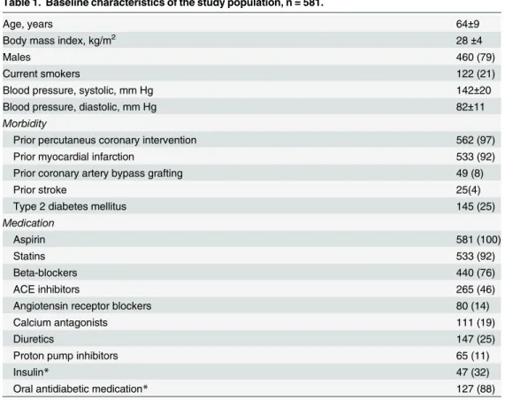
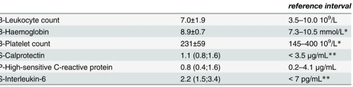
Calprotectin was positively, though weakly, correlated with AA-induced platelet aggregation according to Multiplate Analyzer (Fig 1). There was no significant association between calpro-tectin and collagen-induced Multiplate Analyzer platelet aggregation (Fig 2) or VerifyNow
Table 2. Baseline bichemical characteristics of the study population, n = 581.
reference interval
B-Leukocyte count 7.0±1.9 3.5–10.0 109/L
B-Haemoglobin 8.9±0.7 7.3–10.5 mmol/L*
B-Platelet count 231±59 145–400 109/L*
S-Calprotectin 1.1 (0.8;1.6) <3.5μg/mL**
P-High-sensitive C-reactive protein 0.8 (0.4;1.6) 0.2–4.1μg/mL
S-Interleukin-6 2.2 (1.5;3.4) <7 pg/mL**
Data is presented as mean±SD or median (25%;75%). B: blood, S: serum, P: plasma. *Reference interval for both men and women.
**In healthy individuals. No established reference intervals for stable CAD patients.
doi:10.1371/journal.pone.0125992.t002
Table 3. Baseline characteristics of the study population according to platelet aggregation using Mul-tiplate Analyzer, 1.0 mM arachidonic acid, n = 581.
<163 AU*min 163 AU*min p-value
n = 283 n = 298
Age, years 64±9 64±10 0.25
Body mass index, kg/m2 27±4 28±5 0.06
Males 227 (80) 227 (78) 0.48
Current smokers 45 (16) 75 (26) 0.01
Blood pressure, systolic, mm Hg 142±20 141±20 0.65
Blood pressure, diastolic, mm Hg 83±12 82±10 0.09
Morbidity
Prior percutaneus coronary intervention 275 (97) 281 (96) 0.64
Prior myocardial infarction 265 (94) 265 (91) 0.22
Prior coronary artery bypass grafting 23 (8) 26 (9) 0.77
Prior stroke 13 (5) 12 (4) 0.84
Type 2 diabetes mellitus 57 (20) 88 (30) 0.01
Medication
Aspirin 282 (100) 292 (100) 1.00
Statins 256 (91) 272 (93) 0.36
Beta-blockers 216 (77) 219 (75) 0.70
ACE inhibitors 128 (45) 136 (47) 0.80
Angiotensin receptor blockers 43 (15) 36 (12) 0.33
Calcium antagonists 40 (14) 68 (23) 0.01
Diuretics 53 (19) 90 (31) 0.001
Proton pump inhibitors 29 (10) 35 (12) 0.60
Insulin* 16 (28) 30 (34) 0.47
Oral antidiabetic medication* 48 (84) 78 (89) 0.46
Data is presented as mean±SD, n(%) or median 25%;75%). B: blood, S: serum, P: plasma, ACE:
Angiotensin converting enzyme. Patients were divided into two subgroups on the basis of the median (163 AU*min) of AA-induced platelet aggregation using the Multiplate Analyzer.
*Out of 145 patients with type 2 diabetes.
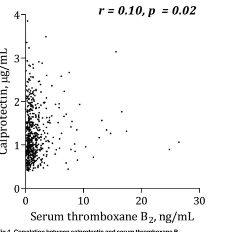
(Fig 3). Calprotectin was positively associated with serum thromboxane B2(Fig 4), sP-selectin
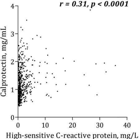
(r = 0.10, p = 0.02), leukocytes (r = 0.33, p<0.0001), hs-CRP (Fig 5) and IL-6 (Fig 6).
After adjusting for traditional risk factors (gender, age, type 2 diabetes, smoking status and body mass index) in a linear multivariate regression analysis, calprotectin no longer predicted increased AA-induced platelet aggregation according to the Multiplate Analyzer (p = 0.10). Similar results were found with collagen-induced platelet aggregation and VerifyNow.
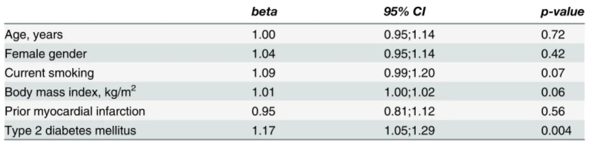
Independent determinants of calprotectin
In order to identify clinical and demographical determinants of calprotectin, we performed lin-ear multivariate regression analysis including age, gender, smoking, body mass index, prior myocardial infarction and type 2 diabetes as independent variables (Table 4). Type 2 diabetes was an independent determinant of calprotectin (p = 0.004), and trends were seen for body mass index (p = 0.06) and smoking (p = 0.07). Calprotectin levels were increased by 17% in pa-tients with type 2 diabetes compared with non-diabetes papa-tients, and by 9% in smokers com-pared with non-smokers, however, the latter did not reach statistical significance (Table 4).
Discussion
This is the first study to investigate the association between calprotectin and platelet aggrega-tion in stable, high-risk CAD patients. The main findings of the study were that 1) increased levels of calprotectin were associated with AA-induced platelet aggregation according to
Fig 1. Correlation between calprotectin and platelet aggregation induced by arachidonic acid using Multiplate Analyzer.
Multiplate Analyzer and serum thromboxane B2, and 2) type 2 diabetes was an independent
determinant of increased calprotectin levels.
Calprotectin and platelet aggregation
We found positive associations between calprotectin levels and AA-induced platelet aggrega-tion according to Multiplate Analyzer. Furthermore, positive correlaaggrega-tions between levels of cal-protectin and serum thromboxane B2and sP-selectin were found. This may suggest that
calprotectin could influence on both platelet activation and subsequent aggregation; however, all correlations were rather weak. In a previous study including part of the present study popu-lation, we demonstrated that high levels of hs-CRP were associated with increased platelet ag-gregation in stable CAD patients [19]. In the present study, we have demonstrated that calprotectin was significantly associated with both hs-CRP and IL-6.
Inflammation is an important player in thrombosis [25,26]. Calprotectin has been shown to induce a thrombogenic, inflammatory response in endothelial cells by increasing the transcrip-tion of pro-inflammatory chemokines and adhesion molecules [27], which may secondarily ac-tivate platelets, possibly through expression of P-selectin on endothelial cells. However, a direct effect of calprotectin on platelet aggregation has not been established. Recently, in an experi-mental study, Wang et al. reported that the subunit MRP14 or S100A9 is released by platelets and that the subunit is involved in the molecular pathways leading to thrombus formation through CD36 [28]. Other inflammatory markers have also been proposed to influence
Fig 2. Correlation between calprotectin and platelet aggregation induced by collagen using Multiplate Analyzer.
platelets; CRP may indirectly affect platelet reactivity through preceding FcƴRIIa-dependent monocyte-activation [25] and CRP-induction of macrophage-derived tissue factor generation [26]. These pathways may partly explain the association between hs-CRP and reduced anti-platelet effect of aspirin [19], and the association between hs-CRP and calprotectin, the latter being demonstrated in the present study.
Calprotectin and cardiovascular risk factors
Diabetes, obesity and smoking are traditional cardiovascular risk factors, which have previous-ly been shown to associate with elevated levels of calprotectin [29]. In the present study, type 2 diabetes was an independent determinant of increased calprotectin levels, and trends were seen for body mass index and smoking. Thus, our results extend previous findings in healthy indi-viduals showing that calprotectin levels are positively influenced by smoking and body mass index [16].
Diabetes is considered a pro-thrombotic state, probably explained by disturbances in vari-ous steps of thrombus formation, which may lead to increased platelet activation and aggrega-tion [30]. Several mechanisms, including chronic low-grade inflammation, have been
suggested as potential mechanisms leading to platelet hyper reactivity, hyper coagulability and compromised fibrinolysis [31,32]. In our study, calprotectin levels were increased by 17% in CAD patients with type 2 diabetes compared with non-diabetes patients. Our results extend the study by Peng et al. showing elevated calprotectin levels in patients with type 2 diabetes and CAD compared with type 2 diabetes patients without CAD [33]. Additionally, the authors
Fig 3. Correlation between calprotectin and platelet aggregation induced by arachidonic acid using VerifyNow.
found that calprotectin levels correlated positively with the severity of CAD and with carotid intima-media thickness in diabetes patients without clinically overt CAD [33]. Catalan et al. re-ported increased levels of calprotectin in patients with obesity associated type 2 diabetes and found that calprotectin levels decreased following weight loss [34].
In our study, type 2 diabetes was an independent predictor of increased calprotectin levels, whereas the association between calprotectin and increased platelet aggregation was less pro-nounced. The association between diabetes and chronic low-grade inflammation may explain the increased levels of calprotectin in diabetic patients. To the best of our knowledge, no studies have demonstrated a relationship between increased calprotectin levels and a reduced anti-platelet effect of aspirin. Most likely, calprotectin only has a modest influence on anti-platelet aggregation.
Body mass index correlated significantly with increased calprotectin levels, thus extending previous findings in healthy individuals [16,35]. However, when adjusted for gender, age, prior myocardial infarction, diabetes and smoking, only a trend was seen. This may be partially ex-plained by the influence of type 2 diabetes; calprotectin levels have been reported to be higher in obese than in non-obese non-diabetic subjects [35–37], whereas calprotectin levels did not differ between obese and non-obese patients with diabetes [35,36], which is in line with our re-sults (data not shown). This may suggest that the mechanisms involved in augmented calpro-tectin production in obesity and type 2 diabetes overlap.
Calprotectin is primarily released from neutrophils and monocytes/macrophages [29]. Smoking is known to stimulate granulopoiesis and calprotectin production, and previous stud-ies have demonstrated a strong relationship between blood neutrophil counts and calprotectin
Fig 4. Correlation between calprotectin and serum thromboxane B2.
concentrations [16,36]. In the present study, smoking was associated with elevated levels of cal-protectin, however the association became non-significant when adjusted for gender, age, prior myocardial infarction, diabetes and body mass index.
Increased calprotectin concentrations have been associated with atherosclerosis [13,38] and plaque vulnerability [13,39], severity of CAD [33], acute myocardial infarction [11,14,40], and increased risk of future cardiovascular events in both healthy individuals [12,16] and patients with cardiovascular disease [15,40]. Calprotectin has been associated with endotoxin-induced cardiomyocyte dysfunction [41] as well as amplification of inflammation [42]. This interplay may be involved in the development of CAD and acute coronary syndromes [43].
In our study, calprotectin levels were higher in patients without prior myocardial infarction than in patients with prior myocardial infarction. However, the association became non-significant after adjustment for gender, age, diabetes, smoking and body mass index. Thus, our results suggest that in stable CAD patients, prior myocardial infarction is not a major determi-nant of calprotectin levels.
Strengths and limitations
The overall strength of this study is the inclusion of a large study population with stable CAD. Although we studied high-risk CAD patients, our study population is similar to a large fraction of everyday CAD patients, and the study results thus have high external validity. Pre-analytical procedures, aspirin dosing and the interval from aspirin intake to platelet aggregation analysis were strictly standardized. Furthermore, compliance with aspirin was confirmed in all patients by low serum thromboxane B2levels.
Fig 5. Correlation between calprotectin and C-reactive protein.
In this observational study, we were not able to establish causality between calprotectin and platelet aggregation levels. Further limitations include that we did not measure neutrophil count or the subunit MRP 8.
Conclusion
Although weak, the present study demonstrated for the first time positive correlations between calprotectin levels, platelet aggregation and serum thromboxane B2in stable, high-risk CAD
patients treated with low-dose aspirin.
Table 4. Calprotectin and clinical and demographical predictors, n = 581.
beta 95% CI p-value
Age, years 1.00 0.95;1.14 0.72
Female gender 1.04 0.95;1.14 0.42
Current smoking 1.09 0.99;1.20 0.07
Body mass index, kg/m2 1.01 1.00;1.02 0.06
Prior myocardial infarction 0.95 0.81;1.12 0.56
Type 2 diabetes mellitus 1.17 1.05;1.29 0.004
Multiple linear regression analysis. CI: confidence interval.
doi:10.1371/journal.pone.0125992.t004
Fig 6. Correlation between calprotectin and interleukin-6.
Acknowledgments
We gratefully acknowledge the laboratory assistance from technician Vivi Bo Mogensen, who performed ELISA analyses.
Author Contributions
Conceived and designed the experiments: SBL ELG MP SDK AMH. Performed the experi-ments: SBL. Analyzed the data: SBL ELG MP AMH SDK. Contributed reagents/materials/ analysis tools: SBL ELG MP AMH SDK. Wrote the paper: SBL ELG MP AMH SDK.
References
1. Libby P. Inflammation in atherosclerosis. Arterioscler Thromb Vasc Biol 2012; 32:2045–51. doi:10. 1161/ATVBAHA.108.179705PMID:22895665
2. Falk E, Nakano M, Bentzon JF, Finn AV, Virmani R. Update on acute coronary syndromes: the patholo-gists' view. Eur Heart J 2013; 34:719–28. doi:10.1093/eurheartj/ehs411PMID:23242196
3. Baigent C, Blackwell L, Collins R, Emberson J, Godwin J, Peto R, et al. Aspirin in the primary and sec-ondary prevention of vascular disease: collaborative meta-analysis of individual participant data from randomised trials. Lancet 2009; 373:1849–60. doi:10.1016/S0140-6736(09)60503-1PMID:19482214 4. Kuzniatsova N, Shantsila E, Blann A, Lip GY. A contemporary viewpoint on 'aspirin resistance'. Ann
Med 2012; 44:773–83. doi:10.3109/07853890.2011.605388PMID:22380694
5. Wurtz M, Grove EL. Interindividual variability in the efficacy of oral antiplatelet drugs: definitions, mech-anisms and clinical importance. Curr Pharm Des 2012; 18:5344–61. PMID:22724409
6. Averill MM, Kerkhoff C, Bornfeldt KE. S100A8 and S100A9 in cardiovascular biology and disease. Arterioscler Thromb Vasc Biol 2012; 32:223–9. doi:10.1161/ATVBAHA.111.236927PMID:22095980 7. Edgeworth J, Gorman M, Bennett R, Freemont P, Hogg N. Identification of p8,14 as a highly abundant
heterodimeric calcium binding protein complex of myeloid cells. J Biol Chem 1991; 266:7706–13. PMID:2019594
8. Hessian PA, Edgeworth J, Hogg N. MRP-8 and MRP-14, two abundant Ca(2+)-binding proteins of neu-trophils and monocytes. J Leukoc Biol 1993; 53:197–204. PMID:8445331
9. de SD, Fillet M, Ribbens C, Maree R, Meuwis MA, Lutteri L, et al. Monomeric calgranulins measured by SELDI-TOF mass spectrometry and calprotectin measured by ELISA as biomarkers in arthritis. Clin Chem 2008; 54:1066–75. doi:10.1373/clinchem.2007.099549PMID:18436720
10. Ho GT, Lee HM, Brydon G, Ting T, Hare N, Drummond H, et al. Fecal calprotectin predicts the clinical course of acute severe ulcerative colitis. Am J Gastroenterol 2009; 104:673–8. doi:10.1038/ajg.2008. 119PMID:19262524
11. Altwegg LA, Neidhart M, Hersberger M, Muller S, Eberli FR, Corti R, et al. Myeloid-related protein 8/14 complex is released by monocytes and granulocytes at the site of coronary occlusion: a novel, early, and sensitive marker of acute coronary syndromes. Eur Heart J 2007; 28:941–8. PMID:17387139 12. Healy AM, Pickard MD, Pradhan AD, Wang Y, Chen Z, Croce K, et al. Platelet expression profiling and
clinical validation of myeloid-related protein-14 as a novel determinant of cardiovascular events. Circu-lation 2006; 113:2278–84. PMID:16682612
13. Ionita MG, Vink A, Dijke IE, Laman JD, Peeters W, van der Kraak PH, et al. High levels of myeloid-related protein 14 in human atherosclerotic plaques correlate with the characteristics of rupture-prone lesions. Arterioscler Thromb Vasc Biol 2009; 29:1220–7. doi:10.1161/ATVBAHA.109.190314PMID: 19520974
14. Katashima T, Naruko T, Terasaki F, Fujita M, Otsuka K, Murakami S, et al. Enhanced expression of the S100A8/A9 complex in acute myocardial infarction patients. Circ J 2010; 74:741–8. PMID:20190427 15. Morrow DA, Wang Y, Croce K, Sakuma M, Sabatine MS, Gao H, et al. Myeloid-related protein 8/14 and
the risk of cardiovascular death or myocardial infarction after an acute coronary syndrome in the Prava-statin or AtorvaPrava-statin Evaluation and Infection Therapy: Thrombolysis in Myocardial Infarction (PROVE IT-TIMI 22) trial. Am Heart J 2008; 155:49–55. PMID:18082488
17. Bernlochner I, Steinhubl S, Braun S, Morath T, Jaitner J, Stegherr J, et al. Association between inflam-matory biomarkers and platelet aggregation in patients under chronic clopidogrel treatment. Thromb Haemost 2010; 104:1193–200. doi:10.1160/TH10-05-0266PMID:20838744
18. Gremmel T, Perkmann T, Seidinger D, Koppensteiner R, Panzer S, Kopp CW, et al. Differential impact of inflammation on six laboratory assays measuring residual arachidonic acid-inducible platelet reactivi-ty during dual antiplatelet therapy. J Atheroscler Thromb 2013; 20:630–45. PMID:23739624
19. Larsen SB, Grove EL, Kristensen SD, Hvas AM. Reduced antiplatelet effect of aspirin is associated with low-grade inflammation in patients with coronary artery disease. Thromb Haemost 2013; 109:920–9. doi:10.1160/TH12-09-0666PMID:23407706
20. Muller K, Aichele S, Herkommer M, Bigalke B, Stellos K, Htun P, et al. Impact of inflammatory markers on platelet inhibition and cardiovascular outcome including stent thrombosis in patients with symptom-atic coronary artery disease. Atherosclerosis 2010; 213:256–62. doi:10.1016/j.atherosclerosis.2010. 07.023PMID:20728084
21. Siller-Matula JM, Delle-Karth G, Christ G, Neunteufl T, Maurer G, Huber K, et al. Dual responsive-ness to antiplatelet treatment is a stronger predictor of cardiac adverse events than isolated non-responsiveness to clopidogrel or aspirin. Int J Cardiol 2013; 167:430–5. doi:10.1016/j.ijcard.2012.01. 016PMID:22305813
22. Schmidt M, Maeng M, Jakobsen CJ, Madsen M, Thuesen L, Nielsen PH, et al. Existing data sources for clinical epidemiology: The Western Denmark Heart Registry. Clin Epidemiol 2010; 2:137–44. PMID: 20865111
23. Grove EL, Hvas AM, Johnsen HL, Hedegaard SS, Pedersen SB, Mortensen J, et al. A comparison of platelet function tests and thromboxane metabolites to evaluate aspirin response in healthy individuals and patients with coronary artery disease. Thromb Haemost 2010; 103:1245–53. doi: 10.1160/TH09-08-0527PMID:20352155
24. Larsen SB, Neergaard-Petersen S, Grove EL, Kristensen SD, Hvas AM. Increased platelet aggregation and serum thromboxane levels in aspirin-treated patients with prior myocardial infarction. Thromb Hae-most 2012; 108:140–7. doi:10.1160/TH12-01-0026PMID:22534977
25. Hanriot D, Bello G, Ropars A, SeguDevaux C, Poitevin G, Grosjean S, et al. C-reactive protein in-duces pro- and anti-inflammatory effects, including activation of the liver X receptor alpha, on human monocytes. Thromb Haemost 2008; 99:558–69. doi:10.1160/TH07-06-0410PMID:18327405 26. Nakagomi A, Freedman SB, Geczy CL. Interferon-gamma and lipopolysaccharide potentiate monocyte
tissue factor induction by C-reactive protein: relationship with age, sex, and hormone replacement treat-ment. Circulation 2000; 101:1785–91. PMID:10769278
27. Viemann D, Strey A, Janning A, Jurk K, Klimmek K, Vogl T, et al. Myeloid-related proteins 8 and 14 in-duce a specific inflammatory response in human microvascular endothelial cells. Blood 2005; 105:2955–62. PMID:15598812
28. Wang Y, Fang C, Gao H, Bilodeau ML, Zhang Z, Croce K, et al. Platelet-derived S100 family member myeloid-related protein-14 regulates thrombosis. J Clin Invest 2014; 124:2160–71. PMID:24691441 29. Schiopu A, Cotoi OS. S100A8 and S100A9: DAMPs at the Crossroads between Innate Immunity,
Tradi-tional Risk Factors, and Cardiovascular Disease. Mediators Inflamm 2013; 2013:828354. doi:10.1155/ 2013/828354PMID:24453429
30. Grant PJ. Diabetes mellitus as a prothrombotic condition. J Intern Med 2007; 262:157–72. PMID:17645584 31. Grove EL, Gregersen S. Antiplatelet therapy in patients with diabetes mellitus. Curr Vasc Pharmacol
2012; 10:494–505. PMID:22272895
32. Neergaard-Petersen S, Hvas AM, Kristensen SD, Grove EL, Larsen SB, Phoenix F, et al. The influence of type 2 diabetes on fibrin clot properties in patients with CAD. Thromb Haemost 2014; 112:1142–50. doi:10.1160/TH14-05-0468PMID:25187394
33. Peng WH, Jian WX, Li HL, Hou L, Wei YD, Li WM, et al. Increased serum myeloid-related protein 8/14 level is associated with atherosclerosis in type 2 diabetic patients. Cardiovasc Diabetol 2011; 10:41. doi:10.1186/1475-2840-10-41PMID:21592353
34. Catalan V, Gomez-Ambrosi J, Rodriguez A, Ramirez B, Rotellar F, Valenti V, et al. Increased levels of calprotectin in obesity are related to macrophage content: impact on inflammation and effect of weight loss. Mol Med 2011; 17:1157–67. doi:10.2119/molmed.2011.00144PMID:21738950
35. Mortensen OH, Nielsen AR, Erikstrup C, Plomgaard P, Fischer CP, Krogh-Madsen R, et al. Calprotectin —a novel marker of obesity. PLoS One 2009; 4:e7419. doi:10.1371/journal.pone.0007419PMID: 19823685
37. Sekimoto R, Kishida K, Nakatsuji H, Nakagawa T, Funahashi T, Shimomura I. High circulating levels of S100A8/A9 complex (calprotectin) in male Japanese with abdominal adiposity and dysregulated ex-pression of S100A8 and S100A9 in adipose tissues of obese mice. Biochem Biophys Res Commun 2012; 419:782–9. doi:10.1016/j.bbrc.2012.02.102PMID:22390934
38. McCormick MM, Rahimi F, Bobryshev YV, Gaus K, Zreiqat H, Cai H, et al. S100A8 and S100A9 in human arterial wall. Implications for atherogenesis. J Biol Chem 2005; 280:41521–9. PMID:16216873 39. Hirata A, Kishida K, Nakatsuji H, Hiuge-Shimizu A, Funahashi T, Shimomura I. High serum S100A8/A9 levels and high cardiovascular complication rate in type 2 diabetics with ultrasonographic low carotid plaque density. Diabetes Res Clin Pract 2012; 97:82–90. doi:10.1016/j.diabres.2012.01.026PMID: 22333479
40. Jensen LJ, Pedersen S, Bjerre M, Mogelvang R, Jensen JS, Flyvbjerg A. Plasma calprotectin predicts mortality in patients with ST segment elevation myocardial infarction treated with primary percutaneous coronary intervention. J Interv Cardiol 2010; 23:123–9. doi:10.1111/j.1540-8183.2010.00532.xPMID: 20337863
41. Boyd JH, Kan B, Roberts H, Wang Y, Walley KR. S100A8 and S100A9 mediate endotoxin-induced car-diomyocyte dysfunction via the receptor for advanced glycation end products. Circ Res 2008;
102:1239–46. doi:10.1161/CIRCRESAHA.107.167544PMID:18403730
42. Ehrchen JM, Sunderkotter C, Foell D, Vogl T, Roth J. The endogenous Toll-like receptor 4 agonist S100A8/S100A9 (calprotectin) as innate amplifier of infection, autoimmunity, and cancer. J Leukoc Biol 2009; 86:557–66. doi:10.1189/jlb.1008647PMID:19451397