Serum Cystatin C Reflects Angiographic
Coronary Collateralization in Stable Coronary
Artery Disease Patients with Chronic Total
Occlusion
Ying Shen1☯, Feng Hua Ding1☯, Rui Yan Zhang1, Qi Zhang1, Lin Lu1,2, Wei Feng Shen1,2
*
1Department of Cardiology, Rui Jin Hospital, Shanghai Jiaotong University School of Medicine, Shanghai 200025, People’s Republic of China,2Institute of Cardiovascular Diseases, Shanghai Jiaotong University School of Medicine, Shanghai 200025, People’s Republic of China
☯These authors contributed equally to this work. *E-mail:[email protected]
Abstract
Objective
We investigated whether and to what extent cystatin C was associated with angiographic coronary collateralization in patients with stable coronary artery disease and chronic total occlusion.
Methods
Serum levels of cystatin C and high-sensitive C-reactive protein (hsCRP) and glomerular fil-tration rate (GFR) were determined in 866 patients with stable angina and angiographic total occlusion of at least one major coronary artery. The degree of collaterals supplying the distal aspect of a total occlusion from the contra-lateral vessel was graded as poor (Rentrop score of 0 or 1) or good coronary collateralization (Rentrop score of 2 or 3).
Results
In total, serum cystatin C was higher in patients with poor collateralization than in those with
good collateralization (1.08±0.32 mg/Lvs. 0.90±0.34 mg/L, P<0.001), and correlated
inversely with Rentrop score (adjusted Spearmen’sr= -0.145, P<0.001). The prevalence
of poor coronary collateralization increased stepwise with increasing cystatin C quartiles
(P for trend<0.001). After adjusting for age, gender, risk factors for coronary artery disease,
GFR and hsCRP, serum cystatin C0.97 mg/L remained independently associated with
poor collateralization (OR 2.374, 95% CI 1.660 ~ 3.396, P<0.001). The diagnostic value of
cystatin C levels for detecting poor coronary collateralization persisted regardless of age, gender, presence or absence of diabetes, hypertension or renal dysfunction.
OPEN ACCESS
Citation:Shen Y, Ding FH, Zhang RY, Zhang Q, Lu
L, Shen WF (2015) Serum Cystatin C Reflects Angiographic Coronary Collateralization in Stable Coronary Artery Disease Patients with Chronic Total Occlusion. PLoS ONE 10(9): e0137253. doi:10.1371/ journal.pone.0137253
Editor:Stephan Henrik Schirmer,
Universitätsklinikum des Saarlandes, GERMANY
Received:June 22, 2015
Accepted:August 13, 2015
Published:September 24, 2015
Copyright:© 2015 Shen et al. This is an open
access article distributed under the terms of the
Creative Commons Attribution License, which permits unrestricted use, distribution, and reproduction in any medium, provided the original author and source are credited.
Data Availability Statement:With respect to PLOS'
data policy, we confirm that the ethically compliant dataset containing data of this study will be made upon request. The Ethics Committee of Rui Jin Hospital, Shanghai Jiaotong University School of Medcine is responsible for overseeing these restrictions. The contact and responsible person is Dr. Kang Chen, director, Research & Development Administration Department, Ruijin Hospital, Shanghai JiaoTong University School of Medicine. Address: 197, Ruijin Er Road, Shanghai 200025, China, Email:
Conclusions
Serum cystatin C reflects angiographic coronary collateralization in patients with stable
cor-onary artery disease, and cystatin C0.97 mg/L indicates a great risk of poor coronary
collaterals.
Introduction
Coronary collateral vessels are interarterial connections that potentially offer an alternative source of blood supply to a vascular territory subtended by occluded coronary arteries [1,2]. Well-formed coronary collaterals are capable of providing a minimum perfusion for jeopar-dized or hibernating myocardium, preserving left ventricular function, and improving clinical prognosis in patients with coronary artery disease [3–5]. Multiple clinical and biochemical fac-tors and inflammatory cytokines have been suggested to promote or to inhibit the formation of coronary collaterals, and collateral growth is also influenced by the severity of coronary artery disease [6–12]. Cystatin C, an endogenous anti-angiogenic factor, was considered as an emerg-ing biomarker in cardiovascular disease and proved to be an important predictor for adverse outcomes among patients with coronary artery disease [13–15]. However, the diagnostic value of serum cystatin C for evaluating coronary collateralization has been largely unclear. Since early detection of poor coronary collateralization may have clinical relevance as cardiovascular mortality associated with coronary artery disease with or without diabetes or chronic kidney disease is significantly higher partly due to impaired coronary collateralization [1–3], it is perti-nent to examine the relationship between serum cystatin C and coronary collateralization in patients with stable coronary artery disease. In this study, we hypothesized that elevated cysta-tin C level is an indicator of poor coronary collateralization in patients with stable coronary artery disease. We selected a unique cohort of patients with stable angina and chronic total occlusion and assessed the presence and degree of coronary collateralization using the Rentrop scoring system, as a severe coronary stenosis especially complete obstruction is a prerequisite for spontaneous collateral recruitment and this angiographic assessment of coronary collaterals is routinely applied in clinical practice [16,17]. Serum levels of high-sensitivity C-reactive pro-tein (hsCRP) were also determined to compare inflammatory condition in these patients.
Subjects and Methods
Ethics Statement
This study was approved by the Institutional Review Board of Rui Jin Hospital, Shanghai Jiao-tong University School of Medicine. Written informed consents were obtained from all patients, and clinical investigation was conducted according to the principle of the Declaration of Helsinki.
Subjects
Initially, we screened a total of 1092 consecutive patients with stable angina and chronic total occlusion (>3 months) of at least one major epicardial coronary artery between March 2009
and February 2015 from the database of Advanced Glycation Endproducts and Development of CAD Program (AGENDA,S1 Document) in Rui Jin Hospital, Shanghai. This program included coronary artery disease patients with or without diabetes, aiming to investigate the mechanisms of atherosclerosis and elements affecting this process including advanced
Funding:The present work is supported by the
National Natural Science Foundation of China (81400327) and Science Technology Committee of Shanghai Municipal Government (14ZR1425800). These funders support the basic and clinical researches in the field of cardiovascular diseases. However, no author received a salary from any of these funders, The funders had no role in study design, data collection and analysis, decision to publish, or preparation of the manuscript.
Competing Interests:The authors have declared
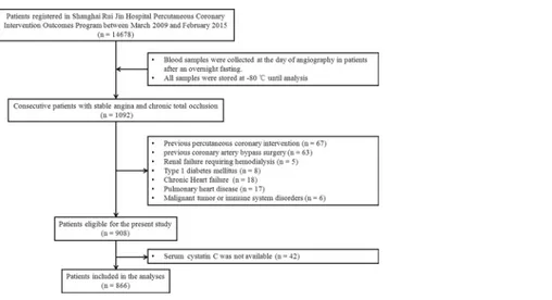
glycation endproducts in diabetes. Two-hundred and twenty six patients were excluded by the following exclusion criteria: (a) percutaneous coronary intervention within the prior 3 months; (b) previous coronary artery bypass grafting; (c) renal failure requiring hemodialysis; (d) chronic heart failure, pulmonary heart disease, malignant tumor or immune system disorders; (e) type 1 diabetes; and (f) unavailability of cystatin C data. Finally, 866 patients fit the inclu-sion criteria and composed the study cohort(Fig 1).
Hypertension, type 2 diabetes and dyslipidemia were defined according to European Society of Hypertension (ESH) / European Society of Cardiology (ESC) guidelines for the management of arterial hypertension [18], criteria of the American Diabetes Association [19], and Third Report of The National Cholesterol Education Program (NCEP) [20], respectively. Stable angina was diagnosed based on the criteria of the American College of Cardiology/ American Heart Association [21].
Angiography and Collateral Grading
Coronary angiography was performed via the femoral or radial access with 6Fr diagnostic cath-eters. The angiographic assessment was done independently by two blinded interventional car-diologists (RYZ, QZ), according to lesion classification scheme of the American College of Cardiology/ American Heart Association [22]. In case of disagreement, the difference in inter-pretation was resolved by a third reviewer (WFS). The Rentrop scoring system was used to grade collateral filling from the contra-lateral vessel (often via connections of the epicardial surface or intraventricular septum) as described previously [17]: 0 = none; 1 = filling of side branches only; 2 = partially filling of the epicardial segment; 3 = complete filling of the epicar-dial segment. Poor coronary collateralization was defined as Rentrop score 0 or 1 and good cor-onary collateralization was defined as Rentrop score 2 or 3, as in previous studies [6–10]. In patients with more than one chronic total occlusion, the vessel with the highest collateral grade was chosen for analysis.
Biochemical Measurement and Estimation of GFR
We obtained blood samples at the day of angiography in all patients after an overnight fasting, and stored them at -80°C until analysis. Assessment of serum levels of creatinine, blood urea nitrogen, uric acid, lipid profiles, glucose, and glycosylated hemoglobin (HbA1c), was made
Fig 1. Flowchart of patient enrollment.
with standard laboratory techniques [7,8]. Serum cystatin C was determined by high sensitive latex-enhanced immune- turbidimetric method with an automatic biochemical analyzer (7600–020; Hitachi Inc, Tokyo, Japan) [23,24], and hsCRP level was assayed by ELISA (Bio-check Laboratories, Toledo, OH, USA). Glomerular filtration rate (GFR) was estimated using the creatinine-based abbreviated Modification of Diet in Renal Disease (MDRD) formula [25].
Statistical Analysis
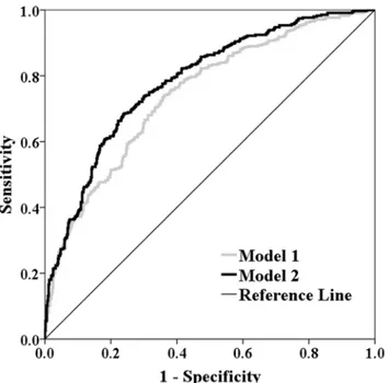
EpiData Entry 3.1 Software was used for data entry and documentation, and EXCEL was adopted to manage database. SPSS 20.0 for Windows (SPSS, Inc., Chicago, IL, USA) was used for statistical analyses. Data are presented as mean ± standard deviation (SD) and number (percentages). For continuous variables, differences between groups were evaluated by t test for normally distributed values; otherwise, the Mann-Whitney U test was applied. For categorical variables, differences between groups were evaluated with the chi-square test. Spearman’s rho test was used to assess the correlation between serum cystatin C and Rentrop scores. The serum levels of cystatin C were divided into 4 groups according to quartile distribution. Uni-variable and multiUni-variable logistic regression analyses after adjustment for age, gender, body mass index (BMI), traditional risk factors for coronary artery disease including smoking, hypertension, hyperlipidemia and diabetes, multi-vessel disease, GFR, and hsCRP were per-formed to detect the relationship between poor collateralization and serum levels of cystatin C. Regression models with above-mentioned potential confounding factors (model 1) and further inclusion of serum cystatin C (model 2) were also developed to explore the independent deter-minants for poor collateralization. Hosmer–Lemeshow X2test was used for model calibration. Receiver-operating characteristic (ROC) analyses were performed with serum cystatin C and predicted probabilities (C statistic) for poor collateralization derived from regression models with and without cystatin C. The areas under the curves (AUC) were compared using DeLong method with MedCalc software for windows (version 11.4, Mariakerke,Belgium). The likeli-hood ratio test and the net reclassification improvements (NRI) and integrated discrimination improvements (IDI) were used to assess the improvement of goodness of fit and predictive per-formance for model 2 compared with model 1 [26]. All analyses used 2-sided tests with an overall significance level of alpha = 0.05.
Results
Baseline Characteristics
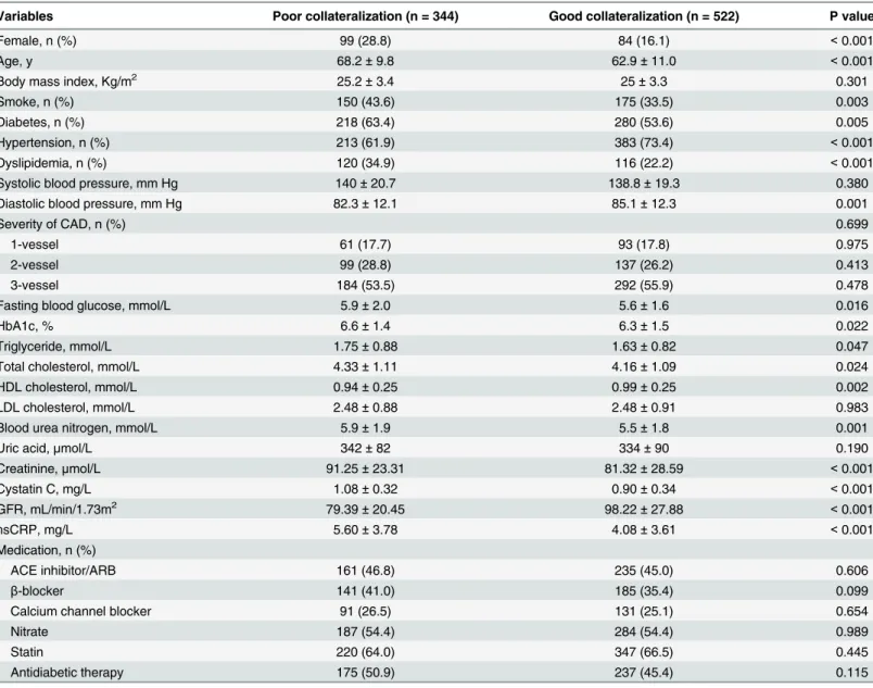
Among overall 866 patients included in the final analysis, poor and good coronary collaterali-zation was detected in 344 and 522 patients, respectively. Patients with poor coronary collatera-lization were older, females and cigarette smokers in higher percentage and had more diabetes and dyslipidemia but were less hypertensive than those with good collateralization (for all com-parisons, P<0.01). Despite similar degree of coronary artery disease and medical treatments,
diastolic blood pressure, high-density lipoprotein cholesterol, and GFR were lower, but serum levels of creatinine and hsCRP were more elevated in those with poor collateralization (Table 1).
Serum Cystatin C and Coronary Collateralization
Serum cystatin C was significantly higher in patients with poor collateralization than in those with good collateralization (1.08 ± 0.32 mg/L vs. 0.90 ± 0.34 mg/L, P<0.001), and correlated
inversely with Rentrop score before (Spearmen’s r = -0.263, P<0.001) and after (Spearmen’s r
disease (including smoking, hypertension, hyperlipidemia and diabetes), multi-vessel disease, GFR and serum level of hsCRP. In addition, the prevalence of poor coronary collaterals increased stepwise from the lowest quartile (<0.76 mg/L) to the highest quartile of serum cystatin C
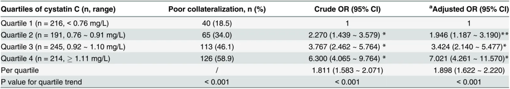
(1.11 mg/L) (P for trend<0.001)(Fig 2). Odds ratio for poor collateralization increased to
6.300 (95% confidence interval [CI] 4.065 ~ 9.764) in the highest compared with those in the lowest quartile of cystatin C level (P<0.001). These associations remained significant after
adjusting for multiple variables (OR: 7.021, 95% CI 4.261 ~ 11.570, P<0.001)(Table 2).
ROC curve analysis showed that AUC was 0.687 (95% CI 0.652 ~ 0.722, P<0.001) and the
optimal cut-point of serum cystatin C was 0.97 mg/L, with a diagnostic sensitivity and specific-ity of 61.3% and 67.2% for the presence of poor coronary collateralization.
Table 1. Baseline characteristics and biochemical assessment in patients with poor and good collateralization.
Variables Poor collateralization (n = 344) Good collateralization (n = 522) P value
Female, n (%) 99 (28.8) 84 (16.1) <0.001
Age, y 68.2±9.8 62.9±11.0 <0.001
Body mass index, Kg/m2 25.2±3.4 25±3.3 0.301
Smoke, n (%) 150 (43.6) 175 (33.5) 0.003
Diabetes, n (%) 218 (63.4) 280 (53.6) 0.005
Hypertension, n (%) 213 (61.9) 383 (73.4) <0.001
Dyslipidemia, n (%) 120 (34.9) 116 (22.2) <0.001
Systolic blood pressure, mm Hg 140±20.7 138.8±19.3 0.380
Diastolic blood pressure, mm Hg 82.3±12.1 85.1±12.3 0.001
Severity of CAD, n (%) 0.699
1-vessel 61 (17.7) 93 (17.8) 0.975
2-vessel 99 (28.8) 137 (26.2) 0.413
3-vessel 184 (53.5) 292 (55.9) 0.478
Fasting blood glucose, mmol/L 5.9±2.0 5.6±1.6 0.016
HbA1c, % 6.6±1.4 6.3±1.5 0.022
Triglyceride, mmol/L 1.75±0.88 1.63±0.82 0.047
Total cholesterol, mmol/L 4.33±1.11 4.16±1.09 0.024
HDL cholesterol, mmol/L 0.94±0.25 0.99±0.25 0.002
LDL cholesterol, mmol/L 2.48±0.88 2.48±0.91 0.983
Blood urea nitrogen, mmol/L 5.9±1.9 5.5±1.8 0.001
Uric acid,μmol/L 342±82 334±90 0.190
Creatinine,μmol/L 91.25±23.31 81.32±28.59 <0.001
Cystatin C, mg/L 1.08±0.32 0.90±0.34 <0.001
GFR, mL/min/1.73m2 79.39±20.45 98.22±27.88 <0.001
hsCRP, mg/L 5.60±3.78 4.08±3.61 <0.001
Medication, n (%)
ACE inhibitor/ARB 161 (46.8) 235 (45.0) 0.606
β-blocker 141 (41.0) 185 (35.4) 0.099
Calcium channel blocker 91 (26.5) 131 (25.1) 0.654
Nitrate 187 (54.4) 284 (54.4) 0.989
Statin 220 (64.0) 347 (66.5) 0.445
Antidiabetic therapy 175 (50.9) 237 (45.4) 0.115
Data are mean±SD or number (%). ACE, angiotensin converting enzyme; ARB, angiotensin receptor blocker; CAD, coronary artery disease; GFR,
glomerularfiltration rate; HbA1c, glycosylated hemoglobin A1c; HDL, high-density lipoprotein; hsCRP, high sensitive C reactive protein; LDL, low-density lipoprotein
Risk for Poor Collateralization
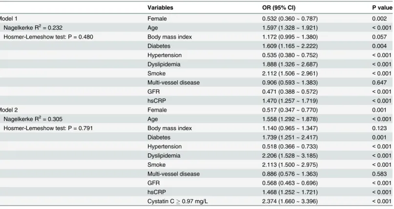
Multivariate logistic regression analysis revealed that age, gender, traditional risk factors for coronary artery disease, GFR and serum level of hsCRP were determinants for poor coronary collateralization (model 1). After adjustment for these variables, serum cystatin C0.97 mg/L remained independently associated with poor collateralization (OR: 2.374, 95% CI 1.660 ~ 3.396, P<0.001) (model 2)(Table 3). The calibrations of both models were good (P = 0.480
and P = 0.791, respectively). Compared with model 1, the addition of serum cystatin C (0.97 mg/L) in model 2 significantly improved the goodness-of-fit and predictive performance with an increase of Nagelkerke R2of 7.3% (P<0.001) and C statistic of 0.039 (95% CI 0.020 ~
0.059, P<0.001)(Fig 3)and a NRI and IDI of 10.5% (95% CI 4.6% ~ 16.4%, P<0.001) and
5.9% (95% CI 4.3% ~ 7.5%, P<0.001), respectively.
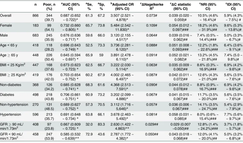
Diagnostic Value of Cystatin C in Different Patient Subgroups
We performed the sensitivity analyses according to age, sex, and presence or absence of over-weight or obesity (BMI25 Kg/m2), diabetes, hypertension and at least mild renal dysfunction (GFR<90 mL/min/1.73m2)(Table 4). The diagnostic value of cystatin C for detecting poor
coronary collateralization was consistent in all patient subgroups, with the AUCs ranging from
Fig 2. Prevalence of poor collateralization in patients across quartiles of serum cystatin C.
doi:10.1371/journal.pone.0137253.g002
Table 2. Odds ratio of poor collateralization according to cystatin C in patients with chronic total occlusion.
Quartiles of cystatin C (n, range) Poor collateralization, n (%) Crude OR (95% CI) aAdjusted OR (95% CI)
Quartile 1 (n = 216,<0.76 mg/L) 40 (18.5) 1 1
Quartile 2 (n = 191, 0.76 ~ 0.91 mg/L) 65 (34.0) 2.270 (1.439 ~ 3.579)* 1.946 (1.187 ~ 3.190)** Quartile 3 (n = 245, 0.92 ~ 1.10 mg/L) 113 (46.1) 3.767 (2.462 ~ 5.764)* 3.424 (2.140 ~ 5.477)* Quartile 4 (n = 214,1.11 mg/L) 126 (58.9) 6.300 (4.065 ~ 9.764)* 7.021 (4.261 ~ 11.570)*
Per quartile / 1.811 (1.583 ~ 2.071) 1.898 (1.622 ~ 2.220)
P value for quartile trend <0.001 <0.001 <0.001
CI, confidence interval; OR, odds ratio. *P<0.001
**P<0.01 vs quartile 1
aMultiple-adjustment for gender, age, body mass index, diabetes, hypertension, dyslipidemia, smoke, multi-vessel disease, glomerular
filtration rate and serum level of high sensitive C reactive protein
0.585 to 0.732 (all P<0.01). After adjustment for potential confounding factors, serum
cysta-tin C0.97 mg/L was independently associated with poor coronary collateralization (OR: 2.630 ~ 5.484, all P<0.01). The calibrations of all models with and without cystatin C were
good (P0.125). Compared with the models with traditional variables, additional inclusion of serum cystatin C0.97 mg/L also significantly improved the goodness-of-fit and predictive performance in all subgroup analyses with an increase of Nagelkerke R2of 2.9% ~ 10.9% (all P<0.01), C statistic of 0.029 ~ 0.058 (all P<0.05), and a NRI and IDI of 8.0% ~ 18.2% (all
P<0.05) and 3.7% ~ 9.6% (all P<0.001), respectively(Table 4).
Discussion
This study showed that elevated serum cystatin C was associated with poor coronary collatera-lization in patients with stable coronary artery disease and chronic total occlusion.
It is well recognized that after coronary artery occlusion, myocardial blood flow distal to occluded segment could be, at least partly, restored by collateral formation mainly through two different processes of blood vessel growth. New capillaries may form via ischemia-induced sprouting from post-capillary venules (angiogenesis), and the development and maturation of these newly growing vessels are regulated by a balance of pro-angiogenic and anti-angiogenic factors. Arteriogenesis denotes a transformation of pre-existing arterioles into functional (mus-cular) collateral arteries and is predominantly stimulated by pressure gradient across the occluded segment and tangential fluid shear stress at the vessel wall. Arteriogenesis, not angio-genesis, is the leading mechanism in restoring blood flow following arterial occlusion. Both processes are related to endothelial and smooth muscle cells and various growth factors and
Table 3. Logistic regression analyses for poor collateralization in patients with chronic total occlusion.
Variables OR (95% CI) P value
Model 1 Female 0.532 (0.360 ~ 0.787) 0.002
Nagelkerke R2= 0.232 Age 1.597 (1.328 ~ 1.921) <0.001
Hosmer-Lemeshow test: P = 0.480 Body mass index 1.172 (0.995 ~ 1.380) 0.057
Diabetes 1.609 (1.165 ~ 2.222) 0.004
Hypertension 0.535 (0.380 ~ 0.752) <0.001
Dyslipidemia 1.888 (1.326 ~ 2.687) <0.001
Smoke 2.112 (1.506 ~ 2.961) <0.001
Multi-vessel disease 0.906 (0.593 ~ 1.383) 0.647
GFR 0.471 (0.388 ~ 0.572) <0.001
hsCRP 1.470 (1.257 ~ 1.719) <0.001
Model 2 Female 0.517 (0.347 ~ 0.770) 0.001
Nagelkerke R2= 0.305 Age 1.558 (1.292 ~ 1.878) <0.001
Hosmer-Lemeshow test: P = 0.791 Body mass index 1.140 (0.965 ~ 1.347) 0.123
Diabetes 1.739 (1.251 ~ 2.417) 0.001
Hypertension 0.518 (0.366 ~ 0.733) <0.001
Dyslipidemia 2.206 (1.528 ~ 3.185) <0.001
Smoke 2.113 (1.500 ~ 2.975) <0.001
Multi-vessel disease 0.886 (0.576 ~ 1.363) 0.583
GFR 0.568 (0.463 ~ 0.696) <0.001
hsCRP 1.468 (1.252 ~ 1.721) <0.001
Cystatin C0.97 mg/L 2.374 (1.660 ~ 3.396) <0.001
CI, confidence interval; GFR, glomerularfiltration rate; hsCRP, high sensitive C reactive protein; OR, odds ratio
adversely affected by inflammatory cytokines [27]. Collateral growth in patients with coronary artery disease is highly heterogeneous and influenced by a number of factors. We found that old age, female gender, traditional risk factors for coronary artery disease, and reduced renal function were associated with poor collateralization, whereas hypertension, especially elevated diastolic blood pressure, correlated with well-formed coronary collaterals. Several studies have suggested that poorly developed coronary collaterals may be related to chronic inflammation of low degree, as evidenced by elevated serum levels of cytokines such as hsCRP [28], tumor necrosis factor (TNF)-α[29], soluble endothelial adhesion molecules [30], monocyte chemoat-tractant protein-1 [31], and C1q/TNF-related protein-1 [8], which inhibit key components of angiogenesis particularly endothelial progenitor cell differentiation, survival and function. Recently, apelin has been shown to promote angiogenesis, and its serum levels were associated with coronary collateral development [32]. Akin et al revealed that white blood cell subtypes, especially the neutrophil-to-lymphocyte ratio, can be used as an indicator of systemic inflam-mation, and an elevated neutrophil- to-lymphocyte ratio was independently associated with impaired coronary collateral formation in patients with stable coronary artery disease [9]. High serum cystatin C was thought to be related directly to both inflammation and atherosclerosis [13]. Some studies have highlighted that inflammatory cytokines stimulate lysosomal cathep-sins, which may be associated with increased cystatin C levels [33]. These effects could cause worse vascular endothelial changes, inflammation and atherosclerosis in patients with coro-nary artery disease. However, the role of cystatin C in the formation of corocoro-nary collaterals remains largely unclear. The present study demonstrated that poor coronary collateralization corresponded significantly with elevated cystatin C levels, which is contrast to a previous report showing no significant difference in serum cystatin C between patients with poor and good coronary collaterals [34]. The exact explanation behind these different results may be, at least
Fig 3. Receiver operating characteristic curves of predicted probabilities derived from regression models for detecting poor collateralization.Model 1 includes variables of age, gender, body mass index, traditional risk factors for coronary artery disease (diabetes, hypertension, dyslipidemia and smoke), multi-vessel disease, GFR and serum level of high sensitive C reactive protein. Model 2 includes variables in model 1 and serum cystatin C0.97 mg/L.
partly, related to a heterogeneous population in their study, which contained a small number of diabetic patients (<20% in the final analysis) and a significant proportion of patients with
acute coronary syndrome (approximately 50%) or non-occluded coronary lesions [34]. It is well documented that hyperglycemia abolished the formation of coronary collateral vessels [35], and diabetes represents an independent risk factor for poor coronary collateralization [10, 36]. In addition, cysteine proteases are associated with formation of necrotic core and rupture of the cap, as well as macrophage apoptosis in atherosclerotic plaques, and the synthesis of cystatin C may be significantly increased during acute myocardial ischemia [37]. Notably, the recruitment of coronary collaterals is predominantly driven by shear force along the pressure
Table 4. The diagnostic value of serum cystatin C for evaluation of poor collateralization in different patients subgroups.
n Poor, n (%)
aAUC (95%
CI)
aSe,
%
aSp,
%
bAdjusted OR
(95% CI)
c
ΔNagelkerke R2
c
ΔC statistic (95% CI)
cNRI (95%
CI)
cIDI (95%
CI)
Overall 866 344
(39.7)
0.687 (0.652 ~ 0.722)*
61.3 67.2 3.507 (2.521 ~ 4.878)*
0.073# 0.039 (0.020 ~ 0.059)#
10.5% (4.6% ~ 16.4%)#
5.9% (4.3% ~ 7.5%) #
Female 183 99
(54.1)
0.732 (0.660 ~ 0.805)*
65.7 73.8 5.484 (2.542 ~ 11.830)*
0.109# 0.054 (0.012 ~ 0.097)###
18.2% (4.6% ~ 31.9%)##
9.6% (5.3% ~ 13.8%)#
Male 683 245
(35.9)
0.676 (0.636 ~ 0.717)*
59.6 66.0 3.120 (2.155 ~ 4.517)*
0.064# 0.039 (0.016 ~ 0.062)##
7.4% (0.5% ~ 14.4%)###
5.0% (3.3% ~ 6.7%)# Age<65 y 418 118
(28.2)
0.696 (0.643 ~ 0.749)*
52.5 73.3 3.736 (2.281 ~ 6.120)*
0.088# 0.051 (0.008 ~ 0.093)###
12.2% (1.8% ~ 22.6%)###
6.4% (3.6% ~ 9.1%)#
Age65 y 448 226
(50.4)
0.645 (0.594 ~ 0.697)*
65.9 59 3.891 (2.478 ~ 6.110)*
0.091# 0.052 (0.021 ~ 0.082)#
13.2% (4.5% ~ 21.8%)##
7.4% (5.0 ~ 9.8%)# BMI<25 Kg/m2 447 168
(37.6)
0.673 (0.623 ~ 0.723)*
62.5 66.7 3.222 (2.030 ~ 5.114)*
0.063# 0.035 (0.009 ~ 0.062)##
8.6% (0.3% ~ 16.9%)###
6.9% (4.2% ~ 9.6%)# BMI25 Kg/m2 419 176
(42.0)
0.703 (0.654 ~ 0.752)*
60.2 67.9 4.002 (2.465 ~ 6.497)*
0.087# 0.042 (0.011 ~ 0.072)##
12.6% (4.3% ~ 21.0%)##
5.6% (3.5% ~ 7.6%)# Non-diabetes 368 126
(34.2)
0.685 (0.628 ~ 0.741)*
68.3 61.6 4.368 (2.513 ~ 7.591)*
0.090# 0.044 (0.012 ~ 0.078)##
8.8% (0.9% ~ 16.7%)###
6.9% (4.2% ~ 9.6%)#
Diabetes 498 218
(43.8)
0.706 (0.661 ~ 0.751)*
60.9 73.2 3.202 (2.099 ~ 4.886)*
0.067# 0.041 (0.015 ~ 0.067)##
11.7% (3.5% ~ 20.0%)##
5.6% (3.5% ~ 7.6%)# Non-hypertension 270 131
(48.5)
0.689 (0.627 ~ 0.752)*
57.3 70.5 3.112 (1.716 ~ 5,646)*
0.057# 0.036 (0.006 ~ 0.067)###
14.1% (3.5% ~ 24.7%)##
5.4% (2.9% ~ 7.9%)# Hypertension 596 213
(35.7)
0.691 (0.648 ~ 0.734)*
63.8 66.1 3.678 (2.463 ~ 5.492)*
0.081# 0.058 (0.031 ~ 0.085)#
8.0% (0.6% ~ 15.4%)###
7.7% (5.6% ~ 9.7%)# GFR90 mL/
min/1.73m2 408 (23.8)97 0.667 (0.609~ 0.725)
*
32.0 83.3 2.630 (1.422 ~ 4.863)**
0.029## 0.029 (0.007 ~0.050)##
12.8% (1.4% ~ 24.2%)###
3.7% (1.8% ~ 5.7%)# GFR<90 mL/
min/1.73m2 458 (53.9)247 0.585 (0.532~ 0.639)
**
72.9 43.6 2.787 (1.772 ~ 4.382)*
0.050## 0.043 (0.018 ~ 0.068)##
12.0% (4.1% ~ 20.0%)##
5.0% (3.2% ~ 6.8%)#
AUC, area under curve; BMI, body mass index; CI, confidence interval; IDI, integrated discrimination improvement; GFR, glomerularfiltration rate; NRI,
net reclassification improvement; OR, odds ratio
a AUC,Se and Sp denote area under the curve for serum cystatin C in detecting poor collateralization in different patients subgroup and corresponding sensitivity and specificity with a cut-off point of 0.97 mg/L
b odds ratio (95% CI) of serum cystatin C0.97 mg/L after adjustment for gender, age, BMI, diabetes, hypertension, dyslipidemia, multi-vessel disease and serum level of high sensitive C reactive protein (hsCRP).
c Improvement of goodness-of-fit and predictive performance of additional inclusion of serum cystatin C0.97 mg/L to the model with traditional variables including gender, age, BMI, diabetes, hypertension, dyslipidemia, multi-vessel disease and hsCRP
*P<0.001
**P<0.01 for AUC or adjusted OR of cystatin C in diagnosing poor collateralization
#P<0.001
##P<0.01
###P<0.05 for comparisons between logistic regression models with and without serum cystatin C0.97 mg/L
gradient that develops when the native vessel is tightly stenotic or even completely occluded [16,38]. In view of these conditions and for avoiding possible confounding factors, we analyzed the relationship between serum cystatin C and coronary collateralization in a unique cohort of patients with stable angina and chronic total occlusion.
The main finding of this study is that serum cystatin C correlated inversely with Rentrop score even after adjusting for multiple variables including GFR and levels of hsCRP. The preva-lence of poor coronary collateralization increased stepwise with increasing cystatin C quartiles. An optimal cut-off point of serum cystatin C level of 0.97 mg/L provided a diagnostic sensitiv-ity and specificsensitiv-ity of 61.3% and 67.2% for the presence of poor coronary collateralization. Patients with a cystatin C0.97mg/L had 2.37-fold increased risk of poor collateralization. Interestingly, additional inclusion of serum cystatin C0.97 mg/L in the model significantly improved the diagnostic accuracy for poor coronary collateralization in all subgroup popula-tion based on age, gender, presence or absence of diabetes, hypertension, or at least mild renal dysfunction by 8.0–18.2%. These observations support a notion that anti-angiogenic cystatin C is an indicator of coronary collateral formation in patients with stable coronary artery disease and chronic total occlusion. The results of the present study are also in line with our previous findings that cystatin C-based equation was superior to creatinine-based formula for estimat-ing GFR and identifyestimat-ing poor collateralization in type 2 diabetic patients with coronary artery disease [24]. Because available data on cystatin C-based GFR equation remain very limited, and measurement of cystatin C is inexpensive and easily available, the possible use of serum cystatin C as a marker for risk of poor coronary collateralization would be desirable and clini-cally meaningful in the management of stable coronary artery disease patients.
It is evident that the effect of cystatin C levels on coronary collateralization reflects the net result of several pathophysiological processes. Cystatin Cper sedisplays anti-angiogenic
char-acteristics by reducing endothelial cell tubule formation and cysteinyl cathepsin activities [37, 39]. Furthermore, cystatin C as a cysteine proteinase inhibitor is associated with cardiovascular risk factors as well as inflammation, which may promote vascular endothelial damage and cause poor collateralization [13,15]. Notably, approximately 60% of patients had type 2 diabe-tes in the present study. Previous reports including ours have shown that an interaction between advanced glycation endproducts (AGE) and receptor for AGE (RAGE) plays a critical role in the acceleration of coronary atherosclerosis and abnormal collateral vessel formation [10,35,36]. Likewise, renal dysfunction which commonly occurs in patients with severe coro-nary artery disease and is partly reflected by elevated serum levels of cystatin C, adversely affect several components necessary for collateral growth through various regulatory mechanisms and gene expression [40–44].
The development of coronary collaterals may be also influenced by medications. It has been shown that statin treatment was associated with good collateralization assessed by the Rentrop classification due partly to reduced apoptosis [45], whereas angiotensin-converting enzyme inhibitor (ACEI) therapy contributed to poor collateral development in patients with coronary occlusion via inhibiting the expression of angiotensin II-induced growth factors such as vascu-lar endothelial growth factor, platelet-derived growth factor, and fibroblast growth factor [46]. Recently, van der Hoeven et al reported a positive relation between chronic use ofβblockers and well-formed collaterals assessed by collateral flow index [47]. The use ofβblockers reduces heart rate and improves fluid shear stress at the endothelial wall which stimulates coronary col-lateral growth. In addition,βblockers could decrease catecholamine-mediated inflammatory response. In this study we did not observe an association between medical treatments with stat-ins, ACEI orβblockers and angiographic grade of coronary collateralization.
invasive parameters of collateral function and does not actually rate the collaterals themselves, but their effect in filling the occluded arterial segment [17]. Furthermore, adequate angio-graphic plane and optimal axial alignment of the catheter during contrast injection and acqui-sition time of collateral filling are important for assessing collateral circulation using the Rentrop method. Measurement of coronary flow index which requires simultaneous record-ings of central aortic pressure and the distal pressure within the occluded segment of the coro-nary artery represents the“gold standard”for evaluating the competence and functional significance of coronary collaterals. However, coronary flow index can only be determined dur-ing coronary intervention, and meticulous care should be taken to keep the guiddur-ing catheter away from the coronary orifice during aortic pressure recording and to ensure the absence of antegrade flow leakage after pressure wire passage.
One of the limitations is that the study design was one of retrospective analysis for the point of coronary collateral investigation; thus, it allows us to detect association but not to predict outcome. Nevertheless, these results may reflect true associations in the real world setting as prescription data were captured from a standard database. Another limitation is that our study did not include other possible factors influencing collateral development in patients with coro-nary artery disease especially inflammatory cytokines [8–12,28–31], although the effects of the established clinical variables were evaluated by multivariate analysis. The relative strength of a model by incorporating all potential biomarkers for predicting poor coronary collateralization deserves further investigations.
In conclusion, the present study is the first to demonstrate an association between elevated serum cystatin C and reduced angiographic coronary collateralization in a large cohort of sta-ble coronary disease patients with chronic total occlusion. The optimal cut-point of cystatin C in serum of0.97 mg/L indicates a great increased risk of poor coronary collateralization. These findings may provide new insights on risk assessment and management strategy for this unique high-risk population.
Supporting Information
S1 Document. Protocol Registration Receipt: Advanced Glycation Endproducts and Devel-opment of CAD (AGENDA) (NCT02089360).
(DOCX)
Author Contributions
Conceived and designed the experiments: YS. Performed the experiments: RYZ QZ LL. Ana-lyzed the data: FHD. Contributed reagents/materials/analysis tools: LL. Wrote the paper: WFS.
References
1. Traupe T, Gloekler S, de Marchi SF, Werner GS, Seiler C (2010) Assessment of the human coronary collateral circulation. Circulation 122:1210–1220. doi:10.1161/CIRCULATIONAHA.109.930651PMID:
20855668
2. Hsu PC, Su HM, Lee HC, Juo SH, Lin TH, Voon WC, et al. (2014) Coronary collateral circulation in patients of coronary ectasia with significant coronary artery disease. PLoS One 9:e87001. doi:10. 1371/journal.pone.0087001PMID:24475209
3. Celik T, Iyisoy A, Yuksel CU, Celik M, Isik E (2010) The prognostic significance of coronary collaterals in patients with ischemic heart disease: an essential response to ischemia. Int J Cardiol 138:101–103.
doi:10.1016/j.ijcard.2008.06.012PMID:18657333
4. Meier P, Hemingway H, Lansky AJ, Knapp G, Pitt B, Seiler C. (2012) The impact of the coronary collat-eral circulation on mortality: a meta-analysis. Eur Heart J 33:614–621. doi:10.1093/eurheartj/ehr308
5. Shen Y, Wu F, Pan C, Zhu T, Zhang Q, Zhang RY, et al. (2014) Clinical relevance of angiographic coro-nary collaterals during primary corocoro-nary intervention for acute ST-elevation myocardial infarction. Chin Med J (Engl) 127:66–71.
6. Sun Z, Shen Y, Lu L, Zhang RY, Pu LJ, Zhang Q, et al. (2013) Clinical and angiographic features asso-ciated with coronary collateralization in stable angina patients with chronic total occlusion. J Zhejiang Univ Sci B 14:705–712. doi:10.1631/jzus.BQICC704PMID:23897789
7. Shen Y, Ding FH, Wu F, Lu L, Zhang RY, Zhang Q, et al. (2015) Association of blood pressure and cor-onary collateralization in type 2 diabetic and nondiabetic patients with stable angina and chronic total occlusion. J Hypertens 33:621–626. doi:10.1097/HJH.0000000000000455PMID:25490709
8. Shen Y, Lu L, Liu ZH, Wu F, Zhu JZ, Sun Z, et al. (2014) Increased serum level of CTRP1 is associated with low coronary collateralization in stable angina patients with chronic total occlusion. Int J Cardiol 174:203–206. doi:10.1016/j.ijcard.2014.03.205PMID:24746545
9. Akın F, Ayça B, Çelik Ö,Şahin C (2015) Predictors of poor coronary collateral development in patients with stable coronary artery disease: Neutrophil-to-lymphocyte ratio and platelets. Anatol J Cardiol 15:218–223. doi:10.5152/akd.2014.5263PMID:25880175
10. Shen Y, Lu L, Ding FH, Sun Z, Zhang RY, Zhang Q, et al. (2013) Association of increased serum gly-cated albumin levels with low coronary collateralization in type 2 diabetic patients with stable angina and chronic total occlusion. Cardiovasc Diabetol 12:165. doi:10.1186/1475-2840-12-165PMID:
24209601
11. van den Hengel LG, Hellingman AA, Nossent AY, van Oeveren-Rietdijk AM, de Vries MR, Spek CA, et al. (2013) Protease-activated receptor (PAR)2, but not PAR1, is involved in collateral formation and anti-inflammatory monocyte polarization in a mouse hind limb ischemia model. PLoS One 8:e61923. doi:10.1371/journal.pone.0061923PMID:23637930
12. Keeley EC, Moorman JR, Liu L, Gimple LW, Lipson LC, Ragosta M, et al. (2011) Plasma chemokine levels are associated with the presence and extent of angiographic coronary collaterals in chronic ischemic heart disease. PLoS One 6:e21174. doi:10.1371/journal.pone.0021174PMID:21731663
13. Angelidis C, Deftereos S, Giannopoulos G, Anatoliotakis N, Bouras G, Hatzis G, et al. (2013) Cystatin C: an emerging biomarker in cardiovascular disease. Curr Top Med Chem 13:164–179. PMID:
23470076
14. Liu F, Shen J, Zhao J, Zeng H, Li L, Zhao J, et al. (2013) Cystatin C: a strong marker for lower limb ischemia in Chinese type 2 diabetic patients? PLoS One 8:e66907. doi:10.1371/journal.pone. 0066907PMID:23843968
15. Ferraro S, Marano G, Biganzoli EM, Boracchi P, Bongo AS (2011) Prognostic value of cystatin C in acute coronary syndromes: enhancer of atherosclerosis and promising therapeutic target. Clin Chem Lab Med 49:1397–1404. doi:10.1515/CCLM.2011.607PMID:21605013
16. Levin DC (1974) Pathways and functional significance of the coronary collateral circulation. Circulation 50:831–837. PMID:4425386
17. Rentrop KP, Cohen M, Blanke H, Phillips RA (1985) Changes in collateral channel filling immediately after controlled coronary artery occlusion by an angioplasty balloon in human subjects. J Am Coll Car-diol 5:587–592. PMID:3156171
18. Task Force Members (2013) 2013 ESH/ESC Guidelines for the management of arterial hypertension: the Task Force for the management of arterial hypertension of the European Society of Hypertension (ESH) and of the European Society of Cardiology (ESC). J Hypertens 31:1281–1357. doi:10.1097/01. hjh.0000431740.32696.ccPMID:23817082
19. Expert Committee on the Diagnosis and Classification of Diabetes Mellitus (2003) Report of the expert committee on the diagnosis and classification of diabetes mellitus. Diabetes Care 26 Suppl 1:S5–20. PMID:12502614
20. Expert Panel on Detection, Evaluation, and Treatment of High Blood Cholesterol in Adults (2001) Exec-utive Summary of The Third Report of The National Cholesterol Education Program (NCEP) Expert Panel on Detection, Evaluation, And Treatment of High Blood Cholesterol In Adults (Adult Treatment Panel III). JAMA 285:2486–2497. PMID:11368702
21. Gibbons RJ, Abrams J, Chatterjee K, Daley J, Deedwania PC, Douglas JS, et al. (2007) 2007 chronic angina focused update of the ACC/AHA 2002 guidelines for the management of patients with chronic stable angina: a report of the American College of Cardiology/American Heart Association Task Force on Practice Guidelines Writing Group to develop the focused update of the 2002 guidelines for the man-agement of patients with chronic stable angina. J Am Coll Cardiol 50:2264–2274. PMID:18061078
23. Feng JF, Qiu L, Zhang L, Li XM, Yang YW, Zeng P, et al. (2013) Multicenter study of creatinine- and/or cystatin C-based equations for estimation of glomerular filtration rates in Chinese patients with chronic kidney disease. PLoS One 8:e57240. doi:10.1371/journal.pone.0057240PMID:23526939
24. Shen Y, Ding FH, Wu F, Sun Z, Zhang RY, Zhang Q, et al. (2014) Cystatin C versus creatinine- based definition of renal dysfunction for predicting poor coronary collateralization in type 2 diabetic patients with stable coronary artery disease. J Diabetes & Metab 5:453–459.
25. Ma YC, Zuo L, Chen JH, Luo Q, Yu XQ, Li Y, et al. (2006) Modified glomerular filtration rate estimating equation for Chinese patients with chronic kidney disease. J Am Soc Nephrol 17:2937–2944. PMID:
16988059
26. Pencina MJ, D'Agostino RB Sr, D'Agostino RB Jr, Vasan RS (2008) Evaluating the added predictive ability of a new marker: from area under the ROC curve to reclassification and beyond. Stat Med 27:157–172. PMID:17569110
27. Seiler C, Stoller M, Pitt B, Meier P (2013) The human coronary collateral circulation: development and clinical importance. Eur Heart J 34:2674–2682. doi:10.1093/eurheartj/eht195PMID:23739241
28. Kerner A, Gruberg L, Goldberg A, Roguin A, Lavie P, Lavie L, et al. (2007) Relation of C-reactive protein to coronary collaterals in patients with stable angina pectoris and coronary artery disease. Am J Cardiol 99: 509–512. PMID:17293195
29. Seiler C, Pohl T, Billinger M, Meier B (2003) Tumour necrosis factor alpha concentration and collateral flow in patients with coronary artery disease and normal systolic left ventricular function. Heart 89: 96–
97. PMID:12482804
30. Guray U, Erbay AR, Guray Y, Yılmaz MB, BoyacıAA, Sasmaz H, et al. (2004) Poor coronary collateral circulation is associated with higher concentrations of soluble adhesion molecules in patients with sin-gle vessel disease. Coron Artery Dis 15: 413–417. PMID:15492590
31. Sahinarslan A, Kocaman SA, Topal S, Ercin U, Bukan N, Yalcin R, et al. (2010) Relation between serum monocyte chemoattractant protein-1 and coronary collateral development. Coron Artery Dis 21:455–459. doi:10.1097/MCA.0b013e32833fd29bPMID:20859200
32. Akboga MK, Akyel A, Sahinarslan A, Demirtas CY, Yayla C, Boyaci B, et al. (2014) Relationship between plasma apelin level and coronary collateral circulation. Atherosclerosis 235:289–294. doi:10. 1016/j.atherosclerosis.2014.04.029PMID:24905139
33. Li W, Kornmark L, Jonasson L, Forssell C, Yuan XM (2009) Cathepsin L is significantly associated with apoptosis and plaque destabilization in human atherosclerosis. Atherosclerosis 202:92–102. doi:10. 1016/j.atherosclerosis.2008.03.027PMID:18495127
34. Zhang J, Wang P, Huang YB, Li J, Zhu J, Luo X, et al. (2010) Plasma cathepsin L and its related pro/ antiangiogenic factors play useful roles in predicting rich coronary collaterals in patients with coronary heart disease. J Int Med Res 38:1389–1403. PMID:20926012
35. Abaci A1, Oğuzhan A, Kahraman S, Eryol NK, Unal S, Arinc H, et al. (1999) Effect of diabetes mellitus on formation of coronary collateral vessels. Circulation 99:2239–2242. PMID:10226087
36. Rocic P (2012) Why is coronary collateral growth impaired in type II diabetes and the metabolic syn-drome? Vascul Pharmacol 57:179–186. doi:10.1016/j.vph.2012.02.001PMID:22342811
37. Taglieri N, Koenig W, Kaski JC (2009) Cystatin C and cardiovascular risk. Clin Chem 55:1932–1943.
doi:10.1373/clinchem.2009.128397PMID:19713275
38. Schaper W (2009) Collateral circulation. Past and present. Basic Res Cardiol 104:5–21. doi:10.1007/ s00395-008-0760-xPMID:19101749
39. Lv BJ, Lindholt JS, Cheng X, Wang J, Shi GP (2012) Plasma cathepsin S and cystatin C levels and risk of abdominal aortic aneurysm: a randomized population-based study. PLoS One 7:e41813. doi:10. 1371/journal.pone.0041813PMID:22844527
40. Ying Y, Yang K, Liu Y, Chen QJ, Shen WF, Lu L, et al. (2011) A uremic solute, P-cresol, inhibits the pro-liferation of endothelial progenitor cells via the p38 pathway. Circ J 75:2252–2259. PMID:21747198
41. Hsu PC, Juo SH, Su HM, Chen SC, Tsai WC, Lai WT, et al. (2012) Predictor of poor coronary collaterals in chronic kidney disease population with significant coronary artery disease. BMC Nephrol 13:98. PMID:22935602
42. Xie SL, Li HY, Deng BQ, Luo NS, Geng DF, Wang JF, et al. (2011) Poor coronary collateral vessel development in patients with mild to moderate renal insufficiency. Clin Res Cardiol 100:227–233. doi:
10.1007/s00392-010-0233-8PMID:20865265
43. Kadi H, Ceyhan K, Sogut E, Koc F, Celik A, Onalan O, et al. (2011) Mildly decreased glomerular filtra-tion rate is associated with poor coronary collateral circulafiltra-tion in patients with coronary artery disease. Clin Cardiol 34:617–621. doi:10.1002/clc.20951PMID:21887692
45. Dincer I, Ongun A, Turhan S, Ozdol C, Kumbasar D, Erol C. (2006) Association between the dosage and duration of statin treatment with coronary collateral development. Coron Artery Dis 17:561–565.
PMID:16905969
46. Altin T, Kilickap M, Tutar E, Turhan S, Atmaca Y, Gulec S, et al. (2007). The relationship of chronic angiotensin converting enzyme inhibitor use and coronary collateral vessel development. Int Heart J 48:435–442. PMID:17827815