Rev Bras Cir Plást. 2012;27(3):490-2 490
Wolfenson M et al.
This study was performed at the Clínica Multiplástica do Recife, Recife, PE, Brazil.
Submitted to SGP (Sistema de Gestão de Publicações/Manager Publications System) of RBCP (Revista Brasileira de Cirurgia Plástica/Brazilian Journal of Plastic Surgery).
Article received: December 22, 2010 Article accepted: August 14, 2011
1. Plastic surgeon, full member of the Sociedade Brasileira de Cirurgia Plástica/Brazilian Society of Plastic Surgery (SBCP), Director of the Clínica Mul tiplástica do Recife, Recife, PE, Brazil.
2. Plastic surgeon, full member of the SBCP, Master in Plastic Surgery, Recife, PE, Brazil. 3. Student at the Faculty of Medical Sciences of the Universidade de Pernambuco, Recife, PE, Brazil
Franco T et al. Vendramin FS et al.
CASE REPORT
Breakage of the tip of the cannula in the adipose tissue
during liposuction
Quebra de ponta de cânula, no plano gorduroso, durante lipoaspiração
Moisés Wolfenson1
Claudio RonCatti2
edvaldo alfRedoda silva
JúnioR3
ABSTRACT
During liposuction, 3 cm of the distal segment of the cannula broke within the patient’s adiposetissue.Weusedanimageintensiiertoeasilyandquicklyaddressthiscomplication withoutthedevelopmentofanysequelaeforthepatient.
Keywords:Lipectomy/adverseeffects.Equipmentfailure.
RESUMO
Durante procedimento de lipoaspiração, uma cânula teve seu segmento distal de 3 cm quebradonoplanoadiposo.Osautoresapresentam,nestetrabalho,acondutaadotadapara soluçãodocaso,demaneirasimpleserápida,comempregodeumintensiicadordeimagens, semgrandesdiiculdades.
Descritores: Lipectomia/efeitosadversos.Falhadeequipamento.
INTRODUCTION
Described in 1979, liposuction was an important break through in plastic surgery and has become the most com monly performed surgical procedure of this specialty1.
Despite the high demand for liposuction and the popular acceptance of the procedure, its associated complications – which may be severe – have been targeted by the media.
It is worth noting that, even in the hands of an experienced plastic surgeon, this surgery presents risks. Some of these are rare, such as the breakage of the tip of the cannula within the adipose tissue that is described in this article. We report the solution that we adopted during the procedure to solve this complication;thatis,theuseofanimageintensiier.
CASE REPORT
A 35yearold female student (Caucasian) underwent wet liposuctionofthelanks,thighs,andbackunderepiduralanes thesia. She was maintained in the ventral decubitus position.
During liposuction of the back and upon cannula removal, the surgeon realized that its tip was missing. After ensuring thatthebrokentiphadnotfallenontheloororonthesurgical table, the possibility of breakage of the tip within the pa tient’s adipose tissue was considered.
Palpation of the broken tip of the cannula in the adipose tissue was hampered by the fact that the surgery had already taken half the time originally predicted, in addition to the inconveniencecausedbytheiniltrationofasolutioncontai ning epinephrine.
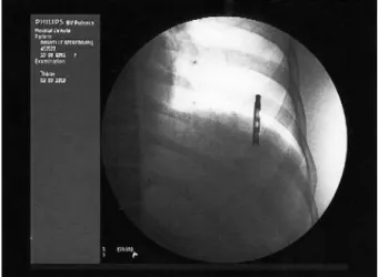
Itwasirstdecidedtoperformradiography,whichindi cated the tip in the adipose tissue. However, the twodimen sional image was not useful for assessing the precise location of the tip of the cannula, because it was impossible to evaluate the depth of the image.
Rev Bras Cir Plást. 2012;27(3):490-2 491
Breakage of the tip of the cannula in the adipose tissue during liposuction
Figure 1 –Image intensiier.
Figure 2 –A straight hemostatic forceps reaches
the broken segment of the cannula.
Figure 3 – Proile view of the broken segment
of the cannula against the ribs.
the procedure performed in the area examined, via a video screenandinrealtime.Byusingthisequipmentandwiththe aid of an Allis forceps inserted into the original liposuction incision, the tip of the cannula was “retrieved and removed from the adipose tissue (Figures 2 and 3).
The surgery proceeded normally and according to the preoperative plan, without any further complications. The patient was discharged the following day and recovered completely after the surgery.
DISCUSSION
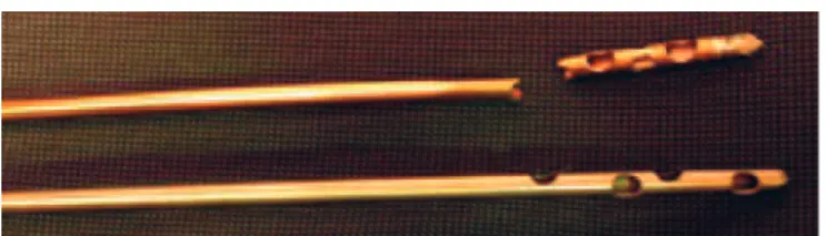
Cannulae for aspirating subcutaneous adipose tissue are available in several materials, diameters, and lengths, and with different types of tips and oriices. The instrument used in this procedure was a steel cannula, 25 cm long, with adiameterof4mmand5oriicesof1.5mm×2mmeach (Figures 4 and 5). Cannulae similar to that used in this study have been employed by the surgical team for more than 5 years without any inconvenience. This particular cannula had been used for only 3 months and had not shown any sign of deterioration.
The entire operation was performed within the norms requiredbythe1711/2003ResolutionoftheBrazilianFe deral Council of Medicine, which establishes safety criteria that should be observed during liposuction. These criteria ensure that the patient has the right to make an informed decision and the physicians are aware of the limits and indi cations for the procedure.
It is believed that it is not necessary to use cannulae with a diameter larger than 4 mm, as at least 1 study showed a similar rate of aspiration by using 4 or 5 mm cannulae. However, the possibility of complications is higher with largerdiameter cannulae2.
The cannula described in this study probably had de fects, as narrow openings in the form of “slots”, in the region of the lumen that, because they were in the interior of the cannula, were not detected by either the manufacturer’s qualitycontrolevaluationorthe medical team. This case demonstrates that, even with experienced surgeons using materials in per fect condition, unexpected situations might occur. Many professionals rely more on their personal expe riences in troubleshooting than on scientific articles, often because of the lack of studies discussing these complica tions. No case reports similar to this study were found in the literature.
Rev Bras Cir Plást. 2012;27(3):490-2 492
Wolfenson M et al.
demonstrates the necessity of performing surgical proce duresinwellequippedhospitalstoensurethesafetyofthe patient and the medical team. Without an image intensifier available during the operation, the “retrieval” of the broken tipofthecannulawouldhavebeenquitedifficult.
REFERENCES
1.GomesRS.Critériosdesegurançaemlipoaspiração.ArqCatarinMed.
2003;32(4):3546.
2.ViterboF,OchoaJS.Vibroliposuction:astudyofrateofaspiration.
Aes thetic Plast Surg. 2002;26(2):11822.
3. Hughes CE 3rd. Reduction of lipoplasty risks and mortality: an ASAPS
survey. Aesthet Surg J. 2001;21(2):1207.
4. Karmo FR, Milan MF, Silbergleit A. Blood loss in major liposuction procedures: a comparison study using suctionassisted versus ultraso nically assisted lipoplasty. Plast Reconstr Surg. 2001;108(1):2417.
Figure 4 –Steel cannula, 25 cm × 4 mm.
Figure 5 – Broken tip of the cannula, 3 cm in length.
Correspondence to: Moisés Wolfenson