Early Neoatherosclerosis as a Cause of
Second-Generation Drug-Eluting Stent Restenosis
Daniel Chamié
1, J. Ribamar Costa Jr.
2, Alexandre Abizaid
3Rev Bras Cardiol Invasiva. 2012;20(3):333-6
ABSTRACT
A case of everolimus-eluting stent restenosis caused by
neoatherogenesis is reported. Optical coherence tomography
indicated the presence of a supericial arch with high optical
intensity in the in-stent mid-segment, followed by signiicant
signal attenuation with poorly deined borders, indicating the
presence of lipid iniltration and/or a necrotic core, similar to
that observed in
de novo
coronary lesions. Signs suggesting
macrophage/foam cell iniltration were observed inside the
ibrous cap, indicating the presence of local inlammatory
activity. The development of new in-stent atherosclerosis at the
site of pre-existing neointimal tissue (neoatherosclerosis) was
recently identiied as an additional cause of coronary stent
failure. The present report is one of the irst to demonstrate
the inding of neoatherosclerosis as a second-generation
drug-eluting stent failure.
DESCRIPTORS:
Coronary artery disease. Coronary restenosis.
Angioplasty. Drug-eluting stents.
© 2012 Elsevier Editora Ltda. and Sociedade Brasileira de Hemodinâmica e Cardiologia Intervencionista. All rights reserved.
1 Physician, interventional cardiologist of the Invasive Cardiology Service
of the Instituto Dante Pazzanese de Cardiologia. São Paulo, SP, Brazil.
2 Physician, interventional cardiologist of the Invasive Cardiology Service
of the Instituto Dante Pazzanese de Cardiologia. São Paulo, SP, Brazil.
3 Associate Professor. Director of the Invasive Cardiology Service of
the Instituto Dante Pazzanese de Cardiologia. São Paulo, SP, Brazil.
Correspondence to: Daniel Chamié. Av. Dr. Dante Pazzanese, 500 – Vila Mariana – São Paulo, SP, Brasil – CEP 04012-909
E-mail: [email protected]
Received on: 8/7/2012 • Accepted on: 7/9/2012
Case Report
RESUMO
Neoaterosclerose Precoce como Causa de Reestenose
de Stent Farmacológico de Segunda Geração
Relatamos um caso de reestenose de stent eluidor de everolimus
causada por neoaterogênese. A tomograia de coerência óptica
revelou, no segmento médio intrastent, presença de arco supericial
com alta intensidade óptica, seguido por signiicativa atenuação
do sinal luminoso, com limites mal deinidos, indicando
pre-sença de iniltração lipídica e/ou núcleo necrótico, semelhante
ao observado em lesões coronárias de novo. Sinais sugerindo
iniltração de macrófagos/foam cells puderam ser observados no
interior da capa ibrosa, denotando presença de atividade
inla-matória local. O surgimento de nova aterosclerose intrastent, no
local de um tecido neointimal já formado (neoaterosclerose), tem
sido recentemente identiicado como causa adicional de falência
de stents coronários. O presente relato é um dos primeiros a
demonstrar o achado de neoaterosclerose como falha de um
stent farmacológico de segunda geração.
DESCRITORES:
Doença da artéria coronariana. Reestenose
coronária. Angioplastia. Stents farmacológicos.
T
he case of a 58-year-old male patient is reported,
an ex-smoker, with hypertension and prior
coro-nary artery bypass grafting surgery, who presented
with acute myocardial infarction without ST-segment
elevation on January 1, 2012, and was submitted to
percutaneous coronary intervention on January 18, 2012,
with implantation of an everolimus-eluting stent in the
middle third of the left circumlex artery.
Six months after the procedure, the patient returned
complaining of stable angina class II according to the
classiication of the Canadian Cardiovascular Society (CCS).
A new angiography showed focal in-stent
reste-nosis (Figure 1). An optical coherence tomography
was performed (Figures 2 and 3) and demonstrated a
heterogeneous vascular response pattern throughout the
previously treated segment. The distal segment of the
stent had a satisfactory vascular healing pattern, with a
thin layer of neointimal hyperplasia with circumferential
distribution and regular borders, in which the tissue
had a homogeneous pattern of high optical intensity
1Chamié et al.
Neoatherosclerosis as a Cause of Second-Generation Drug-Eluting Stent Restenosis
Rev Bras Cardiol Invasiva. 2012;20(3):333-6 334
Figure 1 – Coronary angiography of the procedure and at 6 months. In A, angiography shows an eccentric lesion in the middle third of the left cir-cumlex artery. The arrow points to the stenosis. In B, the inal angiographic result after implantation of an everolimus-eluting stent of 3.5 x 23 mm, post-dilated with a non-compliant balloon of 4 x 12 mm up to 16 atm. A satisfactory angiographic result without residual stenosis in the in-stent segment, no signs of injury at the borders and preserved distal epicardial low (Thrombolysis In Myocardial Infarction – TIMI 3) were observed. Yellow arrows delimit the stent borders. In C, coronary angiography six months after the procedure with binary angiographic restenosis (stenosis diameter of 62%), focal, restricted to the in-stent segment is shown. Yellow arrows delimit the stent borders, and the green arrow points to the site of restenosis.
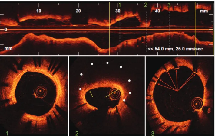
Figure 2 – Optical coherence tomography. In the top panel, longitudinal reconstruction of the left circumflex artery. The stent limits are iden-tified by the yellow vertical bars. The white dotted vertical bars identify three representative images of the intrastent distal segment, site of restenosis and intrastent proximal segment corresponding to images of cross-sections of the vessel shown in the lower panel. Bottom Panel 1: distal segment of the stent showing a region with normal neointimal hyperplasia characterised by homogeneous circumferential distribution, a regular outline and high optical intensity. Bottom Panel 2: site of restenosis with eccentric distribution of neointimal tissue that has charac-teristics similar to those of a lipid plaque in de novo lesions. The white dots indicate the position of the stent struts that were not visualised
due to important attenuation of the optical signal. The yellow arrows indicate areas of increased superficial brightness within the fibrous cap, suggesting the infiltration of macrophages/foam cells. Bottom Panel 3: proximal stent segment showing heterogeneous vascular healing with the presence of struts not covered by neointimal tissue (arrows).
Chamié et al. Neoatherosclerosis as a Cause of Second-Generation Drug-Eluting Stent Restenosis Rev Bras Cardiol Invasiva.
2012;20(3):333-6
335
stents.
4-6A series of 299 autopsy cases demonstrated
that the incidence of neoatherosclerosis is greater in
lesions treated with irst-generation drug-eluting stents
(DES; 31%) compared with bare-metal stents (BMS; 16%),
and the time to its onset is shorter after implantation
of a DES (420 days – 361 days to 683 days) when
compared to BMS (2,160 days – 1,800 days to 2,880
days).
7To date, descriptions of encountering
neoath-erosclerosis in second-generation DES are scarce. The
present report is one of the irst to report the inding of
neoatherosclerosis as a second-generation stent failure.
The prematurity of this phenomenon is noteworthy and
deserves further investigation.
CONFLICTS OF INTEREST
The authors declare no conlicts of interest.
REFERENCES
1. Nakano M, Vorpahl M, Otsuka F, Taniwaki M, Yazdani SK, Finn AV, et al. Ex vivo assessment of vascular response to coronary stents by optical frequency domain imaging. JACC Cardiovasc Imaging. 2012;5(1):71-82.
2. Yabushita H, Bouma BE, Houser SL, Aretz HT, Jang IK, Schlendorf KH, et al. Characterization of human atherosclerosis by optical coherence tomography. Circulation. 2002;106(13): 1640-5. 3. Kang SJ, Mintz GS, Akasaka T, Park DW, Lee JY, Kim WJ, et al.
Optical coherence tomographic analysis of in-stent neoathe-rosclerosis after drug-eluting stent implantation. Circulation. 2011;123(25):2954-63.
of neointimal tissue and presence of several struts with
no tissue covering them (Figure 2, panel 3).
In the medium intrastent segment (Figure 2, panel
2), significant neointimal proliferation with expressive
involvement of the luminal area was observed. A high
optical intensity shallow arch, followed by significant
attenuation of the light signal in which boundaries were
poorly defined was observed, suggesting the existence
of fatty infiltration and/or a necrotic core similar to
de novo
coronary lesions,
2showing new intrastent
atherosclerosis.
3The optical signal attenuation was so
expressive that it prevented the identification of the
structure of the metallic stent struts. Signs of local
inflammatory activity could be inferred by visualising a
‘flecked’ glow within the fibrous cap, with an optical
intensity greater than the surrounding fibrous tissue,
suggesting the infiltration of clusters of macrophages/
foam cells.
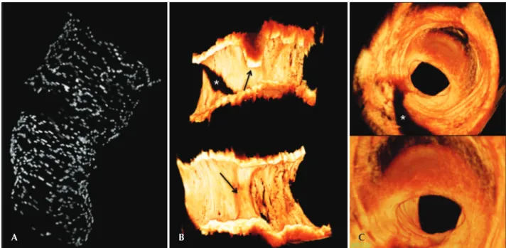
In the three-dimensional reconstruction of the optical
coherence tomography images, there was a discontinuity
image in the central region of the stent structure (restenosis
site) resulting from the hyperattenuation of the optical
signal by the neoatherogenic tissue (Figure 3A).
The development of new intrastent atherosclerosis
at the site of already formed neointimal tissue
(‘neoathe-rosclerosis’) has recently been identiied as an additional
cause of failure (restenosis or thrombosis) of coronary
Figure 3 – Three-dimensional reconstruction of optical coherence tomography images. In A, three-dimensional reconstruction of the stent revealing discontinuity of the stent structure at the site of restenosis through important attenuation of the optical signal promoted by fatty iniltration of the neointima. In B, longitudinal images of the open vessel in two orthogonal planes showing the eccentricity and the three-dimensional distribution of the restenosis point. The black arrows indicate the site of restenosis. The asterisk indicates the shadow caused by the presence of the 0.014-inch guidewire. In C, ly-through visualisation of the coronary vessel showing the spatial distribution of neointimal tissue at the site of restenosis. The darker neointimal tissue at the point of restenosis compared with other regions of the vessel is the result of the attenuation of the light signal by lipid iniltration. The asterisk indicates the shadow caused by the presence of the guidewire.
Chamié et al.
Neoatherosclerosis as a Cause of Second-Generation Drug-Eluting Stent Restenosis
Rev Bras Cardiol Invasiva. 2012;20(3):333-6 336
4. Bennett J, Coosemans M, Adriaenssens T. Very late bare metal stent thrombosis due to neoatherosclerotic plaque rupture: an optical coherence tomography inding. Heart. 2012;98(19):1470. 5. Karanasos A, Ligthart JM, Regar E. In-stent neoatherosclerosis:
a cause of late stent thrombosis in a patient with “full metal jacket” 15 years after implantation: insights from optical coher-ence tomography. JACC Cardiovasc Interv. 2012;5(7):799-800.
6. Park SJ, Kang SJ, Virmani R, Nakano M, Ueda Y. In-stent neo-atherosclerosis: a inal common pathway of late stent failure. J Am Coll Cardiol. 2012;59(23):2051-7.