Original Article
Braz J Oral Sci.January | March 2013 - Volume 12, Number 1
Keratocystic odontogenic tumor related to
nevoid basal cell carcinoma syndrome:
clinicopathological study
Luana Eschholz Bomfin
1, Ana Paula M. Vivas
1, Andre Caroli Rocha
2, Maria Isabel W. Achatz
3,
Clovis Antonio L. Pinto
4, Fabio Abreu Alves
51 Department of Stomatology, São Paulo University, São Paulo, SP, Brazil 2 Department of Stomatology, A. C. Camargo Hospital, São Paulo, SP, Brazil 3 Department of Oncogenetics, A. C. Camargo Hospital, São Paulo, SP, Brazil 4 Department of Anatomical Pathology, A. C. Camargo Hospital, São Paulo, SP, Brazil 5 Department of Stomatology, University of São Paulo, Department of Stomatology, A. C. Camargo Hospital, São Paulo, SP, Brazil
Correspondence to:
Fábio Abreu Alves Rua Prof. Antônio Prudente 211, Liberdade, CEP: 01509-900 - São Paulo, SP, Brasil Phone: +55 11 21895129 Fax: +55 11 21895133 E-mail: [email protected]
Abstract
Aim: To assess clinicopathological features of patients with keratocystic odontogenic tumor (KCOT) associated with nevoid basal cell carcinoma syndrome (NBCCS) in a single Brazilian institution.
Methods: After histopathological analyses of KCOT related to NBCCS, the medical charts of 14 patients were assessed. These patients presented a total of 31 primary and 8 recurrent KCOT.
Results: Out of 14 patients, 8 presented a single KCOT, 4 showed synchronous tumors, 1 had 3 metachronous lesions and another patient had 2 synchronous lesions at initial evaluation and then developed other 3 metachronous lesions. Besides the 31 primary KCOTs, 18 lesions were located in mandible and 13 in maxilla. Most tumors presented unilocular pattern and association with a tooth. Conclusions: KCOT is a frequent manifestation of NBCCS and can be its first sign, mainly in young patients. In contrast to a previously published series, most patients presented a single lesion.
Keywords: Gorlin syndrome,keratocyst, keratocystic odontogenic tumor, nevoid basal cell carcinoma syndrome.
Introduction
Nevoid basal cell carcinoma syndrome (NBCCS) was first described in 1960 by Gorlin and Goltz and was characterized by multiple basal cell carcinomas
(BCC), odontogenic keratocysts (OKC) and bifid ribs1.
A multidisciplinary colloquium was recently organized and its aims were to better define the physical findings associated with NBCCS. The participants reviewed the diagnostic criteria of the syndrome and there was no consensus for a formal recommendation. Consequently, a suspected diagnosis of NBCCS should be considered based on the findings of less stringent criteria of: (1) one major criterion and molecular confirmation; (2) two major criteria; or (3) one major and
Received for publication: January 14, 2013
24
24
24
24
24
two minor criteria. In addition, medulloblastoma (MB) should be considered a major criterion as it may lead to increased early detection of the syndrome. Both major and minor criteria
according to this colloquium are shown in Table 12.
Basal Cell Carcinoma (BCC) and OKC are the most
common incident manifestations of the syndrome3.In 2004,
Agaram et al. showed significant loss of heterozygosity of
tumor suppressor genes in sporadic lesions4. Consequently,
these authors hypothesized the neoplastic nature of the lesion and the World Health Organization later recommended changing the name of the lesion from OKC to keratocystic odontogenic tumor (KCOT).
There are few reported cases of KCOT associated with NBCCS from Brazil and most data feature biological behavior. In addition, we have found only 13 well-documented patients have been found in English language literature with such association in the Brazilian population in the last 20 years5-10. The purpose of this paper is to describe clinical and histopathological aspects of KCOTs associated with NBCCS and report other manifestations associated with the syndrome. Furthermore, this study shows the largest clinical data about KCOT associated with NBCCS ever compiled in the Brazilian population.
Material and methods
Characterization of the Study
This study consisted of a retrospective analysis of patients with KCOT diagnosed between 1970 and 2009. A total of 74 patients presented KOCT, 14 being related to NBCCS. Clinical criteria for NBCCS diagnosis were based
on Bree et al.2 The present study was approved by the Ethics
Committee of AC Camargo Hospital (number 1322/2009).
Clinical data
The medical charts of 14 patients presenting KCOT related to NBCCS were evaluated. These patients presented a total of 31 primary and 8 recurrent KCOT. Data including
Major criteria
1. Basal cell carcinomas (BCC) prior to 20years old or excessive numbers of BCC out of proportion to prior sun exposure and skin type 2. Odontogenic Keratocystic of the jaw prior to 20 years of age
3. Palmar or plantar pitting
4. Lamellar calcification of the falx cerebri 5. Medulloblastoma, typically desmoplastic 6. First degree relative with NBCCS
Minor Criteria
1. Rib anomalies
2. Macrocephaly determined after adjustment for height
3. Other specific skeletal malformations and radiologic changes (i.e., vertebral anomalies, kyphoscoliosis, short fourth metacarpals, postaxial polydactyly). 4. Cleft/lip palate
5. Ovarian/cardiac fibroma 6. Lymphomesenteric cysts
7. Ocular abnormalities (i.e., strabismus, hipertelorisms, congenital cataracts, glaucoma, coloboma)
Table 1. Major and minor criteria stated by First International Colloquium on NBCCS Bree et al2.
age, gender, race, signs, symptoms, radiographic features, treatment and recurrence were analyzed.
Histopathological analysis
The hematoxylin/eosin (HE)-stained slides of KCOTs were retrieved and submitted to histopathological exam. All slides were reviewed by an oral pathologist and findings were confirmed by a second general pathologist (Pinto CAL).
Statistical analysis
Frequency distribution tables were built in order to record patient clinical data, tumors and histopathological features. The recurrence rate was calculated by Kaplan-Meier estimator. All data analysis was performed using R version 2.13.0 software (http://www.r-project.org)
Results
Clinical features of 14 patients
Six (42.86%) patients with KCOT were referred by the Cutaneous Oncology Department due to previous diagnosis of NBCCS and the remaining 8 (57.14%) had KCOT as first manifestation of the syndrome.
The patient’s age at KCOT diagnosis ranged from 8 to 66 years (mean age of 31 years), and most of the lesions occurred in the second decade of life. Nine out of 14 patients (64.29%) were female and 12 patients (85.71%) were Caucasian (Table 2).
Among the 14 patients, 8 patients presented a single KCOT, 4 presented KCOT synchronous tumors (total of 15 lesions), 1 had 3 KCOT metachronous lesions and 1 patient had 2 synchronous lesions at the initial evaluation and developed other 3 metachronous lesions during the follow-up period (Table 2).
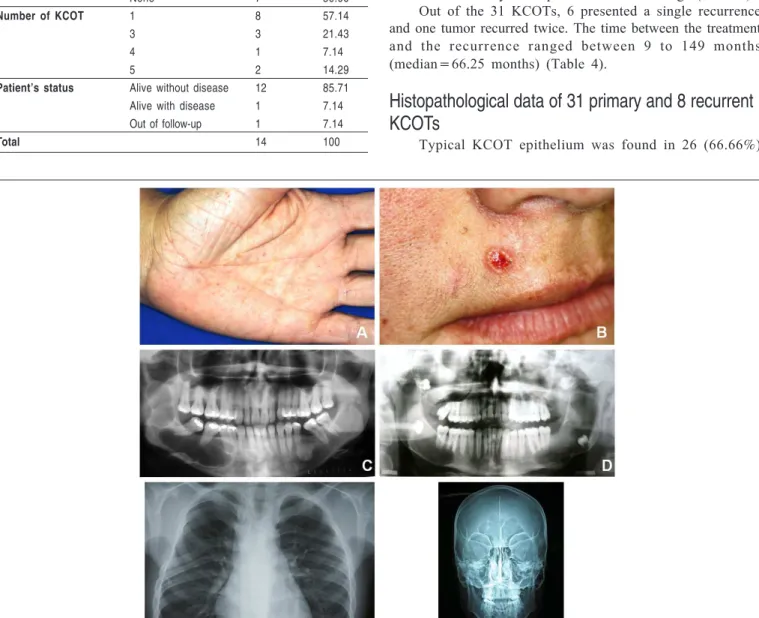
In addition to KCOTs, the most common clinical manifestations were basal cell carcinoma (CBC), palmar pits, abnormal ribs and vertebrae, and calcification of the falx cerebri (Figure 1). The NBCCS clinical manifestations are listed in Table 3.
25
25
25
25
25
Variable Category n %
Age 0-9 17.14
10-19 5 35.71
20-29 2 14.29
30-39 4 28.57
40-69 2 14.28
Gender Female 9 64.29
Male 5 35.71
Race Caucasian 12 85.71
Not Caucasian 2 14.29
Main complaint Swelling 5 35.71
None 9 64.29
Extra-oral examination None 10 71.43
Facial asymmetry 3 21.43
Fistula 1 7.14
Intra-oral examination Lump 6 42.86
Fistula 1 7.14
None 7 50.00
Number of KCOT 1 8 57.14
3 3 21.43
4 1 7.14
5 2 14.29
Patient’s status Alive without disease 12 85.71
Alive with disease 1 7.14
Out of follow-up 1 7.14
Total 14 100
Table 2. Clinical features of 14 patients with NBCCS.
Fig. 1: Some of clinical manifestations of NBCCS. A. Multiple palmar pits (patient 13). B. Basal cell carcinoma located in fluff region (patient 13). C. One KCOT located in ramus and posterior mandible and with multilocular pattern (patient 4). D. Presence of four synchronous KCOT with unilocular pattern involving 3rd impacted molar
(patient 11). E. Chest radiograph demonstrating bifid ribs (patient 12). F. Antero-posterior skull radiograph showing calcification of the falx cerebri (patient 2).
Clinicopathological features of 31 primary KCOT in
14 patients
At initial evaluation, 5 (35.71%) patients complained of swelling and 9 (64.29%) had no symptoms. The time of complaint ranged between 15 days and 6 months (mean=2 months). On extra-oral examination, 3 patients (21.43%) presented facial asymmetry, 1 patient extra-oral fistula (7.14%) and no alterations were observed in 10 patients (71.43%). On intra-oral examination, 6 patients (42.86%) presented lumps, 1 patient (7.14%) a fistula and 7 patients (42.86%) presented no alterations.
Radiographic analysis of 31 primary KCOTs demonstrated that 18 (58.06%) lesions were located in the mandible and 13 (41.94%) in the maxilla. Most of the tumors presented a unilocular pattern and had tooth association (Figure 1). The size of the lesions ranged from 2.5 to 10 cm (mean = 6.14 cm). According to KCOT treatment, enucleation associated with curettage was performed in 30 cases (96.78%) and the other case was treated by marsupialization and curettage (Table 4).
Out of the 31 KCOTs, 6 presented a single recurrence and one tumor recurred twice. The time between the treatment and the recurrence ranged between 9 to 149 months (median=66.25 months) (Table 4).
Histopathological data of 31 primary and 8 recurrent
KCOTs
Typical KCOT epithelium was found in 26 (66.66%)
26
26
26
26
26
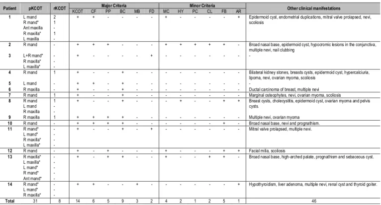
Patient pKCOT rKCOT Major Criteria Minor Criteria Other clinical manifestations
KCOT CF PP BC MB FD MC HY PC CL FB AR
1 L mand R mand* Ant maxilla R maxilla* L maxilla 2 1 -1
-+ + - - - - + - - - - + Epidermoid cyst, endometrial duplications, mitral valve prolapsed, nevi, scoliosis
2 R mand - + + + - - - + + + + + - Broad nasal base, epidermoid cyst, hypocromic lesions in the conjunctiva, multiple nevi, nail clubbing
3 L+R mand* R maxilla* L maxilla* -+ - - - - + - - -
-4 R mand 1 + - - + - - - Bilateral kidney stones, breasts cysts, epidermoid cyst, hypercalciuria, lipoma, nevi, ovarian myoma, scoliosis
5 L mand - + + - + - - -
-6 R maxilla - + - - + - - - Ductal carcinoma of breast, multiple nevi
7 R mand 1 + - - + - - - Marginal osteophytes, nevi, ovarian myoma, scoliosis
8 R mand L mand R maxilla
1
-+ - - + - - - + - - + + Breast cysts, cholecystitis, epidermoid cyst, ovarian myoma and pelvis cysts.
9 R maxilla 1 + + + + - - - Multiple nevi, ovarian myoma
10 R mand - + + + + - - - + - Broad nasal base, nevi and prognathism.
11 R mand* L mand* R maxilla* L maxilla*
-+ - - + - + - - - Mitral valve prolapsed, multiple nevi.
12 R mand - + - + - - - + - - - + + Facial milia, scoliosis
13 R maxilla* L maxilla* L mand* R mand* Ant mand*
-+ - + + - - + - - + + - Broad nasal base, high-arched palate, prognathism and sebaceous cyst.
14 R mand* L mand* R maxilla*
-+ + - - + - - - + Hypothyroidism, liver adenoma, multiple nevi, renal cyst and thyroid goiter.
Total 31 8 14 6 5 9 3 2 4 2 1 2 5 1 46
Table 3. Clinical features of the 14 patients with NBCCS distributed according to main major and minor NBCCS criteria2.
pKCOT=primary keratocystic odontogenic tumor, rKCOT=recurrent keratocystic odontogenic tumor, CF=Calcification of the falx cerebri, PP=Palmar pits, BC=Basal cell carcinoma, MB=Meduloblastoma, FD=First degree relative with NBCCS, MC=Macrocephaly, SC=Scoliosis, HY=Hypertelorism, PC=Pectus cavitatum, CL=Clinodactilia, FB=Frontal bossing, AR=Abnormal ribs, L=left, R=right, mand=mandible, * synchronous KCOT, +presence, - absence.
tumors and 13 lesions had typical areas of KCOT and areas of epithelial hyperplasia. Furthermore, detachment of the epithelium was seen in 27 (69.23%) cases. Interestingly, only one case presented evident epithelium dysplasia.
Variable Category n %
Location Mandible Posterior 7 22.58
Posterior + ramus 6 19.35
Ramus 3 9.68
Anterior 2 6.45
Total 18 58.06
Maxilla Posterior 12 38.71
Anterior 1 3.23
Total 13 41.94
Radiographic pattern Multilocular 6 19.35
Unilocular 15 48.39
Not informed 10 32.26
Tooth association Yes 13 54.17
No 11 45.83
Not informed 7 22.58
Treatment
Enucleation 30 96.78
and curettage
Marsupialization and 1 3.23
curettage
Recurrence No 24 77.42
Yes 7 22.58
Table 4. Clinical and radiographic features of the 31
primary KCOT.
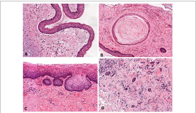
In relation to the connective tissue, 21 cases (53.84%) demonstrated mild inflammation in 11 tumors (28.20%), moderate in 5 (12.82%), and severe in 5 cases (12.82%). Satellite cysts, remnants of odontogenic epithelium, budding and dystrophic calcification were found in 11 (28.20%), 14 (35.89%), 9 (23.07%), 6 cases (15.38%), respectively (Figure 2).
Discussion
BCCs and KCOT are the main features observed in
patients with NBCCS3. Similarly, Kimonis et al. evaluated
clinical and radiological data of 105 persons with NBCCS. Pits, BCCs, jaw cysts and falx calcification were the most common anomalies, and according to their results the authors suggested some major and minor criteria for NBCCS
diagnosis11. Comparing these criteria with the First
International Colloquium on NBCCS criteria2, there were two
important alterations. The latter suggested changing rib anomalies to minor criterion and MB to major criterion. Interestingly, Amlashi et al. evaluated 76 patients with MBs and three of them had syndromic MBs. Additionally, the authors reviewed the literature and found other 33 patients with syndromic MBs. The mean age of syndromic MBs was 4 years (earlier than sporadic MBs) and most syndromic patients were younger than 2 years12. Only one of these patients developed MB at 3 years. At 17 years he presented 3 synchronous KCOTs, and at 18 years calcification of the falx cerebri. It is worthy of note that calcification of the falx cerebri had been previously investigated in this patient.
27
27
27
27
27
Fig. 2:A. Typical epithelium lining of a KCOT (H.E., 10.3X). B. Daughter cyst observed in cystic capsule (H.E., 4.8X). C. Budding of the basal cell layer of the lining epithelium (H.E., 9.8X). D. Remnants of odontogenic epithelium in the cystic wall (H.E., 9.3X).
Kimonis et al.evaluated the falx cerebri calcification in 82
individuals with NBCCS11. This calcification was present in
23 out of 29 (79%) individuals over the age of 40, 20 out of 26 (77%) individuals between the ages of 20 and 40 and 10 out of 27 (37%) individuals under the age of 20.
In the stomatological system, besides KCOT, other benign and malignant tumors have been described in NBCCS patients such as ameloblastoma, myxoma, fibrosarcoma, squamous cell carcinoma, adenoid cystic carcinoma and lymphoma. Furthermore, development defects such as cleft lip/palate, dental ectopy/heterotopy, impacted teeth, dental agenesis, malocclusion, mandibular prognathism, high-arched palate, skeletal open bite and hyperplasia of mandibular coronoid process have also been reported10,13-17. Interestingly, Ponti et al.18 evaluated 41 ameloblastomas and two of them were related to NBCCS. In addition, PTCH 1 germline mutations were also detected in both cases and negative in the others. The authors suggested including ameloblastoma as a criterion for syndrome identification. The present series reveals that only one patient presented high-arched palate associated with prognatism.
In the present study, 74 patients with KCOT were reviewed and 14 (19.17%) of them also presented NBCCS. KCOT was the first sign of the syndrome in 8 (57.14%)
patients. In a similar study, Lo Muzio et al.19 evaluated 37
individuals with NBCCS, and 34 of them had KCOT (92%). In approximately 70% of these patients, the first manifestation
of the syndrome was KCOT.In general, most of the patients
with NBCCS are females and KCOT occurs in the second
decade of life with a mean age ranging from 17 to 26 years
19-21. In the present series, 9 out of 14 patients were females
and there were two peaks of age, in the second and fourth decades. As a consequence, the mean age was 31 years, differing from the above-mentioned studies.
In the largest series in English literature, Woolgar et al. evaluated 164 KCOTs in syndromic patients and 379 KCOTs
not associated with NBCCS20. It was observed that the
posterior area of the mandible was the main affected site, followed by the maxillary molar region in both groups. Since the syndromic patients almost always have more than one tumor, it is to be expected that more maxillary tumors are present in these patients. Such data were also demonstrated in the present study, in which multiple lesions were found in 6 patients and accounted for 23 tumors (12 in the mandible and 11 in the maxilla). Interestingly, 8 patients presented a single lesion, 6 affecting the mandible and 2 the maxilla. In general, the mandible was the main location of the lesions (18 cases - 58.06%), 13 being in the body/ramus and 5 in the anterior area. In addition, there were 12 cases (38.71%) in the posterior and 1 case in the anterior region of the maxilla.
Other studies have also shown multiple lesions affecting syndromic patients. Kimonis et al. reported that 78 (74%) NBCCS patients presented KCOT with number of tumors
ranging from 1 to 2811.However, the authors did not clarify
which tumors were primary or recurrent. Furthermore, it was also shown that 5 individuals had more than 10 KCOTs in their lifetime. Ahn et al. reviewed 33 well-documented case
28
28
28
28
28
reports of NBCCS published between 1981 and 2002. Out of the total, 30 patients (90.9%) had KCOT and the number of lesions per patient ranged from 1 to 6 (mean 2.7 lesions)21. In a recent NBCCS case series reported in Indian patients, all 6
patients developed multiple KCOT (range 3 to 6)22.
Differently, most of the patients in this study (8 out of 14) had a single KCOT, and the other 6 patients presented 23 tumors (range 3 to 5). The above-mentioned research involving Brazilian patients with KCOT related to NBCCS reports only 13 patients, and there was no information on 3 patients, 3 had a single lesion, 4 had 2 lesions, 2 had 3 lesions and one patient had 5 lesions5-10.
Regarding the treatment of KCOT, Zecha et al. demonstrated that 58 patients who did not have an NBCCS diagnosis and were treated with enucleation alone had recurrence in 20.7% of the cases23. A lower rate was described by Boffano et al. accounting for 11.9% of 261 tumors treated by enucleation and curettage24. In this study, the association of enucleation and curettage was performed in 30 (96.78%) cases (13 patients). Recurrence was observed in 7 tumors (22.58%), which corresponded to 5 patients. One other patient was at first treated by marsupialization and after 10 months by curettage. This patient has been asymptomatic for 5 years. Recurrences usually manifest within the first 5 to 7 years.
However, Zhao et al.demonstrated recurrence after 13 years
of follow-up25.Similarly, our study demonstrated 2 patients
who had recurrence after 10 years.
In summary, KCOT related to NBCCS can affect patients at a younger age than sporadic KCOT and multiple tumors are commonly found. Interestingly, in this series, 8 out of 14 patients (57%) had a single lesion. Early diagnosis of the syndrome and a long follow-up period is important due to the severity of clinical manifestations. Moreover, a multidisciplinary team is required, including dentists, dermatologists, geneticists and neurologists to improve the diagnosis and quality of life.
Acknowledgments
We wish to thank Ana Laura Miziara for text revision.
Funding
This study was supported by São Paulo State Research Foundation (FAPESP).
References
1. Gorlin RJ, Goltz RW. Multiple nevoid basal-cell epithelioma, jaw cysts
and bifid rib. A syndrome. N Engl J Med. 1960; 62: 908-12.
2. 2- Bree AF, Shah MR; BCNS Colloquium Group. Consensus statement
from the first international colloquium on basal cell nevus syndrome (BCNS). Am J Med Genet A. 2011; 155A: 2091-7.
3. Evans DG, Ladusans EJ, Rimmer S, Burnell LD, Thakker N, Farndon
PA. Complications of the naevoid basal cell carcinoma syndrome: results of a population based study. J Med Genet. 1993; 30: 460-4.
4. Agaram NP, Collins BM, Barnes L, Lomago D, Aldeeb D, Swalsky P et
al. Molecular analysis to demonstrate that odontogenic keratocysts are neoplastic. Arch Pathol Lab Med. 2004; 128: 313-7.
5. Tincani AJ, Martins AS, Andrade RG, Franco JR EFM, Camargo MAB,
Martins AS. Nevoid Basal-Cell Carcinoma Syndrome: literature review and case report in a family. Sao Paulo Med J. 1995; 113: 917-21.
6. Melo ES, Kawamura JY, Alves CA, Nunes FD, Jorge WA, Cavalcanti
MG. Imaging modality correlations of an odontogenic keratocyst in the nevoid basal cell carcinoma syndrome: a family case report. Oral Surg Oral Med Oral Pathol Oral Radiol Endod. 2004; 98: 232-6.
7. Lopes NN, Caran EM, Lee ML, Silva NS, Rocha AC, Macedo CR.
Gorlin-Goltz syndrome and neoplasms: a case study. J Clin Pediatr Dent. 2010; 35: 203-6.
8. Visioli F, Martins CA, Heitz C, Rados PV, Sant’Ana Filho M. Is nevoid
basal cell carcinoma syndrome really so rare? Proposal for an investigative protocol based on a case series. J Oral Maxillofac Surg. 2010; 68: 903-8.
9. Casaroto AR, Loures DC, Moreschi E, Veltrini VC, Trento CL, Gottardo
VD et al. Early diagnosis of Gorlin-Goltz syndrome: case report. Head Face Med. 2011; 2: 1-5.
10. Pereira CM, Lopes AP, Meneghini AJ, Silva AF, Botelho TL. Oral diffuse B-cell non-Hodgkin’s lymphoma associated to Gorlin-Goltz syndrome: a case report with one year follow-up. Indian J Pathol Microbiol. 2011; 54: 388-90.
11. Kimonis VE, Goldstein AM, Pastakia B, Yang ML, Kase R, DiGiovanna
JJ et al. Clinical manifestations in 105 persons with nevoid basal cell carcinoma syndrome. Am J Med Genet. 1997; 69: 299-308.
12. Amlashi SF, Riffaud L, Brassier G, Morandi X. Nevoid basal cell carcinoma syndrome: relation with desmoplastic medulloblastoma in infancy. A population-based study and review of the literature. Cancer. 2003; 98: 618-24. 13. Gorlin RJ. Nevoid basal-cell carcinoma syndrome. Medicine (Baltimore).
1987; 66: 98-113.
14. Hasegawa K, Amagasa T, Shioda S, Kayano T. Basal cell nevus syndrome with squamous cell carcinoma of the maxilla: report of a case. J Oral Maxillofac Surg. 1989; 47: 629-33.
15. Yilmaz B, Goldberg LH, Schechter NR, Kemp BL, Ruiz H. Basal cell nevus syndrome concurrent with adenoid cystic carcinoma of salivary gland. J Am Acad Dermatol. 2003; 48: S64-6.
16. Eslami B, Lorente C, Kieff D, Caruso PA, Faquin WC. Ameloblastoma associated with the nevoid basal cell carcinoma (Gorlin) syndrome. Oral Surg Oral Med Oral Pathol Oral Radiol Endod. 2008; 105: e10-3. 17. Lo Muzio L. Nevoid basal cell carcinoma syndrome (Gorlin syndrome).
Orphanet J Rare Dis. 2008; 25: 32.
18. Ponti G, Pastorino L, Pollio A, Nasti S, Pellacani G, Mignogna MD et al. Ameloblastoma: a neglected criterion for nevoid basal cell carcinoma (Gorlin) syndrome. Fam Cancer. 2012; 11: 411-8.
19. Lo Muzio L, Nocini PF, Savoia A, Consolo U, Procaccini M, Zelante L et al. Nevoid basal cell carcinoma syndrome. Clinical findings in 37 Italian affected individuals. Clin Genet. 1999; 55: 34-40.
20. Woolgar JA, Rippin JW, Browne RM. The odontogenic keratocyst and its occurrence in the nevoid basal cell carcinoma syndrome. Oral Surg Oral Med Oral Pathol. 1987; 64: 727-30.
21. Ahn SG, Lim YS, Kim DK, Kim SG, Lee SH, Yoon JH. Nevoid basal cell carcinoma syndrome: a retrospective analysis of 33 affected Korean individuals. Int J Oral Maxillofac Surg. 2004; 33: 458-62.
22. Gupta SR, Jaetli V, Mohanty S, Sharma R, Gupta A. Nevoid basal cell carcinoma syndrome in Indian patients: a clinical and radiological study of 6 cases and review of literature. Oral Surg Oral Med Oral Pathol Oral Radiol. 2012; 113: 99-110.
23. Zecha JA, Mendes RA, Lindeboom VB, van der Waal I. Recurrence rate of keratocystic odontogenic tumor after conservative surgical treatment without adjunctive therapies - A 35-year single institution experience. Oral Oncol. 2010; 46: 740-2.
29
29
29
29
29
24. Boffano P, Ruga E, Gallesio C. Keratocystic odontogenic tumor (keratocyst): preliminary retrospective review of epidemiologic, clinical, and radiologic features of 261 lesions from University of Turin. J Oral Maxillofac Surg. 2010; 68: 2994-9.
25. Zhao YF, Wei JX, Wang SP. Treatment of odontogenic keratocysts: a follow-up of 255 Chinese patients. Oral Surg Oral Med Oral Pathol Oral Radiol Endod. 2002; 94: 151-6.