Keratocystic odontogenic tumor: role of
cone beam computed tomography and
magnetic resonance imaging
Thiago de Oliveira Gamba, DDS, MSc n Isadora Luana Flores, DDS, MSc, PhD n Antonione Bezerra Pinto, MS Andre Luiz Costa, DDS, MSc, PhD n Mari Eli Moraes, DDS, MSc, PhD n Sérgio Lúcio Lopes, DDS, MSc, PhD Keratocystic odontogenic tumors (KCOTs) are known for unique and
varied behavior, high recurrence rates, and distinctive histopathologic findings. Differential diagnosis and management of KCOTs may be challenging because other jaw lesions may present similar charac-teristics. Careful interpretation of cone beam computed tomograms and magnetic resonance images has great significance for precise assessment of KCOTs and their relationships to adjacent anatomic structures as well as for treatment planning. This case report describes
a KCOT that developed in the left angle and mandibular ramus in association with a semierupted third molar.
Received: May 26, 2014 Accepted: October 6, 2014
Key words: cone beam computed tomography, keratocystic odontogenic tumor, magnetic resonance imaging, odontogenic keratocyst, oral diagnosis
T
he keratocystic odontogenic tumor (KCOT), previously designated by the term odontogenic keratocyst, is one of the most common odontogenic lesions.1 According to the World Health Organization (WHO), it is widely accepted that KCOT originates from odontogenic epithelium, the chief sources of which are remnants of dental lamina and basal cells of the oral epithelium.1,2 The WHO reclas-sified KCOT as a benign developmental odontogenic neoplasia in 2005.2 This new designation reflects its aggressive clinicalbehavior and high local recurrence, which are associated with high cellular prolifera-tion rate and increased expression of anti-apoptosis genes in the basal layer.2,3
Despite their destructive potential, a substantial number of KCOTs are found incidentally during routine radiographic examination, where they may appear as a unilocular radiolucency with well-defined sclerotic margins or a multilocular radio-lucent lesion with scalloped borders.3-5 Clinically, most KCOTs involve no signs and symptoms until they cause bone
displacement, cortical disruption, pain, and drainage (if infection is present).2,3,6 KCOTs affect a wide range of ages and present a male predilection; posterior portions of mandible are the most frequently involved areas.2,3,6 This lesion can mimic other odon-togenic and nonodonodon-togenic lesions.2,3,6
Conventional radiographs are usually adequate for initial assessment of the posi-tion and size of KCOTs.7 However, digital imaging modalities have been used for more accurate diagnosis of odontogenic tumors, including KCOT.8 Cone beam computed
Fig 1. Panoramic radiograph showing the radiolucent unilocular lesion and its relationship to the semierupted transverse mandibular left third molar.
Digital Radiology
tomography (CBCT) and magnetic reso-nance imaging (MRI) are valuable comple-mentary examinations for assessment of additional information, such as borders of large lesions, bone expansion, cortical perfo-ration, contents, heterogeneity, intratumoral vascularity, and soft tissue involvement.2,6,8
The aim of the present report is to describe a case of KCOT and provide an overview of CBCT and MRI findings to assist dentists in performing accurate analysis of these tumors.
Case report
A 30-year-old white woman presented with the chief complaint of pain, halito-sis, and oral drainage that had lasted for 1 week. Her past medical history did not reveal any preexisting major disease, and the results of the extraoral examination were unremarkable. Intraoral examina-tion revealed a semierupted mandibular left third molar that was partially covered by erythematous and edematous tissue. A fetid odor, drainage, and trismus com-pleted the intraoral findings, and a clinical diagnosis of pericoronitis was suggested.
However, panoramic radiography dis-closed an extensive radiolucent unilocular lesion with sclerotic borders. The lesion involved the mandibular left first and second molars up to the inferior mandible cortical and extended to the left ascending ramus of the mandible (Fig 1). There was no radiographic sign of root resorption of the first and second molars. The mandibu-lar left third momandibu-lar was in a transalveomandibu-lar position, and the mandibular canal was surrounded by the lesion. Due to the lesion location, presence of a semierupted tooth, and age of the patient, dentigerous cyst and KCOT were the first differential diagnoses, but unicystic ameloblastoma was not ruled out. Because of the emer-gency clinical situation, the mandibular left third molar was extracted after admin-istration of local anesthesia. Prophylactic antibiotic therapy was provided before sur-gery, and an amoxicillin regimen (500 mg, 3 times a day) was continued for 1 week.
The patient returned for her 1-week recall appointment with a complaint of pain. A CBCT scan was performed, and the multiplanar axial and coronal recon-structed images showed a dense soft tissue lesion and expansion and thinning of the lingual cortex in the region of the posterior
body, angle, and ramus of the mandible on the left side. Disruption of the lingual cortex was observed in some areas of the mandibular body and ramus. These images also revealed inferior displacement of the left mandibular canal by the lesion. There were some hypodense areas in the middle of the lesion, corresponding to postsurgi-cal trauma from mandibular third molar extraction (Fig 2 and 3). Unfortunately, it was not possible to distinguish the true
borders of the lesion because soft tissue definition is limited in CBCTs.
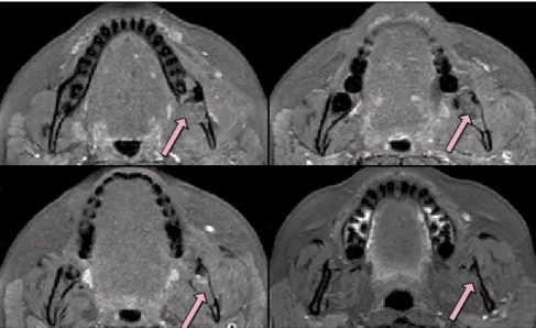
To establish the true limits of the lesion and its relationship with the surround-ing tissues and thus determine the best treatment approach, MRI examinations were performed with T1-weighted image (T1WI) and T2-weighted image (T2WI) sequences. Axial T1WIs revealed an inter-mediate signal image occupying the entire mandibular left body and ramus, rupture
www.agd.org General Dentistry January/February 2016 3
Fig 2. Inferosuperior axial cone beam computed tomograms (CBCTs) providing evidence of the expansion, thinning, and disruption of the lingual cortex (arrows).
Fig 3. Anteroposterior coronal CBCTs. A and B. Area of expansion (arrows). C. Hypodense area inside the lesion (arrow). D. Disruption of the lingual cortex (arrows).
A
C D
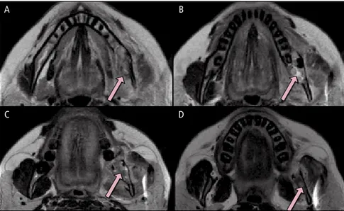
of the lingual cortical bone, and extension of the mass into the floor of the mouth. It was possible to observe well-defined borders of the lesion with no invasion of muscle tissue. The T2WIs showed the same aspects as the T1WIs and evidence of hypersignal areas inside the lesion that cor-responded to the location of acute second-ary infection (pericoronitis) before third molar extraction (Fig 4 and 5).
An incisional biopsy was performed, and the microscopic examination of the surgical specimen revealed a parakera-tinized KCOT with an epithelial lining, 6 to 8 cells thick, associated with the hyperchromatic palisaded basal cell layer and corrugated parakeratotic surface. The patient was referred to a maxillofacial sur-geon; because of the extent of the lesion, a marsupialization was performed. A contin-uous follow-up schedule was established to confirm complete enucleation of the cyst.
Discussion
Radiologic diagnosis of a lesion depends on association of many criteria, includ-ing the location, demarcation, cortical involvement, periosteal and soft tissue alterations, and density, in relation to the adjacent structures within the jaw.9 Although panoramic radiography is the primary choice of radiologic examination for dentists, superimposition of craniofa-cial structures generates images with limi-tations, distortions, and magnifications.10 In contrast, CBCT generates images that are both dimensionally faithful and anatomically accurate for diagnosis of oral and maxillofacial pathoses, such as KCOT.10 Moreover, the risk of injury to adjacent anatomic structures, such as teeth and lingual nerves, and the risk of mandibular fracture are clearly identified through CBCT images.7,11,12 Another advantage of CBCT examination is the reduced radiation dose compared to fan beam computed tomography (FBCT), associated with the high spatial resolu-tion of osseous structures.7,11,12 Therefore, accurate 3-dimensional information about the extent, borders, and exact measure-ments of KCOTs can be obtained.11 In the present case, CBCTs revealed disruption of lingual cortical bone and mandibular canal involvement, contributing to the selection of initial conservative treatment through marsupialization.
Although CBCT clarifies the lesion extent and provides details about osseous expansion and wall thinning, limitations of soft tissue identification restrict wide clinical application of CBCT; soft tissue boundaries are identified more consis-tently on FBCT than in CBCT.13 In this scenario, MRI is a noninvasive method that offers superior images of the internal composition of a lesion, and its inherent soft tissue discrimination facilitates thin-section acquisitions showing involvement, intensity, and structure of the lesion.11,12 Furthermore, electronic postprocessing software, such as image inversion, allows delineation of lesions, reducing or even eliminating the need for administration of contrast as was used in the present case.14
Investigation of lesion contents can be crucial for distinguishing KCOTs from other odontogenic lesions.15 For example, in KCOTs, intermediate or high signal intensity on T1WI sequence and heter-ogenous low to high signal intensity on T2WI sequence can reflect the presence of material in the cystic lumen that consists of desquamated keratin, cholesterol crystal, or components of secondary infection on microscopic examination.15,16 Differences in keratin contents also are reflected in dif-ferent MRI patterns among KCOTs and orthokeratinized odontogenic cysts, which are independent entities according to
WHO criteria.2,8 Moreover, MRI is con-sidered useful for differentiating between KCOT and other odontogenic lesions, such as dentigerous cyst, glandular odon-togenic cyst, and radicular cyst, which tend to present homogenous intermediate signal intensity on T1WI and homog-enous high signal intensity on T2WI sequence.16 Finally, significant differences in signal intensity on T1WI and T2WI sequences and short TI (inversion time) inversion recovery images, caused by cystic components, provide characteristic find-ings that allow KCOT to be distinguished from ameloblastoma.
In the present case, findings on the MRIs were in accordance with aspects mentioned in the literature and demonstrated the soft tissue components asssociated with KCOT.8 Moreover, the precise secondary extension of the lesion to the floor of the mouth only can be clearly visualized through MRI, because it provides high-definition contrast of the anatomical structures of adjacent soft tissue in relation to the lesion.17,18 Nevertheless, detection of osseous abnor-malities via MRI is limited, and CBCT is considered the standard examination for complete and reliable assessment of bone.19,20 In the present case, both CBCT and MRI examinations played a key role in diagnostic imaging, enabling evaluation of soft and hard tissue aspects of the KCOT.
Fig 4. Inferosuperior axial T1-weighted magnetic resonance images (MRIs) showing the intermediate signal intensity of the lesion (arrows) and its real limits.
Digital Radiology Keratocystic odontogenic tumor: role of cone beam computed tomography and magnetic resonance imaging
Conclusion
Cone beam computed tomography and MRI represent complementary techniques to panoramic examination, revealing some unique pathologic aspects of KCOTs. Magnetic resonance imaging is less com-monly applied in the dental field because of its high cost and limited access outside of hospitals and medical facilities. On the other hand, CBCT is widely available in dental offices. However, because diagnosis and treatment planning of odontogenic and nonodontogenic lesions should be based on analysis in 3 dimensions, and KCOT is a common jaw lesion, the com-bination of CBCT and MRI examinations should be used to facilitate assessment.
Author information
Dr Gamba is a doctoral student,Department of Oral Diagnosis, Piracicaba School of Dentistry, University of Campinas, Brazil. Dr Flores is an adjunct professor, Oral Pathology Area, Dentistry Department, Federal University of Juiz de Fora, Governador Valadares Campus,
Brazil. Dr Pinto is a professor, Latosensu Institute, Teresina, Brazil. Dr Costa is an associate professor, Department of Orthodontics, City University of São Paulo, Brazil. Drs Moraes and Lopes are associate professors, Department of Diagnosis and Surgery, São José dos Campos School of Dentistry, São Paulo State University, Brazil.
References
1. Jafaripozve N, Jafaripozve S, Khorasgani MA. Keratho-cyst odontogenic tumor: importance of selection the best treatment modality and a periodical follow-up to prevent from recurrence: a case report and literature review. Int J Prev Med. 2013;4(8):967-970. 2. Barnes L, Eveson JW, Reichart P, Sidransky D, eds.
World Health Organization Classification of Tumours. Pathology and Genetics of Head and Neck Tumours. Lyon, France: IARC Press; 2005.
3. Grasmuck EA, Nelson BL. Keratocystic odontogenic tumor. Head Neck Pathol. 2010;4(1):94-96. 4. Kaneda T, Minami M, Kurabayashi T. Benign
odonto-genic tumors of the mandible and maxilla. Neuroim-aging Clin N Am. 2003;13(3):495-507.
5. Buckley PC, Seldin EB, Dodson TB, August M. Multiloc-ularity as a radiographic marker of the keratocystic odontogenic tumor. J Oral Maxillofac Surg. 2012; 70(2):320-324.
6. Press SG. Odontogenic tumors of the maxillary sinus. Curr Opin Otolaryngol Head Neck Surg. 2008;16(1): 47-54.
7. Koçak-Berberoğlu H, Çakarer S, Brkić A, Gürkan-Koseoglu B, Altuğ-Aydil B, Keskin C. Three-dimen-sional cone-beam computed tomography for diagnosis of keratocystic odontogenic tumours; eval-uation of four cases. Med Oral Patol Oral Cir Bucal. 2012;17(6):e1000-e1005.
8. Bronoosh P, Shakibafar AR, Houshyar M, Nafarzade S. Imaging findings in a case of Gorlin-Goltz syndrome: a survey using advanced modalities. Imaging Sci Dent. 2011;41(4):171-175.
9. Bernaerts A, Vanhoenacker FM, Hintjens J, Chapelle K, De Schepper AM. Imaging approach for differential diagnosis of jaw lesions: a quick reference guide. JBR-BTR. 2006;89(1):43-46.
10. Guttenberg SA. Oral and maxillofacial pathology in three dimensions. Dent Clin North Am. 2008;52(4):843-873. 11. Brauer HU, Diaz C, Manegold-Brauer G. Radiographic assessment of a keratocystic odontogenic tumour us-ing cone-beam computed tomography. Eur Arch Paedi-atr Dent. 2013;14(3):173-177.
12. American Dental Association Council on Scientific Af-fairs. The use of cone-beam computed tomography in dentistry: an advisory statement from the American Dental Association Council on Scientific Affairs. J Am Dent Assoc. 2012;143(8):899-902.
13. Weiss E, Wu J, Sleeman W, et al. Clinical evaluation of soft tissue organ boundary visualization on cone-beam computed tomographic imaging. Int J Radiat Oncol Biol Phys. 2010;78(3):929-936.
14. van Rensburg LJ, Paquette M, Morkel JA, Nortje CJ. Correlative MRI and CT imaging of the odontogenic keratocyst: a review of twenty-one cases. Oral Maxillo-fac Surg Clin North Am. 2003;15(3):363-382. 15. Hisatomi M, Yanagi Y, Konouchi H, et al. Diagnostic
value of dynamic contrast-enhanced MRI for unilocular cystic-type ameloblastomas with homogeneously bright high signal intensity on T2-weighted or STIR MR images. Oral Oncol. 2011;47(2):147-152. 16. Hisatomi M, Asaumi J, Konouchi H, Shigehara H,
Yanagi Y, Kishi K. MR imaging of epithelial cysts of the oral and maxillofacial region. Eur J Radiol. 2003;48(2): 178-182.
17. Faye N, Lafitte F, Williams M, et al. The masticator space: from anatomy to pathology. J Neuroradiol. 2009;36(3):121-130.
18. Muñoz A, Castillo M, Melchor MA, Gutiérrez R. Acute neck infections: prospective comparison be-tween CT and MRI in 47 patients. J Comput Assist Tomogr. 2001;25(5):733-741.
19. Boeddinghaus R, Whyte A. Current concepts in maxil-lofacial imaging. Eur J Radiol. 2008;66(3):396-418. 20. Alkhader M, Ohbayashi N, Tetsumura A, et al.
Diag-nostic performance of magnetic resonance imaging for detecting osseous abnormalities of the temporoman-dibular joint and its correlation with cone beam com-puted tomography. Dentomaxillofac Radiol. 2010; 39(5):270-276.
A
C D
B
Fig 5. Inferosuperior axial T2-weighted MRIs. A, C, and D. Lesion (arrows). B. Hypersignal area of the lesion (arrow).
www.agd.org General Dentistry January/February 2016 5