Rafaella Bastos LEITE(a)
Roberta Barroso CAVALCANTE(b)
Renato Luiz Maia NOGUEIRA(c)
Lélia Batista de SOUZA(d)
Leão PEREIRA PINTO(d)
Cassiano Francisco Weege NONAKA(a)
(a) Universidade Estadual da Paraíba - UEPB, Dental School, Department of Dentistry, Campina Grande, PB, Brazil.
(b) Universidade de Fortaleza - Unifor, Dental School, Department of Oral Pathology, Fortaleza, CE, Brazil.
(c) Universidade Federal do Ceará – UFC, Dental School, Department of Oral Surgery, Fortaleza, CE, Brazil.
(d) Universidade Federal do Rio Grande do Norte – UFRN, Dental School, Department of Oral Pathology, Natal, RN, Brazil.
Analysis of GLUT-1, GLUT-3, and
angiogenic index in syndromic
and non-syndromic keratocystic
odontogenic tumors
Abstract: The aim of this study was to evaluate the immunoexpression
of glucose transporters 1 (GLUT-1) and 3 (GLUT-3) in keratocystic odontogenic tumors associated with Gorlin syndrome (SKOTs) and non-syndromic keratocystic odontogenic tumors (NSKOTs), and to establish correlations with the angiogenic index. Seventeen primary NSKOTs, seven recurrent NSKOTs, and 17 SKOTs were selected for the study. The percentage of immunopositive cells for GLUT-1 and GLUT-3 in the epithelial component of the tumors was assessed. The angiogenic index was determined by microvessel count. The results were analyzed statistically using the nonparametric Kruskal-Wallis test and Spearman’s correlation test. High epithelial immunoexpression of GLUT-1 was observed in most tumors (p = 0.360). There was a higher frequency of negative cases for GLUT-3 in all groups. The few GLUT-3-positive tumors exhibited low
expression of this protein in epithelial cells. No signiicant difference
in the angiogenic index was observed between groups (p = 0.778).
GLUT-1 expression did not correlate signiicantly with the angiogenic
index (p> 0.05). The results suggest that the more aggressive biological behavior of SKOTs when compared to NSKOTs may not be related to GLUT-1 or GLUT-3 expression. GLUT-1 may play an important role in glucose uptake by epithelial cells of KOTs and this process is unlikely related to the angiogenic index. GLUT-1 could be a potential target for future development of therapeutic strategies for KOTs.
Keywords: Odontogenic Cysts; Odontogenic Tumors; Glucose
Transporter Proteins, Facilitative.
Introduction
Keratocystic odontogenic tumor (KOT), which is classiied by the World
Health Organization (WHO) as a benign cystic neoplasm,1 is an important
odontogenic tumor because of its potentially aggressive biological behavior characterized by its tendency to recur and of its association with Gorlin syndrome in some cases.2,3,4
Compared to KOTs associated with Gorlin syndrome, non-syndromic KOTs (NSKOTs) have been suggested to exhibit a less aggressive behavior Declaration of Interests: The authors
certify that they have no commercial or associative interest that represents a conflict of interest in connection with the manuscript.
Corresponding Author: Cassiano Francisco Weege Nonaka E-mail: [email protected]
https://doi.org/10.1590/1807-3107BOR-2017.vol31.0034
Submitted: Set 28, 2016
characterized by lower growth and iniltration capacity
and lesser tendency to recur.5,6,7 Studies investigating
extracellular matrix composition8 and the expression
of proteases9,10 and proteins involved in cell cycle
regulation and bone remodeling,6,11 as well as those
on the characteristics of fibroblasts found in the tumor stroma,11 emphasize the existence of a more
aggressive biological behavior of KOTs associated with Gorlin syndrome when compared to NSKOTs. The expression of proteins involved in the transport of glucose and in the formation of new blood vessels is important for increasing the uptake of nutrients by metabolically active cells.12 Among the
different proteins involved in glucose uptake into the intracellular medium, glucose transporters 1 (GLUT-1) and 3 (GLUT-3) have been identified in normal human tissues as well as in malignant neoplasms.13,14
In the latter, the expression of these GLUTs has been associated with a more aggressive biological behavior of the tumor characterized by the presence of lymph node metastases, disease recurrence, and lower survival rates of the patients.12,15,16,17 Despite these
important indings, we found only few studies in the
English literature (PubMed database) that investigated the expression of GLUTs in odontogenic lesions.18,19,20
The formation of new blood vessels from pre-existing vascular structures, a process called angiogenesis, involves the participation of different intracellular signaling pathways with proangiogenic and antiangiogenic functions.21,22 This complex
process, which can be measured by determining the angiogenic index with antibodies against endothelial cell epitopes such as CD34, has been recognized as an important event in the development and progression of odontogenic cysts and tumors, including KOTs.23,24,25
Although a crucial process in tumor progression, angiogenesis may not accompany the growth of tumor cells, with the consequent emergence of hypoxic areas in the tumor, a fact particularly highlighted in the case of malignant tumors.26,27 In this respect,
neoplastic cells increase the uptake and metabolism of glucose in order to adapt to and survive under the hypoxic conditions of the microenvironment.12,26
Based on these indings, the objective of the present
study was to evaluate the immunohistochemical expression of GLUT-1 and GLUT-3 in NSKOTs and
KOTs associated with Gorlin syndrome (SKOTs) and to establish correlations with the angiogenic index in order to provide a better understanding of the differences in the biological behavior of these tumors.
Methodology
Forty-one parafin-embedded KOT specimens,
including 17 primary NSKOTs, seven recurrent NSKOTs and 17 SKOTs, obtained from the archives of the Laboratories of Oral Pathology of the University of Fortaleza (Unifor) and of the Department of Dentistry, State University of Paraíba (UEPB), were selected for this study according to the criteria described below.
The sample size was deined based on the number
of cases available in the archives of the reported laboratories. In all cases, the histopathological diagnosis was made according to the Third WHO
Classiication of odontogenic tumors.28 All patients
with Gorlin syndrome were diagnosed according to the criteria proposed by Evans et al.29 and had
multiple KOTs. The patients with NSKOTs had single tumors and were submitted to clinical and radiographic examination to exclude the presence of other manifestations of Gorlin syndrome.
Only KOT cases with suficient biological material
for immunohistochemistry were included in the
sample. KOTs identiied as secondarily inlamed
after histopathological examination were excluded. The study was approved by the local ethics committee (protocol number 631.261).
Immunohistochemistry
The material ixed in 10% formalin and embedded
in paraffin was cut into 3-µm thick histological sections and mounted on glass slides prepared with organosilane adhesive. The tissue sections
were deparafinized and immersed in 3% hydrogen
(AdvanceTM HRP, Dako, Carpinteria, CA, USA) at room
temperature. Peroxidase activity was visualized by immersing the sections in diaminobenzidine (Liquid DAB+, Dako, Carpinteria, CA, USA), which resulted in a brown reaction product. Finally, the sections were counterstained with Mayer’s hematoxylin and mounted with a coverslip.
Erythrocytes and any inlammatory cells inside
blood vessels were used as positive internal controls for GLUT-1 and GLUT-3, respectively. For the anti-CD34 antibody, the positive control consisted of pyogenic granuloma sections. As negative control, the samples were treated as described above, except that the primary antibody was replaced with a solution of bovine serum albumin in PBS.
Immunostaining analysis
After processing of the histological sections and immunohistochemical treatment, each specimen was analyzed under a light microscope (Leica DM 500, Leica Microsystems Vertrieb GmbH, Wetzlar, Germany) by two previously trained examiners who were unaware of whether the case was a syndromic or non-syndromic primary or recurrent tumor.
GLUT-1 and GLUT-3 expression was analyzed in the epithelial component of the tumors using a method adapted from Nonaka et al.23 The epithelial
component of the tumors was analyzed throughout
its extension at 100× magniication and the percentage
of positive cells was established using the following
scores: 0 (negative), 1 (≤ 25% positive cells), 2 (26%–50% positive cells), 3 (51%–75% positive cells), and 4 (≥ 76% positive cells).
The angiogenic index was determined by microvessel count (MVC) using a method adapted from Maeda et al.30 At 100× magniication, ive ields
showing the highest anti-CD34 immunoreactivity were selected immediately beneath the epithelial lining,
and microvessels were quantiied in each ield at
200× magniication. The values obtained per ield were
summed to establish the total number of microvessels. The latter was used to calculate the mean number of microvessels per case. Single immunopositive cells, as well as clusters of immunopositive cells, were
deined as microvessels, irrespective of the presence
of a conspicuous lumen. Additionally, groups of single endothelial cells that could represent different
sections of the same microvessel were deined as
distinct microvessels.31
Statistical analysis
The results were analyzed statistically using the Statistical Package for the Social Sciences (version 17.0; SPSS, Inc., Chicago, USA). The nonparametric Kruskal-Wallis test was used to compare the median GLUT-1 immunoexpression scores in the epithelial component between primary NSKOTs, recurrent NSKOTs, and SKOTs. The results of the epithelial expression of GLUT-3 were only submitted to descriptive statistical analysis because of the small number of immunopositive cases.
For distribution analysis of the MVC data, the Kolmogorov-Smirnov test was applied, revealing absence of a normal distribution. Therefore, the median angiogenic indices were compared between primary NSKOTs, recurrent NSKOTs, and SKOTs using the nonparametric Kruskal-Wallis test. Possible correlations between GLUT-1 immunoexpression scores and the angiogenic index of the tumors were evaluated by Spearman’s correlation test.
A 5% signiicance level (p < 0.05) was adopted for
all tests used in this study.
Results
GLUT-1 and GLUT-3 immunoexpression
Positive immunoexpression of GLUT-1 was observed in the epithelial component of all cases.
Table. Catalog number/clone, specificity, manufacturer, dilution, antigen retrieval, and incubation of primary antibodies.
Catalog number/clone Specificity Manufacturer Dilution Antigen retrieval Incubation
GTX 15309 GLUT-1 GeneTex Inc., San Antonio, TX 1:400 Citrate, pH 6.0, Pascal, 3 min 60 min
GTX 15311 GLUT-3 GeneTex Inc., San Antonio, TX 1:400 Citrate, pH 6.0, Pascal, 3 min 60 min
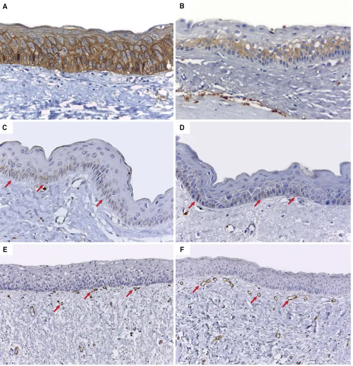
In the SKOT group, cases classiied as score 4 were the most frequent (n = 10; 58.8%) (Figure 1A), followed by scores 3 (n = 4; 23.5%), 1 (n = 2; 11.8%), and 2 (n = 1; 5.9%).
There was a predominance of score 4 cases among
primary NSKOTs (n = 11; 64.7%), followed by scores 3 (n = 5; 29.4%) and 2 (n = 1; 5.9%) (Figure 1B). In the group of recurrent NSKOTs, cases classiied as scores 4 (n = 3; 42.9%) and 2 (n = 2; 28.6%) were slightly more
Figure 1. A) Epithelial immunoexpression of GLUT-1 in more than 76% of cells (score 4) in SKOT (Advance, 400×). B) Immunoexpression of GLUT-1 in 26%-50% of epithelial cells (score 2) in primary NSKOT (Advance, 400×). C) Epithelial immunoexpression of GLUT-3 (arrows) in less than 25% of cells (score 1) in recurrent NSKOT (Advance, 400×). D) Immunoreactivity to GLUT-3 (arrows) in less than 25% of epithelial cells (score 1) in SKOT (Advance, 400×). E) Vessels immunoreactive to anti-CD34 antibody (arrows) in SKOT (Advance, 200×). F) Variably sized vessels labeled by anti-CD34 antibody (arrows) in primary NSKOT (Advance, 200×).
A B
D
F E
frequent when compared to cases classiied as scores 3 (n = 1; 14.3%) and 1 (n = 1; 14.3%). The nonparametric Kruskal-Wallis test revealed no signiicant difference
between groups (p = 0.360) (Figure 2).
Analysis of the epithelial immunoexpression of GLUT-3 revealed a higher frequency of negative cases in all groups. In the few GLUT-3-positive cases, expression of this protein in the epithelial
component was low and all cases were classiied
as score 1. Positive GLUT-3 immunostaining was
observed in only three (17.6%) of the 17 primary NSKOTs and in two (28.6%) of the seven recurrent NSKOTs (Figure 1C). Only four (23.5%) of the 17
SKOTs exhibited GLUT-3 immunoexpression in the epithelial component (Figure 1D).
Angiogenic index
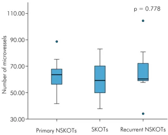
Anti-CD34 immunostaining was observed in single endothelial cells, in clusters of endothelial cells without a distinct vascular lumen, and in vessels with conspicuous lumens. Analysis of the angiogenic index based on MVC revealed a mean number of 59.38
(range: 37.8–83.0) vessels in SKOTs (Figure 1E). The mean vessel number was 62.81 (range: 41.6–88.6) in
primary NSKOTs (Figure 1F). Finally, a mean number
of 65.88 (range: 34.0–104.4) vessels was observed in
recurrent NSKOTs. The nonparametric
Kruskal-Wallis test revealed no signiicant difference between
groups (p = 0.778) (Figure 3).
No signiicant correlations were observed between
GLUT-1 immunoexpression scores and the angiogenic index in SKOTs (p = 0.064; r = 0.459), primary NSKOTs (p = 0.303; r = 0.265) or recurrent NSKOTs (p = 0.984; r = -0.009).
Discussion
Similarly to other tumors, KOTs are characterized by the loss of heterozygosity in tumor suppressor genes such as P16, TSLC1, FHIT, and PTCH.2,32
Mutations in the PTCH gene, which are related to the development of Gorlin syndrome,7,33 have been
identiied as the most important genetic alteration
in KOTs.33 Although NSKOTs and SKOTs apparently
share the same pathogenesis – which consists of the
aberrant activation of the SHH signaling pathway as a result of mutations in the PTCH gene,34,35 studies
have suggested a lower growth and infiltration capacity, as well as a lesser tendency to recur, for non-syndromic tumors.5,6,7
The expression of proteins involved in glucose transport and angiogenesis is important for increasing the uptake of nutrients by metabolically active cells.12
In this respect, GLUT-1 and GLUT-3 are important proteins involved in the transport of glucose into the intracellular medium. The overexpression of these proteins has been associated with a more aggressive biological behavior of different malignant tumors,
Primary NSKOTs SKOTs Recurrent NSKOTs p = 0.778
Number of microvessels
110.00
90.00
70.00
50.00
30.00
Figure 3. Box plot chart illustrating the number of microvessels according to groups of KOT.
Primary NSKOTs SKOTs Recurrent NSKOTs p = 0.360
Scores of GLUT-1 immunoexpression
4
3
2
1
0
including carcinomas of the oral cavity, larynx, lung, and breast, which is characterized by the presence of lymph node metastases, disease recurrence, and lower survival rates of the patients.12,15,16,17
In the present study, positive immunoexpression of GLUT-1 in the epithelial component was observed in all KOT cases, with a higher frequency of score 4
(≥ 76% positive cells). These indings indicate a high
expression of GLUT-1 in the epithelial component of these tumors and suggest an important role of this protein in glucose uptake by epithelial tumor
cells. On the other hand, no signiicant differences in
GLUT-1 immunoexpression were observed between NSKOTs and SKOTs, suggesting that the expression of this glucose transporter is not implicated in the differences in the biological behavior of these tumors.
With respect to GLUT-3, a higher frequency of negative cases was observed in all KOT groups. Furthermore, the few GLUT-3-positive cases exhibited low expression of this glucose transporter in the epithelial component, classified as score 1
(≤ 25% positive cells). Similar indings have been
reported in a study with dentigerous cysts, ameloblastomas, and KOTs.20 Taken together, these
indings suggest that the use of GLUT-3 for glucose
uptake is a rare mechanism in KOTs, which is used by a small number of cells in the epithelial component of these tumors. Furthermore, the results indicate that the differences in the biological behavior of NSKOTs and SKOTs are not related to GLUT-3 expression.
There are only few studies18,19,20 investigating
the expression of GLUTs in odontogenic lesions (PubMed database). Rumayor et al.18 examined
the immunohistochemical and ultrastructural characteristics of ghost cells in calcifying cystic odontoge n ic t u mor s, pi lom at r i xom a s, a nd craniopharyngiomas and observed restricted GLUT-1 immunoexpression in viable cells immediately
adjacent to ghost cells. According to these authors,
since cell metabolism probably slows down during the transformation of ghost cells, GLUT-1 expression in these transitional cells could be related to hypoxia.
On the basis of the indings of Rumayor et al.,18
higher GLUT-1 and GLUT-3 immunoreactivity would be expected for cells of the superficial epithelial layers of KOTs since they are more distant from
connective tissue vascularization and thus in a microenvironment that is more prone to hypoxia. However, all KOTs evaluated in the present study exhibited predominance of GLUT-1 and GLUT-3 immunostaining in deeper layers of the epithelial component and immunoreactivity to these proteins
tended to be absent in cells of the supericial layers.
This pattern of GLUT expression in KOTs may be related to the greater need of glucose uptake by metabolically active cells located in deeper layers of the epithelial component of these tumors. Also within this context, the predominant expression of GLUT-1 and GLUT-3, but particularly of GLUT-1, in deeper epithelial layers may be one of the reasons for the potentially aggressive behavior of these tumors. Taken together, these findings can improve the current knowledge of molecular pathways involved in KOTs. In addition, GLUT-1 could be suggested as a potential target for future development of therapeutic strategies for KOTs.
In line with this suggestion, Sánchez-Romero et al.19
observed high GLUT-1 immunoexpression in the epithelial cells of ameloblastic carcinomas, followed by ameloblastomas and tooth germs. Moreover, the
expression of this glucose transporter was signiicantly
higher in solid ameloblastomas than in unicystic ameloblastomas. According to these authors, GLUT-1 overexpression could be related to aggressiveness in both ameloblastomas and ameloblastic carcinomas.
In view of the limited diffusion capacity of oxygen and glucose from blood vessels,36 angiogenesis
is an essential event for tumor development and progression.21,22 However, the formation of new blood
vessels may not accompany the growth of neoplastic cells and may result in the emergence of hypoxic areas in the tumor.26,27 In this respect, hypoxia-inducible
factor 1 (HIF-1) plays a key role in the adaptation of neoplastic cells to hypoxia by promoting transcriptional activation of a repertoire of genes, such as those encoding GLUT-1 and vascular endothelial growth factor (VEGF).22,26,37 The latter has been described
as an important proangiogenic factor both under physiological and pathological conditions.23,37
and angiogenesis, particularly in tumors. In agreement with this suggestion, studies on endometrial adenocarcinomas and ovarian carcinomas have shown a positive correlation between GLUT-1 expression and microvessel density in the tumors.37,38 On the
other hand, Kitamura et al.39 reported an inverse
correlation between tumor glycolytic metabolism and angiogenesis in hepatocellular carcinomas. Kubo et al.40 observed an association between GLUT-1
immunoexpression and low microvessel density in osteosarcomas and questioned the relationship between glycolytic metabolism and angiogenesis in some malignant tumors. In the present study,
no signiicant correlations were observed between
GLUT-1 expression and the angiogenic index in KOTs. Considering the pattern of GLUT-1 and GLUT-3 immunoexpression in the epithelial component of KOTs observed in the present study, the lack of correlation between GLUT-1 expression and the
angiogenic index is an expected result. As identiied
in the present study, GLUT-1 immunoexpression predominated in the deeper layers of the epithelial component of KOTs. Since these epithelial cells are closer to the connective tissue vascularization, they are found in a microenvironment that is less prone to hypoxia. Accordingly, Sánchez-Romero et al.19
observed a predominance of a prostromal expression pattern of GLUT-1 in ameloblastomas, which may be induced by alternative pathways to hypoxia. Taken
together, these indings support the suggestion that,
in the epithelial component of KOTs, high GLUT-1 expression is not related to a process of adaptation to hypoxia, but to the existence of cells with increased metabolic demands.
Conclusion
The results of this study suggest that the more aggressive biological behavior of KOTs associated with Gorlin syndrome when compared to NSKOTs is not related to GLUT-1 or GLUT-3 expression. High GLUT-1 expression indicates an important role of this protein in glucose uptake by epithelial cells of KOTs and that this process is unlikely related to the angiogenic index. The predominant expression of GLUT-1 in deeper epithelial layers may be related to the existence of cells with increased metabolic demands
in KOTs. Taken together, these indings contribute to
the knowledge of the underlying mechanisms involved in the biological behavior of KOTs and support GLUT-1 as a potential target for future development of therapeutic strategies for these tumors.
1. Philipsen HP. Keratocystic odontogenic tumor. In: Barnes L, Eveson JW, Reichart P, Sidransky D, editors. World Health Organization classification of tumors: pathology and genetics of head and neck tumors. Lyon: IARC Press; 2005. p. 306-7.
2. Agaram NP, Collins BM, Barnes L, Lomago D, Aldeeb D, Swalsky P et al. Molecular analysis to demonstrate that odontogenic keratocysts are neoplastic. Arch Pathol Lab Med. 2004;128(3):313-7.
https://doi.org/10.1043/1543-2165(2004)128<313:MATDTO>2.0.CO;2
3. Figueroa A, Correnti M, Avila M, Andea A, DeVilliers P, Rivera H. Keratocystic odontogenic tumor associated with nevoid basal cell carcinoma syndrome: similar behavior to sporadic type? Otolaryngol Head Neck
Surg. 2010;142(2):179-83. https://doi.org/10.1016/j.
otohns.2009.10.008
4. Kichi E, Enokiya Y, Muramatsu T, Hashimoto S, Inoue T, Abiko Y et al. Cell proliferation, apoptosis, and apoptosis-related factors in odontogenic keratocysts and dentigerous cysts. J Oral Pathol
Med. 2005;34(5):280-6.
https://doi.org/10.1111/j.1600-0714.2005.00314.x
5. Díaz-Fernández JM, Infante-Cossío P, Belmonte-Caro R, Ruiz-Laza L, García-Perla-García A, Gutiérrez-Pérez JL. Basal cell nevus syndrome: presentation of six cases and literature review. Med Oral Patol Oral Cir Bucal. 2005;10 Suppl 1:E57-66.
6. Kimi K, Kumamoto H, Ooya K, Motegi K. Immunohistochemical analysis of cell-cycle- and apoptosis-related factors in lining epithelium of odontogenic keratocysts. J Oral Pathol Med.
2001;30(7):434-42.
7. Manfredi M, Vescovi P, Bonanini M, Porter S. Nevoid basal cell carcinoma syndrome: a review of the literature. Int J Oral Maxillofac Surg. 2004;33(2):117-24. https://doi.
org/10.1054/ijom.2003.0435
8. Amorim RF, Godoy GP, Galvão HC, Souza LB, Freitas RA. Immunohistochemical assessment of extracellular matrix components in syndrome and non-syndrome odontogenic keratocysts. Oral Dis. 2004;10(5):265-70. https://doi.
org/10.1111/j.1601-0825.2004.01023.x
9. Cavalcante RB, Pereira KM, Nonaka CF, Nogueira RL, Souza LB. Immunohistochemical expression of MMPs 1, 7, and 26 in syndrome and nonsyndrome odontogenic keratocysts. Oral Surg Oral Med Oral Pathol Oral Radiol
Endod. 2008;106(1):99-105. https://doi.org/10.1016/j.
tripleo.2007.12.028
10. Leonardi R, Matthews JB, Caltabiano R, Greco M, Lombardo C, Loreto C et al. MMP-13 expression in keratocyst odontogenic tumour associated with NBCCS and sporadic keratocysts. Oral Dis. 2010;16(8):795-800.
https://doi.org/10.1111/j.1601-0825.2010.01690.x
11. Hong YY, Yu FY, Qu JF, Chen F, Li TJ. Fibroblasts regulate variable aggressiveness of syndromic keratocystic and non-syndromic odontogenic tumors. J Dent Res. 2014;93(9):904-10. https://doi.org/10.1177/0022034514542108 12. Macheda ML, Rogers S, Best JD. Molecular and cellular
regulation of glucose transporter (GLUT) proteins in cancer. J Cell Physiol. 2005;202(3):654-62. https://doi.
org/10.1002/jcp.20166
13. Medina RA, Owen GI. Glucose transporters: expression, regulation and cancer. Biol Res. 2002;35(1):9-26. https://doi. org/10.4067/S0716-97602002000100004
14. Thorens B, Mueckler M. Glucose transporters in the 21st Century. Am J Physiol Endocrinol Metab. 2010;298(2):E141-5.
https://doi.org/10.1152/ajpendo.00712.2009
15. Ayala FR, Rocha RM, Carvalho KC, Carvalho AL, Cunha IW, Lourenço SV et al. GLUT1 and GLUT3 as potential prognostic markers for Oral Squamous Cell Carcinoma. Molecules. 2010;15(4):2374-87. https://doi.org/10.3390/ molecules15042374
16. Estilo CL, O-charoenrat P, Talbot S, Socci ND, Carlson DL, Ghossein R et al. Oral tongue cancer gene expression profiling: identification of novel potential prognosticators by oligonucleotide microarray analysis. BMC Cancer. 2009;9(1):11. https://doi.org/10.1186/1471-2407-9-11 17. Roh JL, Cho KJ, Kwon GY, Ryu CH, Chang HW,
Choi SH et al. The prognostic value of hypoxia markers in T2-staged oral tongue cancer. Oral Oncol. 2009;45(1):63-8.
https://doi.org/10.1016/j.oraloncology.2008.03.017
18. Rumayor A, Carlos R, Kirsch HM, Andrade BA,
Romañach MJ, Almeida OP. Ghost cells in pilomatrixoma, craniopharyngioma, and calcifying cystic odontogenic tumor: histological, immunohistochemical, and
ultrastructural study. J Oral Pathol Med. 2015;44(4):284-90.
https://doi.org/10.1111/jop.12234
19. Sánchez-Romero C, Bologna-Molina R, Mosqueda-Taylor A, Almeida OP. Immunohistochemical expression of GLUT-1 and HIF-1α in tooth germ, ameloblastoma, and ameloblastic carcinoma. Int J Surg Pathol. 2016;24(5):410-8. https://doi. org/10.1177/1066896916640359
20. Vasconcelos RC, Moura JMO, Brasileiro Junior VL, Silveira EJ, Souza LB. Immunohistochemical expression of GLUT-1, GLUT-3, and carbonic anhydrase IX in benign odontogenic lesions. J Oral Pathol Med. 2016;45(9):712-7. https://doi.
org/10.1111/jop.12427
21. Gordon MS, Mendelson DS, Kato G. Tumor angiogenesis and novel antiangiogenic strategies. Int J Cancer.
2010;126(8):1777-87. https://doi.org/10.1002/ijc.25026
22. Nguyen A, Hoang V, Laquer V, Kelly KM. Angiogenesis in cutaneous disease: part I. J Am Acad Dermatol.
2009;61(6):921-42. https://doi.org/10.1016/j.jaad.2009.05.052
23. Nonaka CF, Maia AP, Nascimento GJ, Freitas RA, Souza LB, Galvão HC. Immunoexpression of vascular endothelial growth factor in periapical granulomas, radicular cysts, and residual radicular cysts. Oral Surg Oral Med Oral Pathol Oral Radiol Endod. 2008;106(6):896-902. https://doi.
org/10.1016/j.tripleo.2008.06.028
24. Alaeddini M, Salah S, Dehghan F, Eshghyar N, Etemad-Moghadam S. Comparison of angiogenesis in keratocystic odontogenic tumours, dentigerous cysts and ameloblastomas. Oral Dis. 2009;15(6):422-7. https://doi.
org/10.1111/j.1601-0825.2009.01566.x
25. Gadbail AR, Mankar Gadbail MP, Hande A, Chaudhary MS, Gondivkar SM, Korde S et al. Tumor angiogenesis: role in locally aggressive biological behavior of ameloblastoma and keratocystic odontogenic tumor. Head Neck. 2013;35(3):329-34. https://doi.org/10.1002/hed.22960 26. Ortega AD, Sánchez-Aragó M, Giner-Sánchez D,
Sánchez-Cenizo L, Willers I, Cuezva JM. Glucose avidity of carcinomas. Cancer Lett. 2009;276(2):125-35. https://doi.
org/10.1016/j.canlet.2008.08.007
27. Pérez-Sayáns M, Suárez-Peñaranda JM, Pilar GD, Barros-Angueira F, Gándara-Rey JM, García-García A. Hypoxia-inducible factors in OSCC. Cancer Lett.
2011;313(1):1-8. https://doi.org/10.1016/j.canlet.2011.08.017
28. Barnes L, Eveson JW, Reichart P, Sidransky D. World Health Organization classification of tumors. Pathology and genetics of head and neck tumors. Lyon: IARC Press; 2005. 29. Evans DG, Ladusans EJ, Rimmer S, Burnell LD, Thakker N,
Farndon PA. Complications of the naevoid basal cell carcinoma syndrome: results of a population based study. J Med Genet. 1993;30(6):460-4. https://doi.org/10.1136/
jmg.30.6.460
30. Maeda K, Chung YS, Takatsuka S, Ogawa Y, Onoda N, Sawada T et al. Tumour angiogenesis and tumour cell proliferation as prognostic indicators in gastric carcinoma. Br J Cancer. 1995;72(2):319-23. https://doi.
31. Weidner N, Semple JP, Welch WR, Folkman J. Tumor angiogenesis and metastasis: correlation in invasive breast carcinoma. N Engl J Med. 1991;324(1):1-8. https://doi. org/10.1056/NEJM199101033240101
32. Henley J, Summerlin DJ, Tomich C, Zhang S, Cheng L. Molecular evidence supporting the neoplastic nature of odontogenic keratocyst: a laser capture microdissection study of 15 cases. Histopathology. 2005;47(6):582-6.
https://doi.org/10.1111/j.1365-2559.2005.02267.x
33. Gomes CC, Diniz MG, Gomez RS. Review of the molecular pathogenesis of the odontogenic keratocyst.
Oral Oncol. 2009;45(12):1011-4. https://doi.org/10.1016/j.
oraloncology.2009.08.003
34. Gu XM, Zhao HS, Sun LS, Li TJ. PTCH mutations in sporadic and Gorlin-syndrome-related odontogenic keratocysts. J Dent Res. 2006;85(9):859-63. https://doi. org/10.1177/154405910608500916
35. Hakim SG, Kosmehl H, Sieg P, Trenkle T, Jacobsen HC, Attila Benedek G et al. Altered
expression of cell-cell adhesion molecules β-catenin/ E-cadherin and related Wnt-signaling pathway in sporadic and syndromal keratocystic odontogenic tumors. Clin Oral Investig. 2011;15(3):321-8. https://doi.org/10.1007/s00784-010-0388-8
36. Bergers G, Benjamin LE. Tumorigenesis and the angiogenic switch. Nat Rev Cancer. 2003;3(6):401-10. https://doi.org/10.1038/nrc1093
37. Ozbudak IH, Karaveli S, Simsek T, Erdogan G, Pestereli E. Neoangiogenesis and expression of hypoxia-inducible factor 1α, vascular endothelial growth factor, and glucose transporter-1 in endometrioid type endometrium adenocarcinomas. Gynecol Oncol. 2008;108(3):603-8.
https://doi.org/10.1016/j.ygyno.2007.11.028
38. Semaan A, Munkarah AR, Arabi H, Bandyopadhyay S, Seward S, Kumar S et al. Expression of GLUT-1 in epithelial ovarian carcinoma: correlation with tumor cell proliferation, angiogenesis, survival and ability to predict optimal cytoreduction. Gynecol Oncol. 2011;121(1):181-6.
https://doi.org/10.1016/j.ygyno.2010.11.019
39. Kitamura K, Hatano E, Higashi T, Narita M, Seo S, Nakamoto Y et al. Proliferative activity in hepatocellular carcinoma is closely correlated with glucose metabolism but not angiogenesis. J Hepatol. 2011;55(4):846-57. https://doi.
org/10.1016/j.jhep.2011.01.038