Si3N4/SiC Nanocomposite Powder from a Preceramic Polymeric
Network Based on Poly(methylsilane) as the SiC precursor
Maurício F. Gozzi, Eduardo Radovanovic, I. Valéria P. Yoshida*
Instituto de Química, Universidade Estadual de Campinas, C.P. 6154, 13083-970 Campinas - SP, Brazil
Received: July 10, 2000; Revised: January 8, 2001
Si3N4/SiC nanocomposite powders were obtained from a preceramic polymeric network based
on poly(methylsilane) as thein situquasi-stoichiometric SiC source. These powders were consti-tuted of nanosized SiC particles homogeneously distributed in the Si3N4particulate matrix.β-SiC
whiskers were grown at 1400 °C in the pores of the matrix. At 1600 °C, theα → βSi3N4phase
transition took place, but no elemental silicon from Si3N4decomposition was detected, evidencing
the protective effect of the SiC phase.
Keywords:composite powders, silicon nitride, silicon carbide
1. Introduction
Silicon nitride-silicon carbide (Si3N4/SiC) composites
have received a great deal of attention in high temperature structural applications, such as turbine and automobile engine components and heat exchangers, due to their po-tentially high strength and toughness, and good creep resis-tance1-6. However, these good characteristics are difficult to achieve by the conventional mixture of SiC and Si3N4
commercial powders.
Several methods have been described to produce small-sized Si3N4and/or SiC powders in a homogeneous mixture,
such as: plasma synthesis, in which Si3N4 particles or
Si-C-N powders are carbon-coated2,3; generationin situof micrometer size β-Si3N4, from Si powder with flowing
N2/H2 gas mixture reaction on SiC particles7; and
poly-meric precursors1,8,9. This last route offers the possibility of improving the compositional homogeneity and tailoring the composition and molecular structure of the ceramic powders. Furthermore, the solubility and the rheology of polymeric precursors provide potential processing routes to binders, to sintering aids and to the formation of thin films and fibers, which are often difficult or impossible to achieve by the more traditional ceramic processing techniques10-12. Small-sized Si3N4and/or SiC powders can be processed
by several methods, to produce ceramic composites with excellent mechanical properties, at ambient and high tem-peratures, and good oxidation resistance2,3,13-16.
In a previous study, a polysilane network prepared from poly(methylsilane), PMS, and tetra-allylsilane, TAS, was
used as a precursor of a quasi-stoichiometric SiC, in a reasonable yield (~60%)17. In this study, Si3N4/SiC
nano-composite powder was obtained from a mixture of PMS/TAS and Si3N4powder, to investigate the evolution
of the microstructure and crystalline phases of the compos-ite powder, in relation to the temperature of pyrolysis. No attempts to obtain the composite in any pre-determined shape was made in the present investigation.
2. Experimental
2.1. Materials
Si3N4powder (Hüls) (325 mesh) was used as received
from the manufacturer. Poly(methylsilane) was synthe-sized by polycondensation of CH3SiHCl2in the presence
of Na, in toluene18, and tetra-allylsilane was prepared through Grignard route19. Si3N4/SiC nanocomposite
pow-der was prepared by the mixture of 4 g of Si3N4powder,
1 g of PMS and 0.5 cm3 of TAS, giving rise to a very viscous slurry, which was poured into an alumina crucible. This procedure was made in a glove box filled with dry argon. The pyrolysis was carried out in an EDG tube furnace (model EDGCON 5P) equipped with an internal alumina tube and a temperature controller, under an argon flow (Air Liquid, 50 mL/min, 99.9 %). The mixture was heated to 180 °C, at 5 °C/min, keeping the sample at this temperature for 60 min; then a second ramp to 350 °C, at 5 °C/min, remaining at this plateau for 90 min, and a third ramp to 1000 °C, at 2 °C/min, followed by holding at this temperature for 120 min. Finally, the sample was cooled
down to room temperature, at 2 °C/min. Three fractions of the resulting powder received distinct thermal treatments in a Thermolyne tube furnace (model F59340-CM): the first one was heated up to 1200 °C, at 10 °C/min, under an argon flow, keeping at this temperature for 2 h; the second fraction received thermal treatment up to 1400 °C, and finally, the third fraction was heated up to 1600 °C, both keeping at the final temperature for 2 h, under the same condition. The ceramic yield was determined by thermo-gravimetric analysis (TGA), at 1000 °C.
2.2. Instrumentation
The composition of the powders was determined by crossing the results obtained by elemental analysis, 29Si MAS NMR spectrum and X-ray fluorescence data. Carbon and nitrogen elemental analysis were performed in an ele-mental analyser (Perkin Elmer 2400) according to the pro-cedure suggested by Ref. 20. Solid-state29Si magic-angle spinning nuclear magnetic resonance (29Si MAS NMR) spectrum was acquired using a pulse angle of 75° and a delay of 1 h, on a Bruker AC300P instrument at 59.6 MHz, with spinning frequency of ~5 kHz. Silicon and metal traces in the samples were estimated by X-ray fluorescence, ap-plying the fundamental parameters method, in a spectrome-ter Spectrace 5000 Tracor X-ray, using cellulose filspectrome-ter. The crystalline phases of the samples were investigated by X-ray diffractometry (XRD) using a diffractometer Schi-madzu, model XD3A, with the CuKαradiation. The grain size values of theβ-SiC, were calculated through the full width of half height measures of the XRD patterns, using the reflection centered at 35.6° (2θ) <111>. Transmission electron microscopy observations (TEM), were performed on a Zeiss CEM 902 microscope, with 80 eV. The samples were grounded to a very fine powder, which was then dispersed in isopropanol and transferred onto a 100 mesh copper grid (parlodium coated) using an eye dropper. Some observations were done in a field emission scanning elec-tron microscope (FESEM) JEOL JSM-6340F. For these investigations, a thin layer of gold previously coated the samples.
3. Results and Discussion
A highly crosslinked polymeric network prepared from the mixture of PMS and TAS was based in a hydrosilylation reaction between SiH bonds, from PMS, and C=C bonds, from TAS, at around 200 °C21. The use of a polymeric network generatedin situas a ceramic precursor hinders the splitting of volatile organosilicon compounds during the pyrolysis process, increasing the ceramic yield12. At around 350 °C, the so-called Kumada’s rearrangement took place, when Si-Si groups were converted into Si-CH2-Si groups
by the insertion of methyl side groups in the main chain of silicon22. The mineralization process (loss of organic and
hydrogen groups) gradually occurred at temperatures above that, and finally, at around 800 °C, the crystallization ofβ-SiC took place21. A typical XRD pattern of the ceramic
product obtained at 1000 °C is shown in Fig. 1, where the mean grain size of the crystal, estimated by the Scherrer’s equation, was around 4 nm.
Si3N4powder used in this work was present in theαand
βcrystalline forms. In addition, a certain amount of crys-talline Si was found in this powder. From the XRD pattern of this sample, the following crystalline composition was estimated23:α-Si3N452%, β-Si3N4 40% and Si 8%. The
total weight composition of this powder was estimated crossing data from X-ray fluorescence,29Si MAS NMR and elemental analysis (taking the amorphous fraction into account): Si3N482%, Si 7%, SiO25%, free carbon 5%, and
metals (mostly Fe, Ni and Cu) 1%.
Besides being the SiC precursor, the PMS/TAS mixture had also a binder role in the preceramic slurry. Conven-iently, after pyrolysis it did not leave any undesirable residue in the final product, as other well-known binders do24. Interesting to note that usually oily or waxy binders perform better than powders25.
The PMS/TAS reactive mixture presented an oily con-sistency. When its proportion was high in the preceramic slurry (PMS/TAS + Si3N4), sedimentation of the nitride
powder occurred, and the fired material was heterogeneous (composition-wise). The Si3N4/PMS/TAS composition
used in this work (see experimental section for details) was adjusted in order to avoid the mentioned sedimentation, and to guarantee the homogeneity of the ceramic powder com-posite.
TEM investigations of several fractions of the compos-ite powder were undertaken. In all of them, a micro/nano-microstructure was observed, with nanosized SiC particles dispersed in the surroundings of theαandβ-Si3N4crystals
(crystalline arrangements were checked by XRD). Figure 2 shows a TEM micrograph of the Si3N4/SiC composite
powder.
Some reports on the mechanical properties of processed Si3N4/SiC composite powders have shown that this
inti-mate and uniform arrangement can lead to a inti-material with excellent mechanical properties1-3,11.
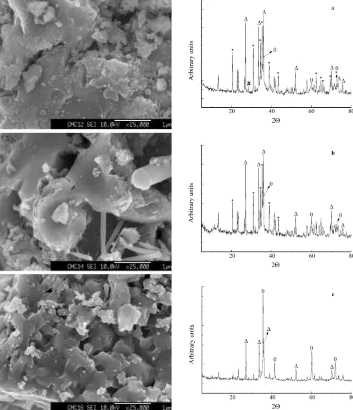
The evolution of the microstructure and crystalline phases of the Si3N4/SiC powder, was monitored after
heat-ing up to 1200 °C, 1400 °C and 1600 °C, for 2 h at each final temperature, under argon atmosphere. Figure 3 shows FESEM micrographs and XRD patterns of the products.
At 1200 °C, the composite powder showed a homoge-neous nature, with no evidence of phase segregation at this amplification. In the respective XRD pattern, phases of β-SiC, Si,α- andβ-Si3N4could be observed. The
estima-tion of the mean grain size ofβ-SiC crystals by the Scher-rer’s equation, was not possible, since the peak used for this calculation at 35.77° (2θ) was overlapped by the peak at 35.24° (2θ), assigned toα-Si3N4.26However, this peak was
thinner than the analogous diffraction of the ceramic ob-tained from PMS/TAS, pyrolysed at 1000 °C, as it was shown in Fig. 1.
At 1400 °C, the SiC phase bridges neighbor Si3N4grains
at their interfaces, and a pronounced formation ofβ-SiC whiskers was observed. The estimated mean size of these whiskers (by FESEM) was 730 nm long and 26 nm wide. Also noticeable was the disappearance of Si signals in the X-ray pattern, suggesting that it was consumed at this temperature. The reaction between the Si impurity, which was present originally in the Si3N4powder, and carbon was
commonly thought to occur in the liquid phase27, or even on the Si activated surface, as:
Si(s,l)+ C(s)→SiC(s) (1)
It is also well known that Si3N4decomposes into liquid
Si when heated at around 1450 °C, under inert atmos-phere28. The result described above suggested that this decomposition became less significant in the presence of SiC phase.
The mechanism of the whisker formation is related to a process in which materials are transported to the vapor phase during the heating process. It has already been re-ported in the literature, that to grow SiC in whisker mor-phology, the presence of reactants in a vapor phase during
the final stage of the reaction is necessary. Otherwise, particulate SiC would result29,30. The reactions involved can be summarized as:
SiO2(s)+ C(s)→SiO(g)+ CO(g) (2)
SiO(g)+ 2C(s)→SiC(s)+ CO(g) (3)
Hence, the overall reaction is:
SiO2(s)+ 3C(s)→SiC(s)+ 2CO(g) (4)
Distributing SiC whiskers uniformly in the ceramic matrixes is not an easy task. However, when it is done, the potential benefit of the whisker reinforcement is possible to achieve31. Considering the homogeneous nature of the Si3N4/SiC nanocomposite powder prepared in this work, it
is reasonable to believe thatβ-SiC whiskers were uniformly distributed in the porous structure of Si3N4grains.
At 1600 °C, a completely different microstructure was observed. SiC whiskers were no longer detectable. It seemed they were embedded in the Si3N4 matrix. The
α-Si3N4 crystals underwent a phase transformation toβ
-Si3N432. The α → β phase transformation occurs via a
solution-precipitation process, and can be hindered by nanosized SiC particles. This effect appears to increase with increasing SiC content33. The transition observed sug-gested that probably there were not enough nanosized SiC particles in the powder composite, to hinder the α → β Si3N4phase transformation. This also explains the
micro-structure observed. The XRD pattern of this composite powder suggests a certain consumption of the Si3N4phase,
once the peak-height ratio of β-SiC to Si3N4 became
greater. At this point, the mean size of theβ-SiC crystals could easily be estimated as being approximately 83 nm.
It is interesting to observe that pure Si3N4presents a
distinct degradation process compared to the powder rein-forced by SiC. While pure Si3N4at 1600 °C under argon
atmosphere is decomposed producing Si, there was no evidence of this element in the Si3N4/SiC composite
pow-der, treated at the same condition. It is very reasonable to suppose that in the reaction of the SiO2 impurity with
carbon (reaction (2)), the CO(g) produced on the Si3N4
particle surface, altered the degradation process of the silicon nitride, to form SiC34:
3CO(g)+ Si3N4(s)→2SiC(s)+ 2N2(g)
+ CO2(g)+ SiO(g) (5)
4. Conclusion
The pyrolysis of the PMS/TAS polymer as thein situ SiC source, in the presence of Si3N4powder, gave rise to
Si3N4/SiC nanocomposite powder with β-SiC uniformly
Growth of β-SiC whiskers was observed when the composite was heated at 1400 °C, under argon atmosphere. At 1600 °C, these SiC whiskers were embedded in the matrix, and theα → β Si3N4phase transformation took
place. There was no evidence of Si formation, opposite to the behavior of pure Si3N4.
The results confirmed the basic concept that the thermal stability behavior of Si3N4/SiC composite powders can be
remarkably improved via the incorporation of nanosized SiC, obtained from polymeric precursor.
Acknowledgments
We gratefully acknowledge financial support from Conselho Nacional de Desenvolvimento Científico e Tec-nológico (CNPq) and Fundação de Amparo à Pesquisa do Estado de São Paulo (FAPESP), Proc. 95/3636-3.
References
1. Sternitze, M.J. Eur. Ceram. Soc., v. 17, p. 1061-1082, 1997.
2. Herrmann, M.; Schuber, C.; Rendtel, A.; Hübner, H. J.Am. Ceram. Soc. Bull., v. 81, p. 1095-1108, 1998. 3. Rendtel, A.; Hübner, H.; Herrmann, M.; Schuber, C.
J.Am. Ceram. Soc. Bull., v. 81, p. 1109-1120, 1998. 4. Kaya, H.Comp. Sci. Tech., v. 59, p. 861-872, 1999. 5. Lange, H.; Wötting, G.; Winter, G.Ang. Chem. Int.
Ed. Engl., v. 30, p. 1579-1597, 1991.
6. Narula, C.K.; Allison, J.E.; Bauer, D.R.; Gandhi, H.S. Chem. Mater., v. 8, p. 984-1003, 1996.
7. Lee, S.Y. J. Am. Ceram. Soc, v. 81, p. 1262-1268, 1998.
7. Bao, X.; Edirisinghe, M.J.Composites: Part A, v. 30, p. 601-610, 1999.
9. Bill, J.; Aldinger, F. Adv. Mater., v. 7, p. 775-787, 1995.
10. Schimidt, W.R.; Sukumar, V.; Hurley Jr, W.J.; Garcia, R.; Doremus, K.H.; Interrante, L.V.; Renlund, G.M. J. Am. Ceram. Soc., v. 73, p. 2412-2418, 1990. 11. Wynne, K.J.; Rice, R.W.Ann. Rev. Mater. Sci., v. 14,
p. 297-334, 1984.
12. Laine, R.M.; Babonneau, F. Chem. Mater., v. 5, p. 260-279, 1993.
13. Weimer, A.W.; Bordia, R.K.Composites: Part B, v. 30, p. 647-655, 1999.
14. Hirano, T.; Niihara, K.Mater. Lett., v. 22, p. 249-254, 1995.
15. Niihara, K.; Izaki, K.; Nakahira, A.J. Jpn. Soc. Pow-der PowPow-der Metall., v. 37, p. 172-176, 1990.
16. Niihara, K.; Suganuma, K.; Nakahira, A.; Izaki, K.J. Mater. Sci. Lett., v. 9, p. 598-599, 1990.
17. Gozzi, M.F.; Gonçalves, M.C.; Yoshida, I.V.P. J. Mater. Sci., v. 34, p. 155-159, 1999.
18. Gozzi, M.F.; Yoshida, I.V.P.Macromolecules, v. 29, p. 7235-7240, 1996.
19. O’Brien, S.; Fishwick, M.; McDermott, B.; Wall-bridge, M.G.H.; Wright, G.A.;Inorg. Synt., v. 13, p. 73-74, 1971.
20. Borda, P.P.; Ledzidins, P. Anal. Chem., v. 52, p. 1777-1778, 1980.
21. Gozzi, M.F.; Yoshida, I.V.P.Eur. Polym. J., v. 33, p. 1301-1306, 1997.
22. Shiina, K.; Kumada, M.J. Org. Chem., v. 23, p. 139, 1958.
23. Gazarra, C.P.; Messier, D.R.Am. Ceram. Soc. Bull., v. 56, p. 777-780, 1977.
24. Smith, J.T.; Quackenbusch, C.L. Am. Ceram. Soc. Bull., v. 59, p.529, 1980.
25. Schwab, S.T.; Blanchard, C.R.; Graef, R.C.J. Mater. Sci., v. 29, p. 6320-6328, 1994.
26. Dajgalik, P.; Galusek, D.J. Mater. Sci .Lett., v. 12, p. 1937-1939, 1993.
27. Tian, J.; Li, J.; Dong, L.J. Am. Ceram. Soc., v. 82, p. 2548-2550, 1999.
28. Rao, R.V.K.; Godkhindi, M.M.J. Mater. Sci., v. 27, p. 2726-2730, 1992.
29. Choi, H.-J.; Lee, J.-G.J. Mater. Sci., v. 30, p. 1982-1986, 1995.
30. Spiandorello, F.M.; Borsa, C.E.; Kiminami, R.H.G.A. Cerâmica, v. 45, p. 193-197, 1999.
31. Baril, D.; Tremblay, S.P.; Fiset, M.J. Mater. Sci., v. 28, p. 5486-5494,1993.
32. Callity, B.D.Elements of X-ray diffraction, Addison-Wesley, Reading, MA, p. 256, 1978.
33. Messier, D.R.; Riley, F.L.; Brook, R.J.J. Mater. Sci., v. 13, p. 199-205,1978.
34. Colquhoun, I.; Thompson, D.P.; Wilson, W.I.; Grieverson, P.; Jack, K.H.I.Proc. Br. Ceram. Soc.v. 22, p. 181-195, 1973.