Hydra vulgaris
Diane Bridge1*, Alexander G. Theofiles1, Rebecca L. Holler1, Emily Marcinkevicius2, Robert E. Steele3, Daniel E. Martı´nez2
1Department of Biology, Elizabethtown College, Elizabethtown, Pennsylvania, United States of America,2Department of Biology, Pomona College, Claremont, California, United States of America,3Department of Biological Chemistry and the Developmental Biology Center, University of California Irvine, Irvine, California, United States of America
Abstract
Background:In the face of changing environmental conditions, the mechanisms underlying stress responses in diverse organisms are of increasing interest. In vertebrates, Drosophila, and Caenorhabditis elegans, FoxO transcription factors mediate cellular responses to stress, including oxidative stress and dietary restriction. Although FoxO genes have been identified in early-arising animal lineages including sponges and cnidarians, little is known about their roles in these organisms.
Methods/Principal Findings:We have examined the regulation of FoxO activity in members of the well-studied cnidarian genusHydra. We find thatHydra FoxOis expressed at high levels in cells of the interstitial lineage, a cell lineage that includes multipotent stem cells that give rise to neurons, stinging cells, secretory cells and gametes. Using transgenicHydrathat express a FoxO-GFP fusion protein in cells of the interstitial lineage, we have determined that heat shock causes localization of the fusion protein to the nucleus. Our results also provide evidence that, as in bilaterian animals,HydraFoxO activity is regulated by both Akt and JNK kinases.
Conclusions:These findings imply that basic mechanisms of FoxO regulation arose before the evolution of bilaterians and raise the possibility that FoxO is involved in stress responses of other cnidarian species, including corals.
Citation:Bridge D, Theofiles AG, Holler RL, Marcinkevicius E, Steele RE, et al. (2010) FoxO and Stress Responses in the CnidarianHydra vulgaris. PLoS ONE 5(7): e11686. doi:10.1371/journal.pone.0011686
Editor:Alexander W. Shingleton, Michigan State University, United States of America
ReceivedFebruary 28, 2010;AcceptedMay 19, 2010;PublishedJuly 21, 2010
Copyright:ß2010 Bridge et al. This is an open-access article distributed under the terms of the Creative Commons Attribution License, which permits unrestricted use, distribution, and reproduction in any medium, provided the original author and source are credited.
Funding:The research described was funded by National Science Foundation grant IBN-0316065 and by an Elizabethtown College faculty grant. The funders had no role in study design, data collection and analysis, decision to publish, or preparation of the manuscript.
Competing Interests:The authors have declared that no competing interests exist.
* E-mail: bridged@etown.edu
Introduction
In bilaterian animals, members of the FoxO family of transcription factors are well-known for their roles in cellular responses to environmental and physiological stress. A single FoxO gene is present inDrosophila(dFOXO) andC. elegans(daf-16), and four are present in mice and humans (FoxO1,FoxO3,FoxO4, andFoxO6). In Drosophila, C. elegans, and mammalian cells, FoxO proteins increase resistance to oxidative stress [1–4]. Transcription of FoxO target genes increases under low nutrient conditions inDrosophila,C. elegans, and mammals [4–6] and during heat shock inC. elegans[7]. FoxO proteins mediate diverse cellular responses to stress. In
Drosophila,C. elegansand mammals, they increase levels of enzymes that detoxify reactive oxygen species (ROS) [1–3,8]. During starvation, FoxO proteins induce autophagy in mouse skeletal muscle and cardiomyocytes, permitting recycling of cellular components [9–11]. dFOXO similarly induces autophagy in the fat body of Drosophila undergoing dietary restriction [12]. In C. elegans and mammals, FoxO proteins increase resistance to DNA damage [13–15]. InC. elegans, DAF-16 regulates transcription of genes involved in pathogen resistance and acts together with the transcription factor heat shock factor 1 to upregulate transcription of small heat shock protein genes [7,16].
Activity of FoxO proteins is regulated by post-transcriptional modification, including phosphorylation. In response to insulin/ IGF-1 and other growth factors, Akt and the related serum- and glucocorticoid-inducible kinase (SGK) phosphorylate FoxO pro-teins (with the exception of FoxO6) at three conserved sites. These phosphorylations promote FoxO binding to 14-3-3 proteins and localization to the cytoplasm [17–20], where FoxO proteins cannot regulate transcription. In contrast, in response to stress the c-Jun N-terminal kinase (JNK) pathway causes FoxO nuclear localization and increased transcriptional activity [21–23]. JNK has been shown to phosphorylate DAF-16 and FoxO4 directly [21,22]. JNK-dependent nuclear localization of mammalian FoxO proteins may also involve phosphorylation of 14-3-3 proteins followed by their disassociation from FoxO proteins [24].
focused on the cnidarian genus Hydra. The Hydra magnipapillata
genome has been sequenced [30], and transgenic Hydra can be produced [31]. In addition, the physiological response to heat shock inHydrahas been examined in some detail [32–34].
Cell and tissue dynamics inHydraspecies are well characterized [35]. The cylindrical body of the adultHydrapolyp consists of two tissue layers, an ectoderm and an endoderm. At one end of the body is a mouth surrounded by a ring of tentacles. At the other end is a basal disk by which the animal adheres to the substrate. In the adult, cells divide continuously in the body column. Cell division in the body column causes displacement of epithelial cells into the tentacles and basal disk [36,37], where cell cycle arrest and terminal differentiation occur [38]. Adult Hydra routinely reproduce asexually, with cells from the body column displaced into buds. The continuous growth and production of buds by adult
Hydracan be explained in part by the presence of three types of multipotent stem cells. These are ectodermal stem cells, endoder-mal stem cells, and interstitial stem cells. Interstitial stem cells are located between epithelial cells in the ectodermal layer and give rise to neurons, secretory cells, gametes, and nematocytes, the stinging cells unique to cnidarians [39]. We have characterized the expression of FoxO in H. vulgaris and the closely related H. magnipapillataand have examined cellular localization of a FoxO-GFP fusion protein in transgenic animals. We find evidence for significant parallels in regulation of FoxO between Hydra and bilaterian animals.
Results
A single FoxO gene is present inH. magnipapillata
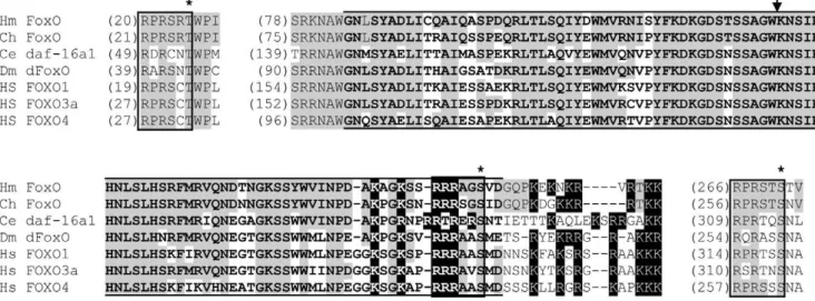
Searches of theH. magnipapillatagenome identified a single FoxO gene. The predicted H. magnipapillata FoxO protein (Figure 1) includes the forkhead winged helix domain characteristic of Fox proteins. A single intron is located within the region encoding the forkhead domain. An intron is present at the same location in FoxO genes from multiple other species, as well as in members of some other Fox gene families [25]. As in known FoxO proteins (with the exception of FoxO6), three consensus Akt/SGK phosphorylation sites are present [4,40–44]. Stretches of basic amino acids
overlapping the end of the forkhead domain match the consensus sequence for a bipartite nuclear localization signal [45].
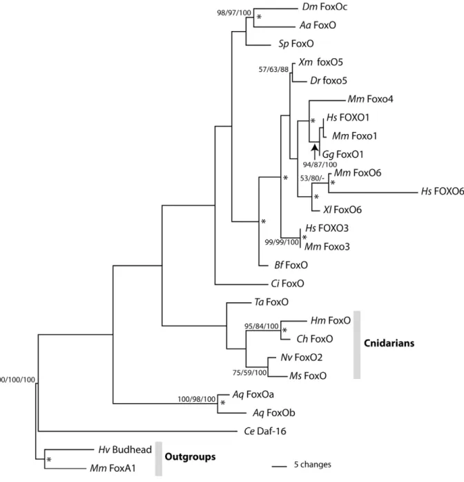
Phylogenetic analyses based on the predicted amino acid sequence of the forkhead domain were performed to confirm that the Hydra gene is a member of the FoxO family. An initial phylogenetic analysis included representatives from mouse of all known Fox gene families. This analysis also included Fox genes fromClytia hemisphaerica[27], a species that, likeHydra, belongs to the cnidarian class Hydrozoa. We found that theH. magnipapillata
gene groups with FoxO genes as expected (data not shown). Phylogenetic analyses including sequences of FoxO forkhead domains from diverse animals were also performed (Figure 2). BLAST searches of the genome ofTrichoplax adherens, a member of the early-evolving animal phylum Placozoa, identified a FoxO gene which was included in analyses. Results of phylogenetic analyses place H. magnipapillata FoxO together with FoxO genes from other cnidarian species. The results imply that H. magnipapillata FoxOis the ortholog ofdaf-16andDrosophila dFOXO. Vertebrate FoxO sequences form a group within the FoxO family, confirming that duplication of FoxO genes has occurred within the vertebrate lineage.
FoxO is expressed in interstitial cells
Hydrainterstitial cells are located between epithelial cells, mainly within the ectoderm. They are present in the body column, but not in the tentacles or basal disk [39]. Interstitial cells include multipotent interstitial stem cells, committed differentiation intermediates derived from them, and unipotent stem cells, also derived from the multipotent interstitial stem cells, which produce eggs or sperm [39]. Interstitial cells give rise to neurons, secretory cells, nematocytes, and gametes [46,47]. During the process of nematocyte formation, cells divide to form nests of nematoblasts connected by cytoplasmic bridges [48]. Cells within the nests then differentiate, separate, and migrate to their final locations. Like interstitial cells, nematoblasts are found in the ectoderm of the body column. Neurons also differentiate within the body column but are present in both tissue layers. Secretory cells are found only in the endoderm. Cells which will give rise to sperm migrate within
Figure 1. Conserved portions of the predictedH. magnipapillataFoxO protein aligned with other FoxO protein sequences.Akt/SGK phosphorylation motifs are enclosed in boxes, with asterisks above phosphorylated residues. The arrow indicates the location of the intron present in
H. magnipapillataFoxO and the other sequences shown. Lines above and below the sequence indicate the forkhead domain. Basic amino acids characteristic of the nuclear localization domain are highlighted in black. Amino acids identical inH. magnipapillataFoxO and another sequence are shaded.
the body column and accumulate under the ectoderm in rounded structures known as testes [49]. Cells with the potential to form eggs proliferate and form a mass under the ectoderm [50]. One oocyte develops within the mass, while the remaining cells transfer cytoplasm to the developing oocyte, initiate apoptosis, and are phagocytosed by the oocyte [50–52].
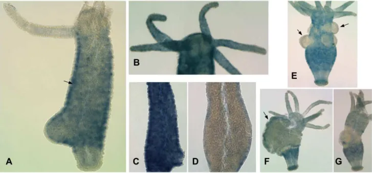
Whole mount in situ hybridization detectedFoxO mRNA in cells of the interstitial cell lineage, with expression stronger in the body column than in the tentacles, head, or basal disk (Figure 3A). Cells expressing FoxO are present in the ectoderm (Figure 3A), indicating thatFoxOis expressed in interstitial cells, nematoblasts, and/or differentiating neurons. Fainter, punctuate expression in
tentacle ectoderm (Figure 3B) suggests that it is also expressed at lower levels in nematocytes and/or neurons.
Because interstitial cells divide more rapidly than epithelial cells, their numbers can be differentially reduced by treatment ofHydra
polyps with hydroxyurea (HU) [53]. To confirm that FoxO is expressed in interstitial cells, we treatedH. magnipapillatawith HU. Counting cells from macerated control and treated animals showed that treatment reduced the ratio of interstitial cells to total cells present by 85 to 90%, depending on the experiment, but did not reduce the ratio of nematoblasts, nematocytes, or neurons to total cells. (In control animals, we found that interstitial cells constituted 22 to 29% of total cells, depending on the experiment.
Figure 2. Results of phylogenetic analyses.Maximum parsimony phylogram of selected FoxO proteins rooted usingMus musculusFoxA1. Numbers at nodes are bootstrap support values calculated by 1000 replicates of Maximum Parsimony/Maximum Likelihood/Neighbor Joining. Bootstrap values under 50 are not shown. Asterisks at nodes indicate Bayesian PP greater than 95%. Species name abbreviations:Aa: Aedes aegypti;
These percentages are consistent with previous results [53,54].) In situ hybridization showed substantially decreasedFoxOexpression in HU-treated compared to control animals (Figure 3C, D). Our results indicate thatFoxOis expressed in interstitial cells, although they do not preclude the possibility that it is also expressed at lower levels in epithelial cells.
We also examinedFoxOexpression during spermatogenesis and oogenesis using the H. vulgaris AEP strain, which readily reproduces sexually in the laboratory. No expression was detected in testes, where proliferation of spermatogonia and their differentiation into sperm take place [55] (Figure 3E). A low level of FoxOexpression was found in developing oocytes (Figure 3F). Following oogenesis, the former egg field is depleted of FoxO -expressing cells (Figure 3G).
FoxO-GFP localization is affected by PI3K-mediated signaling, heat shock, and JNK
To examine the regulation of FoxO cellular localization in
Hydra, we produced lines of transgenicH. vulgariswhich express a FoxO-GFP fusion protein under the control of a Hydra b-actin promoter.Hydraectodermal, endodermal, and interstitial lineage cells represent separate cellular compartments, replenished by separate stem cells. Embryo microinjection can therefore produce animals with stably transgenic cells of one or more of the three cell lineages [31]. Because in situ hybridization indicated thatFoxOis expressed at high levels in cells of the interstitial cell lineage, we examined protein localization primarily in transgenic animals that expressed the FoxO-GFP protein in cells of the interstitial lineage (Figure 4). In interstitial lineage cells, theHydrab-actin promoter used drives expression in stenotele nematocytes, precursors to stenoteles, and ganglionic neurons (R.E. Steele et al., unpublished information). We characterized FoxO-GFP localization in these cell types.
In other organisms studied, insulin/IGF-1 signaling acts through phosphoinositide 3-kinases (PI3K), Akt, and SGK.
Phosphorylation of FoxO by Akt and SGK promotes its cytoplasmic localization, reducing FoxO transcriptional activity [56]. The conserved consensus Akt/SGK phosphorylation sites present in the predictedH. magnipapillataFoxO protein suggest that Akt and/or SGK may be involved in regulating Hydra FoxO
Figure 3. Results of whole mountin situhybridization.A) Expression ofFoxOmRNA in adultH. magnipapillata. The arrow indicates the border between ectoderm and endoderm. B) AdultH. magnipapillatawith longer staining reaction, showing punctuate staining in the tentacles. C) Body column of controlH. magnipapillata. D) Body column of HU-treatedH. magnipapillata. E)H. vulgariswith testes, indicated by arrows. F)H. vulgaris
with developing egg, indicated by an arrow. G)H. vulgarisfollowing egg extrusion. doi:10.1371/journal.pone.0011686.g003
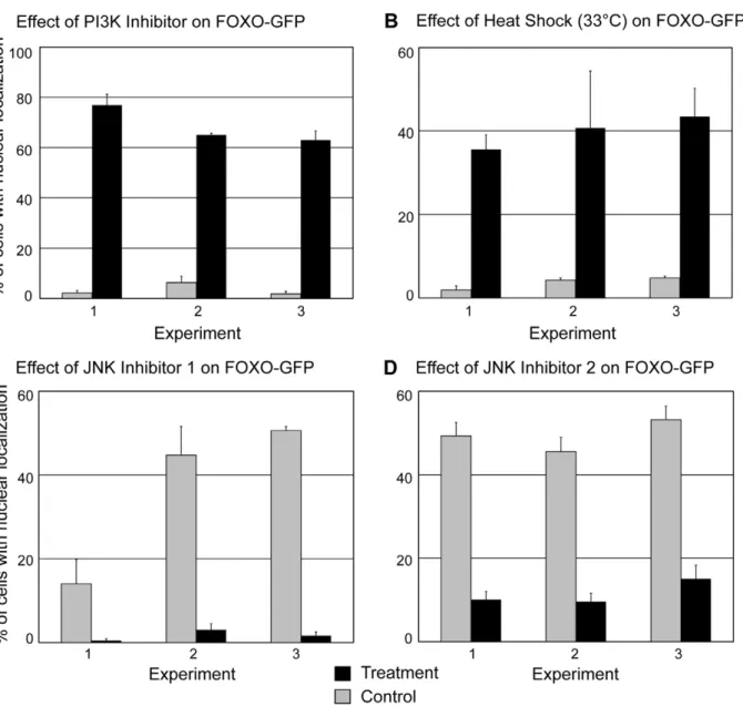
activity. Because PI3K activity leads to activation of Akt and SGK, we treated transgenic animals with an inhibitor of PI3K, LY294002 [57] to address the effect of decreased Akt and SGK activity on Hydra FoxO localization. We found that inhibitor treatment significantly increased the percentage of interstitial lineage cells showing nuclear localization of FoxO-GFP (Figure 5A), suggesting that Akt and/or SGK activity promote the cytoplasmic localization ofHydraFoxO.
To determine whether dietary restriction leads to FoxO nuclear localization in Hydra, we examined FoxO-GFP localization in transgenic animals that were fed daily for two weeks and then starved for two, three, four, or ten days. We found no significant difference in the percentage of interstitial lineage cells showing fusion protein nuclear localization between control animals and
starved animals (N$38 cells examined per animal,P$0.1 in all cases).
To address the effect of heat shock onHydraFoxO localization, transgenic animals were incubated for 90 minutes at 33uC. These conditions cause synthesis of HSP70 inHydra vulgaris[32]. Heat shock significantly increased the percentage of cells showing nuclear localization of FoxO-GFP (Figure 5B). Animals from a second transgenic line, in which ectodermal epithelial cells expressed FoxO-GFP, were subjected to the same heat shock conditions and also showed an increase in fusion protein nuclear localization (data not shown).
Finally, we asked whether the stress-associated kinase JNK promotes the nuclear localization seen in response to heat shock. Transgenic animals were treated for 24 hours with either of two
Figure 5. Effects of experimental treatments on FoxO-GFP localization.Localization was examined in stenotele nematocytes, nematoblasts which are precursors to stenoteles, and ganglionic neurons. A) Effects of PI3K inhibitor treatment—1 hour incubation in 40mM LY294002. N$69 cells
examined per animal. B) Effects of heat shock—90 minutes at 33uC. N$73 cells examined per animal. C) Effects of heat shock on animals treated with JNK inhibitor. Control and treatment animals were subject to heat shock. Treatment involved 24 hour incubation in 2.5mM SP60025 prior to heat
shock in 2.5mM SP60025. N$55 cells examined per animal. D) Effects of heat shock on animals treated with JNK inhibitor. Control and treated animals
were subjected to heat shock. Treatment involved 24 hour incubation in 2mM AS601245 prior to heat shock in 2mM AS601245. N$56 cells examined
per animal.
inhibitors of JNK, SP600125 [58] or AS601245 [59]. Treatment with the concentration of SP600125 used here has been shown to reduce JNK activity in Hydra [60]. Following 24 hours of treatment, animals in inhibitor were placed at 33uC for 90 minutes to induce heat shock. When subjected to heat shock, animals treated with either of the two JNK inhibitors showed significantly less nuclear localization of FoxO-GFP than heat-shocked control animals (Figure 5C, D) These results show that in Hydra, as in bilaterian animals, JNK plays a role in increasing FoxO nuclear localization under conditions of thermal stress.
Discussion
We have identified a single FoxO gene in a member of the cnidarian genusHydra. FoxO genes have been found in members of early-evolving animal phyla, including sponges, placozoans, and cnidarians [25–27]. However, they were not detected in the genome of a member of the choanoflagellates, the sister taxon to metazoans, and have not been found in fungi or plants [25]. Thus, FoxO genes appear to have arisen in the common ancestor of animals.
Hydra FoxOis strongly expressed in interstitial cells in the body columns of adult animals. Hydrainterstitial cells include multip-otent stem cells which maintain populations of stinging cells, secretory cells, and neurons in the adult [39]. Existing data imply that Hydra vulgaris do not show senescence–increased mortality with age [61]. In other organisms, FoxO proteins can reduce damage to cells by increasing levels of antioxidant or heat shock proteins and by promoting genomic stability [6,7]. One possibility is that FoxO functions to reduce damage to interstitial stem cells over the potentially long life of an individual Hydra and its asexually produced offspring. In addition, because Hydra germ cells are not segregated early in development and instead arise from interstitial stem cells [47], FoxO expression in interstitial cells could help to minimize damage that would affect gametes.
FoxO expression has been examined in another cnidarian, the marine hydrozoanClytia hemisphaerica. However, it is not yet clear how similar cell and tissue dynamics are in these two species.
CheFoxOinC. hemisphaericais expressed throughout the polyp as well as in the larval and medusa stages of the life cycle not present in
Hydra. As in theHydrapolyp, expression in both polyp and medusa is strongest in tissues characterized by cell proliferation [27].
Our results provide evidence that, as in bilaterian animals,
HydraFoxO transcriptional activity is negatively regulated by the PI3K/Akt/SGK pathway. Like other FoxO proteins, the predicted H. magnipapillata FoxO protein includes three Akt/ SGK phosphorylation sites. Treatment with a PI3K inhibitor significantly increased nuclear localization of the FoxO-GFP fusion protein (Figure 5A). Our results are consistent with recently reported data which also imply that Hydra FoxO function is affected by the PI3K/Akt/SGK pathway. Lasi et al. [62] induced transient expression ofHydraFoxO fused to GFP by introducing plasmid DNA into epithelial cells using a particle gun. Approx-imately 20–60% of Hydra epithelial cells expressing FoxO-GFP underwent apoptosis [62]. An insulin/IGF-1 receptor gene [63] and three insulin-like peptide genes (unpublished information, Genebank accession numbers GU219979, GU219980, and GU219981) are present in Hydra. Coexpression of one of the
Hydrainsulin-like genes with the FoxO-GFP protein decreased the rate of apoptosis in epithelial cells [62]. This result provides evidence that insulin/IGF-1 signaling, which acts through the PI3K/Akt/SGK pathway, reduces HydraFoxO activity. Regula-tion of FoxO mediated by PI3K/Akt/SGK thus appears to be conserved betweenHydra,Drosophila,C. elegans, and mammals.
Unlike Lasi et al., we seldom observed apoptosis in FoxO-GFP expressing cells, judging by DNA distribution in DAPI-stained cells. This difference in results could be caused by differences in levels of expression of the fusion protein. It could also reflect differences between the epithelial cells examined by Lasi et al. and cells of the interstitial lineage. Because in situ hybridization showed strong FoxO expression in cells of the interstitial cell lineage, in most experiments we characterized FoxO-GFP localization in these cells rather than in epithelial cells.
InHydra, changes in feeding rate alter growth rate. Greater food availability and higher growth rate result in larger animals and more rapid production of new buds [64]. Decreases in food availability result in slower epithelial cell division [64,65], apoptosis of some epithelial cells [65–67] and autophagy in some epithelial cells [68]. FoxO proteins in other organisms can mediate responses to dietary restriction, act to slow cell cycling, and trigger apoptosis [6]. FoxO could therefore potentially be involved in the responses to dietary restriction seen in Hydra. If Hydra insulin/IGF-1 levels decrease under low nutrient conditions, we might expect to see increased FoxO-GFP nuclear localization in starved animals. Interestingly, we did not find that starvation affected FoxO-GFP localization. Our results might still be consistent with a role for FoxO in response to low nutrient conditions. In C. elegans and mammals, under low nutrient conditions AMP-activated protein kinase phosphorylates FoxO proteins and causes increased FoxO transcriptional activity without affecting FoxO cellular localization [5,69].
However, our results might also reflect differences in the effects of low nutrient conditions on epithelial cells and interstitial lineage cells. Apoptosis, autophagy, and changes in cell cycle length in response to dietary restriction have been reported only for epithelial cells [64–68]. Experiments with chimeric animals containing epithelial cells and interstitial cells from differentHydra
strains show that the growth rate of adult Hydra is determined primarily by epithelial cells [70,71]. In contrast, rates of interstitial stem cell division and differentiation appear to be affected by interstitial cell density [53]. The Hydra insulin/IGF-1 receptor gene is known to be expressed in the ectoderm [63]. Under low nutrient conditions, decreased insulin/IGF-1 signaling may lead to increased FoxO function and apoptosis in epithelial cells, while our results may indicate the absence of a corresponding response to low nutrient conditions inHydrainterstitial cells.
In bilaterian animals, FoxO proteins play important roles in cellular resistance to stress. Our data on FoxO-GFP localization provide evidence that FoxO also mediates stress resistance inH. vulgaris. Conditions known to induce heat shock in H. vulgaris
significantly increased nuclear localization of the fusion protein in both interstitial lineage cells (Figure 5B) and ectodermal cells. In other animals, JNK kinases, which are activated in response to environmental stresses including heat shock and oxidative stress, increase nuclear localization and transcriptional activity of FoxO proteins [72,73]. A JNK gene is present inHydra[74]. We found that inHydra vulgaris, JNK inhibitors reduced nuclear localization of FoxO-GFP under heat shock conditions (Figures 5C,D). Our results suggest that, as in bilaterians, JNK increases FoxO activity and cellular resistance to stress inHydra.
stress-induced changes in the transcriptomes of cnidarian species e.g. [75–78]. Since much of the regulation of FoxO protein function is posttranscriptional, our results complement information from such studies. Further understanding of the role of FoxO in stress responses inHydraand other cnidarians could provide insight into the details of these organisms’ physiological responses to stress. Bilaterian FoxO proteins act to integrate different environmental signals, since they mediate responses to multiple stresses including low nutrient levels, oxidative stress, and thermal stress [6,7]. Information about the roles of FoxO proteins in cnidarians may be important in understanding how cnidarian species respond to the combinations of environmental challenges many of them experience.
Materials and Methods
Hydrastrains used and culture conditions
Hydra magnipapillatastrain 105 andHydra vulgarisstrain AEP were maintained under standard culture conditions at 18uC. Animals were fed withArtemia salina(Brine Shrimp Direct) nauplii cultured from cysts for 48 hours at room temperature.
Hydroxyurea treatment
To reduce the interstitial cell population,H. magnipapillatawere incubated in 10 mM hydroxyurea for 24 hours and then in medium without hydroxyurea for 12 hours. This cycle was repeated two additional times [53], followed by four days of culture without hydroxyurea. Animals were fed daily during treatment. Animals were then either macerated or used for whole-mount in situ hybridization. Six treated and six control animals were macerated as described by David [54] except that to disassociate tissue into individual cells, 1.5 ml microcentrifuge tubes containing animals in maceration solution were left for 30 minutes taped to the side of a Vortex Genie mixer set at intermediate speed. Following maceration, numbers of epithelial cells, interstitial cells, nematoblasts, nematocytes, neurons, and gland cells were determined for treated and control animals.
Isolation ofHydra magnipapillataFoxO and phylogenetic analysis
To identify FoxO genes, we searched the assembled H. magnipapillata 105 genome sequence in the Metazome database using tblastn with DAF-16 and dFOXO protein sequences as queries. DNA encodingH. magnipapillataFoxO was isolated from first strand cDNA by PCR using the primers GCGAGA-TATGTTTTTAAATGTCAGTGC and TCCATATAGAAC-TTTCCTGAGTTCATTAG. cDNA was produced from H. magnipapillatapoly-A RNA using the Invitrogen GeneRacer Kit.
Phylogenetic analyses were based on the predicted amino acid sequence of the forkhead domain. Sequences were aligned using ClustalX [79,80] with the following Multiple Alignment Param-eters: Gap Opening Penalty: 10.00, Gap Extension Penalty: 0.20. The most parsimonious tree (Figure 2) was found using PAUP* 4.0 [81] implementing a full heuristic search with 10 random stepwise-addition replicates and TBR branch swapping. Amino acid substitutions were weighted using the protpars matrix of PAUP. Maximum Parsimony, Maximum Likelihood, and Neighbor Joining bootstrap values were calculated based on 1000 replicates. For Maximum Parsimony each bootstrap replicate consisted of a full heuristic search with 10 stepwise-addition random replicates and TBR branch swapping. Maximum likelihood bootstrap was implemented using the Phylip [82] programs SEQBOOT, PROML (with JTT model of amino acid change), and CONSENSE. Bayesian posterior probabilities were calculated
using MrBayes version 3.1.2 [83,84] with two independent runs of 1,000,000 generations each, sampled every 1000 generations with four chains. (Temperature was set at the default value of 0.2; 10% of the first samples were used as burnin.) The average standard deviation split frequencies after 1,000,000 generations was 0.011.
In situhybridization
A 992 base pair long portion of theFoxOcoding sequence was amplified from H. magnipapillata 105 cDNA using the primers CCCAGATGCAAAAGCAGGGAAATC and GCTTTACTG-GTCTAAGTCGCTCGG. This PCR product was cloned into the Promega pGEM-T Easy Vector. To produce the digoxygenin-labeled antisense probe, the plasmid was linearized by digestion with SalI and in vitro transcription was performed using the Roche DIG RNA Labeling Kit and T7 RNA polymerase. To produce the sense probe, the plasmid was linearized with NcoI and transcription was performed using the SP6 polymerase. Whole mount in situ hybridization was performed as described in [85] and [86], except that RNA probes were heated to 65uC for five minutes immediately before use.
TransgenicHydra
We produced an expression construct with theH. magnipapillata
105 FoxO coding sequence fused in frame with a sequence encoding enhanced green fluorescent protein, under the control of theHydrab-actin promoter. Using PCR, an NheI site was added to the 59end of theFoxOcDNA and a HpaI site was added to the 39
end. The resulting cDNA was cut with NheI and HpaI, and then cloned into the expression vector pHyVec4 (GenBank accession DQ385853), which had been cut with NheI and SmaI. pHyVec4 is a modified version of the hoT G plasmid [31]. The structure of the construct was confirmed by DNA sequencing. The FoxO
coding sequence within the construct was checked against the NCBI trace archive sequences forH. magnipapillata 105. Plasmid DNA for embryo injection was purified using the Qiagen Endo-free Gigaprep kit and resuspended in sterile deionized water.
The FoxO-GFP expression construct was microinjected into embryos ofH. vulgarisstrain AEP at the one to eight cell stage as described in Wittlieb et al. [31], using a Narishige IM 300 microinjector. Needles for injection were produced using a Sutter P-30 micropipette puller. Each of the three stably transgenic animals produced was propagated through budding to produce a line of transgenic Hydra. The majority of experiments were conducted with a line termed in[act:FoxO-GFP]1, in which a subset of cells of the interstitial lineage expressed the fusion protein. Specifically, the cells which expressed the protein were precursors to stenotele nematocytes, mature stenotele nemato-cytes, and ganglionic neurons. Fusion protein localization was examined in all of these cell types. The number of cells of each cell type scored was approximately equal in the control and treated animals in each experiment. Fusion protein localization following heat shock was also examined in the line ec[act:FoxO-GFP]1, in which ectodermal cells expressed the fusion protein.
Heat shock, inhibitor and starvation treatments
Animals subjected to heat shock were incubated at 33uC for 90 minutes, conditions known to increase levels of HSP70 [32]. Animals treated with the PI3K inhibitor LY294002 (Sigma) were incubated for 1 hour in the dark at 18uC in 40mM LY294002 in
hydra medium containing 0.2% ethanol. Animals treated with the JNK inhibitor SP60025 (Calbiochem) were incubated in the dark in 2.5mM SP60025 in hydra medium containing 0.0125%
incubated at the same temperatures in N1 -Methyl-1,9-pyrazoloan-throne (Calbiochem) in hydra medium containing 0.0125% DSMO. Animals treated with the JNK inhibitor AS601245 (Calbiochem) were incubated in the dark in 2 uM AS601245 in hydra medium containing 0.15% DMSO. They were first incubated for 24 hours at 18uC and then placed at 33uC for 90 minutes. Control animals were incubated at the same temperatures in 0.15% DSMO/hydra medium. Animals subjected to starvation were fed daily for two weeks and then starved for two, three, four, or ten days. Control animals were fed daily but starved for one day before fixation to reduce background fluorescence. In heat shock, LY294002, SP60025, four-day starvation and ten-day starvation experiments, three treated and three controlHydrawere used for each experiment. In AS601245, two-day starvation and three-day starvation experiments, five treated and five control
Hydra were used. For all treatments, the percentage of cells showing nuclear localization in control and treated animals were compared using the Mann-Whitney U test.
Microscopy
To determine FoxO-GFP localization, animals were processed as follows. Following experimental treatments, animals were fixed
for 1 hour in 4% paraformaldehyde in hydra medium and then washed for 10 minutes in PBS, for 30 minutes in 5mg/mL DAPI
in PBS, and for 5 minutes in PBS. Fixation and washes were performed at 4uC. Cells expressing the fusion protein were visualized at 10006using a Nikon Eclipse 80i microscope and photographed using a SPOT RTke digital camera. Localization was examined in nematoblasts, nematocytes, and neurons.
Acknowledgments
We thank Catherine Dana at the University of California, Irvine, and the Molecular Genetics Core Facility at the Pennsylvania State University College of Medicine for technical assistance.
Author Contributions
Conceived and designed the experiments: DB AGT RLH EM RS DEM. Performed the experiments: DB AGT RLH EM RS DEM. Analyzed the data: RS DEM. Contributed reagents/materials/analysis tools: DB RS DEM. Wrote the paper: DB.
References
1. Honda Y, Honda S (1999) The daf-2gene network for longevity regulates oxidative stress resistance and Mn-superoxide dismutase gene expression in
Caenorhabditis elegans. FASEB J 13: 1385–1393.
2. Kops GJ, Dansen TB, Polderman PE, Saarloos I, Wirtz KW, et al. (2002) Forkhead transcription factor FOXO3a protects quiescent cells from oxidative stress. Nature 419: 316–321.
3. Nemoto S, Finkel T (2002) Redox regulation of forkhead proteins through a
p66shc-dependent signaling pathway. Science 295: 2450–2452.
4. Junger MA, Rintelen F, Stocker H, Wasserman JD, Vegh M, et al. (2003) The
Drosophilaforkhead transcription factor FOXO mediates the reduction in cell number associated with reduced insulin signaling. J Biol 2: 20.
5. Greer EL, Dowlatshahi D, Banko MR, Villen J, Hoang K, et al. (2007) An AMPK-FOXO pathway mediates longevity induced by a novel method of dietary restriction inC. elegans. Curr Biol 17: 1646–1656.
6. Salih DA, Brunet A (2008) FoxO transcription factors in the maintenance of cellular homeostasis during aging. Curr Opin Cell Biol 20: 126–136. 7. Hsu AL, Murphy CT, Kenyon C (2003) Regulation of aging and age-related
disease by DAF-16 and heat-shock factor. Science 300: 1142–1145. 8. Lee KS, Iijima-Ando K, Iijima K, Lee WJ, Lee JH, et al. (2009)
JNK/FOXO-mediated neuronal expression of fly homologue of peroxiredoxin II reduces oxidative stress and extends life span. J Biol Chem 284: 29454–29461. 9. Mammucari C, Milan G, Romanello V, Masiero E, Rudolf R, et al. (2007)
FoxO3 controls autophagy in skeletal muscle in vivo. Cell Metab 6: 458–471. 10. Zhao J, Brault JJ, Schild A, Cao P, Sandri M, et al. (2007) FoxO3 coordinately
activates protein degradation by the autophagic/lysosomal and proteasomal pathways in atrophying muscle cells. Cell Metab 6: 472–483.
11. Sengupta A, Molkentin JD, Yutzey KE (2009) FoxO transcription factors promote autophagy in cardiomyocytes. J Biol Chem 284: 28319–28331. 12. Juhasz G, Puskas LG, Komonyi O, Erdi B, Maroy P, et al. (2007) Gene
expression profiling identifies FKBP39 as an inhibitor of autophagy in larval
Drosophilafat body. Cell Death Differ 14: 1181–1190.
13. Tran H, Brunet A, Grenier JM, Datta SR, Fornace AJ, Jr., et al. (2002) DNA repair pathway stimulated by the forkhead transcription factor FOXO3a through the Gadd45 protein. Science 296: 530–534.
14. Tsai WB, Chung YM, Takahashi Y, Xu Z, Hu MC (2008) Functional interaction between FOXO3a and ATM regulates DNA damage response. Nat Cell Biol 10: 460–467.
15. Curran SP, Wu X, Riedel CG, Ruvkun G (2009) A soma-to-germline transformation in long-lived Caenorhabditis elegans mutants. Nature 459: 1079–1084.
16. Troemel ER, Chu SW, Reinke V, Lee SS, Ausubel FM, et al. (2006) p38 MAPK regulates expression of immune response genes and contributes to longevity inC. elegans. PLoS Genet 2: e183.
17. Biggs WH, 3rd, Meisenhelder J, Hunter T, Cavenee WK, Arden KC (1999) Protein kinase B/Akt-mediated phosphorylation promotes nuclear exclusion of the winged helix transcription factor FKHR1. Proc Natl Acad Sci U S A 96: 7421–7426.
18. Brunet A, Bonni A, Zigmond MJ, Lin MZ, Juo P, et al. (1999) Akt promotes cell survival by phosphorylating and inhibiting a Forkhead transcription factor. Cell 96: 857–868.
19. Kops GJ, Burgering BM (1999) Forkhead transcription factors: new insights into protein kinase B (c-akt) signaling. J Mol Med 77: 656–665.
20. Tang ED, Nunez G, Barr FG, Guan KL (1999) Negative regulation of the forkhead transcription factor FKHR by Akt. J Biol Chem 274: 16741–16746. 21. Essers MA, Weijzen S, de Vries-Smits AM, Saarloos I, de Ruiter ND, et al.
(2004) FOXO transcription factor activation by oxidative stress mediated by the small GTPase Ral and JNK. EMBO J 23: 4802–4812.
22. Oh SW, Mukhopadhyay A, Svrzikapa N, Jiang F, Davis RJ, et al. (2005) JNK regulates lifespan inCaenorhabditis elegansby modulating nuclear translocation of forkhead transcription factor/DAF-16. Proc Natl Acad Sci U S A 102: 4494–4499.
23. Wang MC, Bohmann D, Jasper H (2005) JNK extends life span and limits growth by antagonizing cellular and organism-wide responses to insulin signaling. Cell 121: 115–125.
24. Sunayama J, Tsuruta F, Masuyama N, Gotoh Y (2005) JNK antagonizes Akt-mediated survival signals by phosphorylating 14-3-3. J Cell Biol 170: 295–304. 25. Larroux C, Luke GN, Koopman P, Rokhsar DS, Shimeld SM, et al. (2008) Genesis and expansion of metazoan transcription factor gene classes. Mol Biol Evol 25: 980–996.
26. Magie CR, Pang K, Martindale MQ (2005) Genomic inventory and expression ofSoxandFoxgenes in the cnidarianNematostella vectensis. Dev Genes Evol 215: 618–630.
27. Chevalier S, Martin A, Leclere L, Amiel A, Houliston E (2006) Polarised expression of FoxB and FoxQ2 genes during development of the hydrozoan
Clytia hemisphaerica. Dev Genes Evol 216: 709–720.
28. DeSalvo MK, Voolstra CR, Sunagawa S, Schwarz JA, Stillman JH, et al. (2008) Differential gene expression during thermal stress and bleaching in the Caribbean coralMontastraea faveolata. Mol Ecol 17: 3952–3971.
29. Weis VM, Allemand D (2009) Physiology. What determines coral health? Science 324: 1153–1155.
30. Chapman JA, Kirkness EF, Simakov O, Hampson SE, Mitros T, et al. (2010) The dynamic genome ofHydra. Nature 464: 592–596.
31. Wittlieb J, Khalturin K, Lohmann JU, Anton-Erxleben F, Bosch TC (2006) Transgenic Hydra allow in vivo tracking of individual stem cells during morphogenesis. Proc Natl Acad Sci U S A 103: 6208–6211.
32. Bosch TC, Krylow SM, Bode HR, Steele RE (1988) Thermotolerance and synthesis of heat shock proteins: these responses are present inHydra attenuatabut absent inHydra oligactis. Proc Natl Acad Sci U S A 85: 7927–7931.
33. Gellner K, Praetzel G, Bosch TC (1992) Cloning and expression of a heat-induciblehsp70gene in two species ofHydrawhich differ in their stress response. Eur J Biochem 210: 683–691.
34. Brennecke T, Gellner K, Bosch TC (1998) The lack of a stress response inHydra oligactisis due to reducedhsp70mRNA stability. Eur J Biochem 255: 703–709. 35. Bode PM, Bode HR (1984) Patterning in Hydra. In: Malacinski GM, Bryant SV, eds. Pattern Formation, A Primer in Developmental Biology. New York: Macmillan. pp 213–214.
36. Campbell RD (1967) Tissue dynamics of steady state growth inHydra littoralis. II. Patterns of tissue movement. J Morphol 121: 19–28.
38. Dubel S, Hoffmeister SA, Schaller HC (1987) Differentiation pathways of ectodermal epithelial cells in hydra. Differentiation 35: 181–189.
39. Bode HR (1996) The interstitial cell lineage of hydra: a stem cell system that arose early in evolution. J Cell Sci 109(Pt 6): 1155–1164.
40. Alessi DR, Caudwell FB, Andjelkovic M, Hemmings BA, Cohen P (1996) Molecular basis for the substrate specificity of protein kinase B; comparison with MAPKAP kinase-1 and p70 S6 kinase. FEBS Lett 399: 333–338.
41. Lin K, Hsin H, Libina N, Kenyon C (2001) Regulation of theCaenorhabditis eleganslongevity protein DAF-16 by insulin/IGF-1 and germline signaling. Nat Genet 28: 139–145.
42. Burgering BM, Kops GJ (2002) Cell cycle and death control: long live Forkheads. Trends Biochem Sci 27: 352–360.
43. Jacobs FM, van der Heide LP, Wijchers PJ, Burbach JP, Hoekman MF, et al. (2003) FoxO6, a novel member of the FoxO class of transcription factors with distinct shuttling dynamics. J Biol Chem 278: 35959–35967.
44. Puig O, Marr MT, Ruhf ML, Tjian R (2003) Control of cell number by
DrosophilaFOXO: downstream and feedback regulation of the insulin receptor pathway. Genes Dev 17: 2006–2020.
45. Lange A, Mills RE, Lange CJ, Stewart M, Devine SE, et al. (2007) Classical nuclear localization signals: definition, function, and interaction with importin alpha. J Biol Chem 282: 5101–5105.
46. David CN, Murphy S (1977) Characterization of interstitial stem cells in hydra by cloning. Dev Biol 58: 372–383.
47. Bosch TC, David CN (1987) Stem cells ofHydra magnipapillatacan differentiate into somatic cells and germ line cells. Dev Biol 121: 182–191.
48. David CN, Gierer A (1974) Cell cycle kinetics and development of Hydra attenuata. III. Nerve and nematocyte differentiation. J Cell Sci 16: 359–375. 49. Brien P, Reniers-Decoen M (1950) Etude d’Hydra viridis (Linnaeus) (La
blastoge´ne`se, l’spermatoge´ne`se, l’ovoge´ne`se). Ann Soc R Zool Belg 81: 33–110. 50. Honegger TG, Zurrer D, Tardent P (1989) Oogenesis inHydra carnea: A new model based on light and electron microscopic analyses of oocyte and nurse cell differentiation. Tissue Cell 21: 381–393.
51. Technau U, Miller MA, Bridge D, Steele RE (2003) Arrested apoptosis of nurse cells duringHydraoogenesis and embryogenesis. Dev Biol 260: 191–206. 52. Alexandrova O, Schade M, Bo¨ttger A, David CN (2005) Oogenesis inHydra:
nurse cells transfer cytoplasm directly to the growing oocyte. Dev Biol 281: 91–101.
53. Bode HR, Flick KM, Smith GS (1976) Regulation of interstitial cell differentiation in Hydra attenuata. I. Homeostatic control of interstitial cell population size. J Cell Sci 20: 29–46.
54. David CN (1973) A quantitative method for maceration ofHydratissue. Wilhelm Roux’s Arch 171: 259–268.
55. Miller MA, Steele RE (2000) Lemon encodes an unusual receptor protein-tyrosine kinase expressed during gametogenesis in Hydra. Dev Biol 224: 286–298.
56. Huang H, Tindall DJ (2007) Dynamic FoxO transcription factors. J Cell Sci 120: 2479–2487.
57. Vlahos CJ, Matter WF, Hui KY, Brown RF (1994) A specific inhibitor of phosphatidylinositol 3-kinase, 2-(4-morpholinyl)-8-phenyl-4H-1-benzopyran-4-one (LY294002). J Biol Chem 269: 5241–5248.
58. Bennett BL, Sasaki DT, Murray BW, O’Leary EC, Sakata ST, et al. (2001) SP600125, an anthrapyrazolone inhibitor of Jun N-terminal kinase. Proc Natl Acad Sci U S A 98: 13681–13686.
59. Gaillard P, Jeanclaude-Etter I, Ardissone V, Arkinstall S, Cambet Y, et al. (2005) Design and synthesis of the first generation of novel potent, selective, and in vivo active (benzothiazol-2-yl)acetonitrile inhibitors of the c-Jun N-terminal kinase. J Med Chem 48: 4596–4607.
60. Philipp I, Aufschnaiter R, Ozbek S, Pontasch S, Jenewein M, et al. (2009) Wnt/ beta-catenin and noncanonical Wnt signaling interact in tissue evagination in the simple eumetazoanHydra. Proc Natl Acad Sci U S A 106: 4290–4295. 61. Martinez DE (1998) Mortality patterns suggest lack of senescence in hydra. Exp
Gerontol 33: 217–225.
62. Lasi M, David CN, Bo¨ttger A (2009) Apoptosis in pre-Bilaterians:Hydraas a model. Apoptosis 15: 269–78.
63. Steele RE, Lieu P, Mai NH, Shenk MA, Sarras MP (1996) Response to insulin and the expression pattern of a gene encoding an insulin receptor homologue suggest a role for an insulin-like molecule in regulating growth and patterning in
Hydra. Dev Genes Evol 206: 247–259.
64. Otto JJ, Campbell RD (1977) Tissue economics of hydra: regulation of cell cycle, animal size and development by controlled feeding rates. J Cell Sci 28: 117–132. 65. Bosch TC, David CN (1984) Growth regulation inHydra: relationship between
epithelial cell cycle length and growth rate. Dev Biol 104: 161–171. 66. Cikala M, Wilm B, Hobmayer E, Bottger A, David CN (1999) Identification of
caspases and apoptosis in the simple metazoanHydra. Curr Biol 9: 959–962. 67. Bo¨ttger A, Alexandrova O (2007) Programmed cell death inHydra. Semin
Cancer Biol 17: 134–146.
68. Chera S, Buzgariu W, Ghila L, Galliot B (2009) Autophagy inHydra: a response to starvation and stress in early animal evolution. Biochim Biophys Acta 1793: 1432–1443.
69. Greer EL, Oskoui PR, Banko MR, Maniar JM, Gygi MP, et al. (2007) The energy sensor AMP-activated protein kinase directly regulates the mammalian FOXO3 transcription factor. J Biol Chem 282: 30107–30119.
70. Marcum BA, Campbell RD (1978) Developmental roles of epithelial and interstitial cell lineages in hydra: analysis of chimeras. J Cell Sci 32: 233–247. 71. Sugiyama T, Fujisawa T (1978) Genetic analysis of developmental mechanisms
in hydra. V. Cell lineage and development of chimera hydra. J Cell Sci 32: 215–232.
72. van der Horst A, Burgering BM (2007) Stressing the role of FoxO proteins in lifespan and disease. Nat Rev Mol Cell Biol 8: 440–450.
73. Weston CR, Davis RJ (2002) The JNK signal transduction pathway. Curr Opin Genet Dev 12: 14–21.
74. Philipp I, Holstein TW, Hobmayer B (2005)HvJNK, aHydramember of the c-Jun NH2-terminal kinasegene family, is expressed during nematocyte differenti-ation. Gene Expr Patterns 5: 397–402.
75. Reitzel AM, Sullivan JC, Traylor-Knowles N, Finnerty JR (2008) Genomic survey of candidate stress-response genes in the estuarine anemoneNematostella vectensis. Biol Bull 214: 233–254.
76. Rodriguez-Lanetty M, Harii S, Hoegh-Guldberg O (2009) Early molecular responses of coral larvae to hyperthermal stress. Mol Ecol 18: 5101–5114. 77. Seneca FO, Foreˆt S, Ball EE, Smith-Keune C, Miller DJ, et al. (2009) Patterns of
gene expression in a scleractinian coral undergoing natural bleaching Mar Biotechnol (NY).
78. Voolstra CR, Schnetzer J, Peshkin L, Randall CJ, Szmant AM, et al. (2009) Effects of temperature on gene expression in embryos of the coralMontastraea faveolata. BMC Genomics 10: 627.
79. Thompson JD, Higgins DG, Gibson TJ (1994) CLUSTAL W: improving the sensitivity of progressive multiple sequence alignment through sequence weighting, position-specific gap penalties and weight matrix choice. Nucleic Acids Res 22: 4673–4680.
80. Thompson JD, Gibson TJ, Plewniak F, Jeanmougin F, Higgins DG (1997) The CLUSTAL_X windows interface: flexible strategies for multiple sequence alignment aided by quality analysis tools. Nucleic Acids Res 25: 4876–4882. 81. Swofford DL (1999) PAUP* Phylogenetic analysis using parsimony and other
methods. 4.0 ed: Sinauer Associates.
82. Felsenstein J (1993) PHYLIP (Phylogenetic Inference Package). 3.5c ed: Distributed by the author.
83. Huelsenbeck JP, Ronquist F (2001) MRBAYES: Bayesian inference of phylogenetic trees. Bioinformatics 17: 754–755.
84. Ronquist F, Huelsenbeck JP (2003) MrBayes 3: Bayesian phylogenetic inference under mixed models. Bioinformatics 19: 1572–1574.
85. Grens A, Gee L, Fisher DA, Bode HR (1996)CnNK-2, an NK-2 homeobox gene, has a role in patterning the basal end of the axis in hydra. Dev Biol 180: 473–488.
86. Martı´nez DE, Dirksen M, Bode PM, Jamrich M, Steele RE, Bode HR (1997)