ZDB-Number: 2668735-5 IC Journal No: 8192
Volume 2 Issue 2
Online Available at www.phytojournal.com
Journal of Pharmacognosy and Phytochemistry
Vol. 2 No. 2 2013 www.phytojournal.com Page | 223
Ethno-pharmacognostical Studies on Root Bark of
Rubus
ellipticus
Smith. from Manipur
CL. Ringmichon 1*, Bindu Gopalkrishnan 2 and A.P. Dixit 1
1. Research Laboratory, Department of Botany, K.V. Pendharkar College, Dombivli (E), Maharashtra, 421 203 India.
[E-mail: [email protected]]
2. Research Laboratory, Department of Botany, Mithibai College, Vile Parle (W), Mumbai-56, India.
Rubusellipticus Smith. belongs to family Rosaceae. It is one of the important ethnomedicinal plants of Manipur. The Naga tribe of Manipur uses the root bark of the said plant for curing fever since ancient times. The present study deals with comprehensive pharmacognostical studies on root bark of this plant, it includes macroscopy, microscopy, preliminary phytochemical analysis and physicochemical parameters. Some diagnostic characters are presence of uniserriate root hairs, endodermal cells, stone cells and calcium oxalate crystals. Physicochemical studies revealed total ash (3.35%), acid insoluble ash (1.0%), water soluble ash (0.9%), alcohol soluble extractive (21.12%) water soluble extractive (25.3%) and chloroform extractive (1.3%). This will help in laying down pharmacopeial parameters.
Keyword:Rubusellipticus, Rosaceae, Pharmacognosy
1. Introduction
Manipur is blessed with amazing varieties of flora and fauna. There are various tribes who depend on this natural wealth. One among them is the Naga tribe of Manipur. They dwell in the hilly terrains and totally depend on nature for their livelihood. For curing various ailments they use the medicinal plants from the wild since ages. For the present investigation interviews and direct interactions were made with the local and the medicine men in relation to the plants used in treating fever. Rubus ellipticus Smith.was one among the shortlisted plants used as antipyretics.
Rubus ellipticus Smith. belongs to family
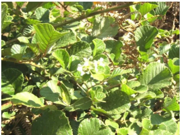
Rosaceae. It is commonly known as ‘Kavathipu’ by the Nagas [1]. It is sub erect straggling shrub with prickles on the branches. The leaves are pinnately trifoliate, flowers white with drupe type of edible fruit (Fig. 1). Besides Manipur the plant
is well distributed in Nilgiri hills, Deccan, Burma
etc [2]. The decoction of root bark is
recommended twice a day for curing fever by the Nagas. The root bark is also used in diarrhea, dysentery, as abortificient, emmenagogue and in fractured bones [3].
Vol. 2 No. 2 2013 www.phytojournal.com Page | 224
2. Material and Methods
2.1 Plant Material: Ethno-medicinal survey
using the suitable questionnaire was conducted. Fresh root bark was collected from a matured plant with prior permission from the forest department of Manipur. The specimen was authenticated from Botanical Survey of India (BSI), Shillong. A voucher specimen has been deposited in Botany Research Laboratory of K.V. Pendharkar College, Thane, India. The accession no. of the sample is (KVP 790). After collection some of the root bark was preserved in FAA solution. Materials were dried in oven at 37 0C and stored in airtight container.
2.2 Macroscopy:The root barks were studied for
its morphological characters using the standard techniques [4].
2.3 Microscopy: Transverse hand cut sections
were taken and made permanent with suitable stains. Quantification and photomicrographs were taken of the permanent preparations. The cell contents were measured using stage and occular micrometer [5, 6].
2.4 Histochemistry: The histochemical studies
for the cell contents were performed using standard methodology [7].
2.5 Powder Study: The powdered drugs were
soaked in aqueous solution of chloral hydrate and mounted in 50% glycerin for microscopical studies [8].
2.6 Proximate Analysis: The physicochemical
parameters like ash values and extractive values were done[9].
2.7 Fluorescence Analysis: The fluorescence
response of powdered drugs exposed to U.V. radiations was studied using the standard procedure [10].
2.8 Preliminary Phytochemical Screening: A
known quantity of dried powder was extracted with chloroform, alcohol and water. These extracts were tested for different constituents [11].
3. Results and Discussion 3.1 Macroscopy:
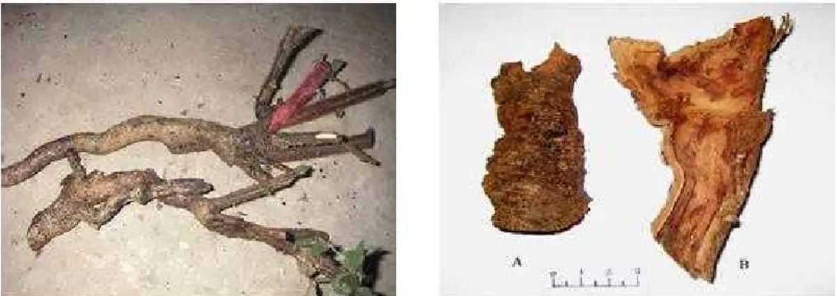
The root bark is 0.8 – 1.0 cm in thickness. It is longitudinally, slightly curved or at times single quilled in shape. Outer surface is grey to dark brown while inner surface is grey to light brown to dark brown or slightly black in colour. It is fibrous in fracture, aromatic odour with strongly bitter and astringent in taste (Figs. 2 and3).
Fig 2: Entire root of Rubusellipticus Smith Fig 3: Root bark (A – Outer surface; B – Inner surface)
3.2Microscopy:
T.S. of root bark shows the following parts-
3.2.1 Epiblema: It is the outermost region with
single layer tangentially elongated cells,
Vol. 2 No. 2 2013 www.phytojournal.com Page | 225
are 3 – 4 layers of thin walled non suberized cells measuring (7.2 – 10.0 – 11.8 µm in length and 12.2 – 17.4 – 20.1 µm in breadth).
3.2.3 Endodermis: Polyderm is followed by a
layer of tangentially elongated cells called endodermal cells measuring (5.2 – 6.4 – 6.9 µm in length and 10. 6 – 11.1 – 12.4 µm in breadth). Casparian band alternate with this cells, measuring (5.7 – 6.2 - µm in length and 10.7 – 11.5 µm in breadth).
3.2.4 Cork: consists of tangentially elongated
suberized compactly arranged cells measuring (7.54 - 10.60 - 12.57µm in length and 12.57 - 17.56 - 20.58 µm in breadth). Few layers of cells are thick walled interrupted with thin wall cells.
3.2.5 Phellogen: is single layer, tangentially
elongated cells measuring (10.60 - 12.52 - 17.51 µm in length and 5.60 - 17.56 - 20.25 µm in breadth).
polyhedral compactly arranged cells, measuring (10.22 - 12.51 - 15.60 µm in length and 22.51 - 25.70 - 30.11 in breadth) filled with simple and compound type of starch grains and tannin filled cells. Each cell contain nucleus.
3.2.7 Secondary Phloem: consists of compactly
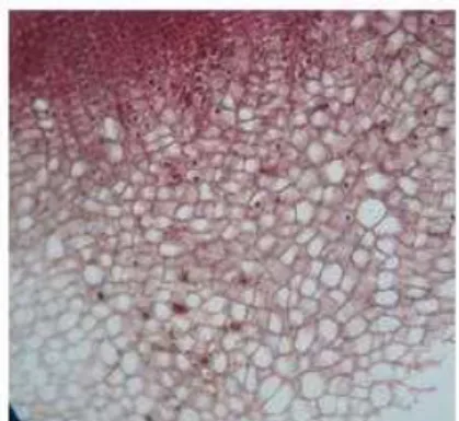
arranged parenchymatous cells measuring (25.50 - 37.52 - 45.56 µm in diameter). Uniserrate to biserrate to multiserrate medullary rays passing through the secondary phloem, each cells measuring (22.54 - 25.55 - 27.57 µm in length and 20.77 - 25.12 - 32.55µm in breadth). Stone cells are embedded in patches within this region (10.76 - 15.12 - 22.50 µm in diameter). Parenchyma cells are filled with simple and compound starch grains, tannin filled cells and few prism shaped calcium oxalate crystals are also found (Figs. 3, 4 and 5).
Fig 3: T.S. of Rubusellipticus bark passing Fig 4: T.S. of Rubusellipticus bark passing
through epiblem & periderm through phelloderm (eh-epidermal hair; pe-periderm; pol-polyderm
en- endodermis; cs- casparin band)
Vol. 2 No. 2 2013 www.phytojournal.com Page | 226
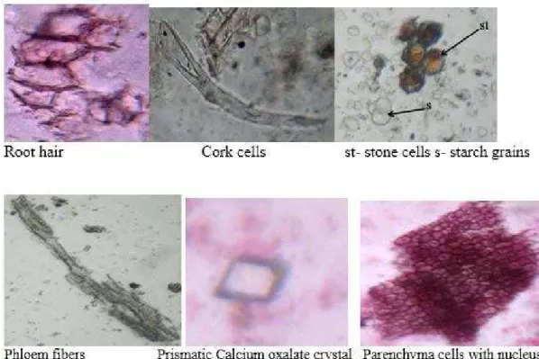
Fig 6: Powder study of Rubusellipticus Smith. root bark
3.4 Histochemical Analysis: the study shows presence of starch, lipids, proteins, tannins, saponins,
glucosides and mucilage. (Table 1).
Table 1: Histochemical analysis
Sr. No. Plant constituent Test Observations
1 Test for Starch + 2 Test for Lipids + 3 Test for Proteins + 4 Test for Tannins + 5 Test for Alkaloids + 6 Test for Saponins + 7 Test for Glucosides + 8 Test for Mucilage + 9 Test for Calcium oxalate crystals +
3.5 Proximate Analysis: The physicochemical parameters like ash values and extractive values are
summarized in (Table 2).
Table 2: Physicochemical evaluation
Ash values
Total ash Not more than 3.35% Acid insoluble ash Not more than 1.0% Water soluble ash Not more than 0.9%
Extractive values
Vol. 2 No. 2 2013 www.phytojournal.com Page | 227
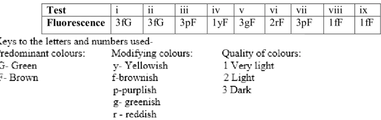
3.6 Fluorescence Analysis:data are summarized in (Table 3).
Table 3: Fluorescence analysis
Test i ii iii iv v vi vii viii ix
Fluorescence 3fG 3fG 3pF 1yF 3gF 2rF 3pF 1fF 1fF
3.7 Preliminary Phytochemical and Physicochemical Evaluation: data are summarized in (Table. 4)
Table 4: Preliminary phytochemical screening
Test for phytoconstituents W C E
Test for Starch + + +
Test for Terpenoids + + +
Test for Proteins + + +
Test for Amino acid + + +
Test for Mucilage + + +
Test for Alkaloids + + +
Test for Anthraquinone glycoside + + +
Test for Cardiac glycoside + + +
Test for Saponin + - -
Test for Tannins + + +
Test for Steroids - - -
Test for Flavonoids - - - Key: W- water extracts, C- Chloroform extract, E- Ethanol extract, + Present, - Absent
4. Conclusion
In nutshell, the above pharmacognostical studies will help in laying a pharmacopeial standard. The ethnomedicinal known plant drug of Manipur will be known in manifold. Detailed phytochemical and pharmacological studies are in progress.
5. Acknowledgments
The authors are thankful to Late Dr. Shraddha N. Shimpi for her constant support during this work. The authors are also thankful to UGC, Delhi for providing Rajiv Gandhi Scholarship to the first author.
6. References
1. Singh NP, Chauhan AS and Mondal MS Flora of
Manipur. Vol.1, BSI, 2000, 391-392.
2. Kumar S. The Medicinal plants of North –East
India. Scientific publishers, Jodhpur, 2002, 127.
3. Kirtikar KR and Basu BD. Indian Medicinal Plants.
Vol. 5, Oriental enterprises, 2001, 1487-1488.
4. Wallis TE. Text Book of Pharmacognosy. Edn 5,
CBS publishers, 1985, 561.
5. Khandelwal K R. Practical Pharmacognosy. Nirali
prakshan. 2004,15.
6. Johanson DAO. Plant Micro technique. Mc. Grew
Vol. 2 No. 2 2013 www.phytojournal.com Page | 228
7. Krishnamurthy KV. Methods in Plant
Histochemistry. S. Vishwanathan Private Ltd. 1988.
8. Chauhan MG and Pillai APG. Microscopic Profile of
Powdered drug used in Indian system of Medicinal- Bark drugs. Vol. 1, Institute of Ayurvedic Medicinal Plant Science, 2005.
9. Anonymous. The Ayurvedic Pharmacopoeia of
India, Government of India, Ministry of Health &
Family Welfare. Vol.1 , The Controller of
Publications, Civil Lines, New Delhi, 2006
10. Chase CR and Pratt R. Fluorescence of Powdered
Vegetable Drugs with Particular Reference to Development of a System of Identification. Journal of the American Pharmaceutical Association (Sci.Ed.). 1949; 38: 324-33.
11. Edeoga HO, Okwu DE and Mbaebie, BO.