doi: 10.1590/S1984-46702012000200010
The freshwater calanoid copepod fauna of the Neotropi-cal region has many species. Currently 18 genera and 82 valid species in Diaptomidae are recorded from the Neotropics (BOXSHALL & HALSEY 2004), most of which are endemic to South America. The geographical distribution of those species is typi-cally restricted by hydrographic basins and latitudinal ranges (BOXSHALL & DEFAYE 2008, SANTOS-SILVA 2008).
Odontodiaptomus was established by KIEFER (1936) and it currently consists of only three species: Odontodiaptomus thomseni (Brehm, 1933) (the type species), Odontodiaptomus michaelseni (Mrázek, 1901) and Odontodiaptomus paulistanus (Wright, 1936). Odontodiaptomus paulistanus has been recorded many times and appears to be widely distributed across the eastern region of the state of São Paulo State in Brazil, with its western limit in the floodplain of the Paraná River, state of Paraná (LANSAC-TÔHAet al. 2009), its southern limit at the Segredo Reservoir on the Iguaçu River (LOPESet al.1997), and its northern limit at the city of Juiz de Fora in the the state of Minas Gerais (WRIGHT 1936, 1937). Odontodiaptomus michaelseni has been recorded from across Uru-guay and Argentina in areas that are close to the mouth of the Plate River, around the confluence of the Paraná and the Uru-guay rivers. By contrast, the only valid record for O. thomseni is that of its original description by BREHM (1933), from Uruguay. REID (1996) included this species in the Red List of threatened species (IUCN – Red List of Threatened Species, 2010) due to the lack of recent records and lack of data on it.
The rediscovery of the species after 77 years is docu-mented here and a redescription, including a detailed
taxo-nomic diagnosis and new images using scanning electron mi-croscopy (SEM) are presented.
MATERIAL AND METHODS
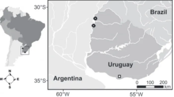
Material was collected on 28 February, 2010, from two sites in the Salto Grande reservoir, located on the border with Uruguay (Salto Province) and Argentina (Entre Rios Province). The sampling sites (Fig. 1) are adjacent to the dam (31°15’31.41”S, 57°55’33.66”W) and upstream from the dam (30°46’27.52”S, 57°47’55.53”W).
Rediscovered after 77 years:
Odontodiaptomus thomseni
– a rare species of
calanoid (Crustacea: Copepoda) from South America
Gilmar Perbiche-Neves
1, Geoff A. Boxshall
2, Carlos E. F. da Rocha
1& Marcos G. Nogueira
31 Departamento de Zoologia, Instituto de Biociências, Universidade de São Paulo. Rua do Matão, Travessa 14, 321, 05508-900 São Paulo, SP, Brazil. Email: gilmarpneves@yahoo.com.br
2 Department of Zoology, The Natural History Museum. Cromwell Road, London SW7 5BD, United Kingdom. 3 Instituto de Biociências, Universidade Estadual Paulista. Rubião Junior, 18618-000 Botucatu, SP, Brazil.
ABSTRACT. The freshwater copepod Odontodiaptomus thomseni (Brehm, 1933) (Calanoida: Diaptomidae) is a rare species that has been reported only once – in its original description (BREHM 1933). The lack of subsequent records led to its inclusion
in the Red List of threatened species (IUCN). Here we present a new record for O.thomseni. It was discovered in Salto Grande reservoir, which is located in the lower stretches of the Uruguay River, between Uruguay and Argentina, at the River Plate basin. In January 2010, three specimens (two males and one female) were found, and these were studied in detail using scanning electron microscopy (SEM). We only had material of Odontodiaptomus paulistanus (Wright, 1936) for comparison, but the position of the lateral spine in right P5 of the male, and the shape and size of lateral wings of the female are especially distinctive. Odontodiaptomus thomseni remains a rare species and we recommend keeping it on the IUCN Red List.
KEY WORDS. Copepod; rare; red list; taxonomy.
dorsal surface (Fig. 3); with sclerotized tooth-like process present on left caudal ramus, and other similar processes on surface of caudal ramus and urosome (Figs 4 and 5); two vestigial setae present on segment 2 of right geniculate antennule; well-de-veloped modified setae present on segments 10, 11 and 13 of right antennule (Fig. 10); hyaline lamella on segment 20 of right antennule; endopod of right leg 5 well developed, 2-seg-mented (Figs 7 and 8); lateral spine of second exopod segment of right leg 5 positioned basally on segment and reaching be-yond middle of segment (Fig. 7); terminal claw longer than segment (Fig. 7). Female: patches of setules present on dorsal surface of prosome; posterolateral wings of prosome strongly asymmetrical (Fig. 25); left lateral wing of genital double-somite with tapering process bearing spine at apex (Fig. 25); second urosomite with wing-like expansion on right side (Fig. 25); scle-rotized tooth-like process on left side of third urosomite (Fig. 26); endopod of leg 5 well developed (Fig. 29).
Redescription. Male. Prosome (Fig. 2): Body length 989 µm, mean width 252 µm. Posterolateral corners of prosome not produced into wing-like expansions; suture between pedigerous somites 4 and 5 complete; groups of long, fine spinules present on dorsal and lateral surfaces of pedigerous somites 3 to 5 (Fig. 3). Urosome: 5-segmented; genital somite symmetrical. Caudal rami (Figs 2, 4 and 5): Semicircular scle-rotized processes present on inner distal margin of left caudal ramus; also with some other sclerotized processes on dorsal, ventral and lateral surfaces of left caudal ramus (Figs 4 and 5) and also in different places. Inner margins of rami with setule rows. Right antennule (Figs 6, 14 and 15.): 22-segmented, gen-iculate, and swollen in mid section; numbers of setae (s), coni-cal setae (cs), long setae (Ls), modified setae (ms), vestigial setae (vs), aesthetascs (ae) and spiny processes (p) on each segment as follows: (1) 1s + 1ae, (2) 2vs + 3s + 1ae, (3) 1vs +1Ls + 1ae, (4) 1s, (5) 1Ls + 1vs +1ae, (6) 1s, (7) 1Ls + 1ae, (8) 1s + 1cs, (9) 1s + 1Ls, (10) 1ms + 1s, (11) 1ms + 1s, (12) 2s + 1ae, (13) 1ms + 1s + 1ae, (14) 1s + 1Ls + 1ae, (15) 1ms + 1Ls + 1p + 1ae, (16) 1ms + 1Ls + 1p + 1ae, (17) 1s + 2ms, (18) 2ms + 1s, (19) 3ms + 1Ls + 1ae, (20) 2s + 2Ls + 1p, (21) 2Ls, (22) 4s + 1ae. Second segment with groove indicating compound origin. Spinous processes on segments 10 and 11 well developed, but smaller than that Dissolved Oxygen (mg.L ) 6.32 8.19
pH 6.90 7.30
Electrical Conductivity (µS.cm-1) 39.30 40.50
Transparency (m) 0.50 0.50
Turbidity (NTU) 35.60 36.40
Total Suspended Solids (mg.L-1) 16.60 11.90
Organic Suspended Solids (mg.L-1) 3.00 2.40
Inorganic Suspended Solids (mg.L-1) 13.60 9.40
Total Nitrogen (mg.L-1) 611.30 651.80
Total Phosphorus (µg.L-1) 36.00 43.30
Chlorophyll-a (mg.L-1) 4.60 4.30
The copepods were sorted and identified under a stereo-microscope and three individuals of Odontodiaptomus thomseni were found, two males and one female. One female and one male were used for SEM, the remaining male has been retained in the collection of the first author.
on segment 13. Segment 14 without spinous process; segments 15 and 16 each with medium-size spine. Modified setae on seg-ments 15 and 16 rhomboidal. Segment 20 with well-developed denticulate hyaline membrane, denticles tiny proximally be-coming more tooth-like distally. Left antennule: 25-segmented, number of setae (s), conical setae (cs), long setae (Ls), vestigial setae (vs) and aesthetascs (ae) on each segment as follows: (seg-ment 1) 1s + 1ae, (2) 3s + 1ae +1vs, (3) 1Ls + 1ae + 1vs, (4) 1s, (5) 1Ls + 1ae +1vs, (6) 1s + 1vs, (7) 1Ls + 1ae, (8) 1s + 1cs, (9) 1s + 1Ls + 1ae, (10) -, (11) 1Ls, (12) 1s + 1cs + 1ae, (13) 1s, (14) 1Ls + 1ae, (15) 1Ls, (16) 1Ls + 1ae, (17) 1s, (18) 1Ls, (19) 1s + 1ae, (20) 1s, (21) 1Ls, (22) 1s + 1Ls, (23) 1Ls + 1ae, (24) 2Ls, (25) 1s + 3Ls + 1ae. Similar to female at Figs 20-22 and 27. Swimming
legs (Figs 11 and 12): Exopods 3-segmented; endopods 3-seg-mented in legs 2-4 and 2-seg3-seg-mented in leg 1; Schmeil´s organ present on second endopod segment of leg 2; endopods of legs 1-4 with row of 4-8 spinules along distal margin; exopods of legs 1 to 4 with 3-5 denticles distally; row of setules present on basis of leg 1. Spine and seta formula of swimming legs as in Tab. II. Similar to female at Fig. 24. Right leg 5 (Figs 7-9, 13 and 16): Coxa with small rounded process carrying apical spine located mid-distally on anterior surface (Fig. 16). Basis ca. 1.3 longer than wide, with rounded process on inner margin and seta on outer margin (Fig. 16). Endopod 2-segmented, length about 2/3 length of lateral spine on second exopod segment; ornamented with fringe of spinules around apex (Figs 7, 8, 13 Figures 2-10. Odontodiaptomus thomseni male: (2) habitus, ventro-lateral; (3) detail of fourth and fifth pedigerous somites, lateral view showing surface ornamentation of long setules; (4) rounded process on right caudal ramus; (5) detail of process on caudal ramus; (6) segments 1 to 6 of right antennule, showing vestigial setae (arrowed); (7) right leg 5 (with arrows in endopod and lateral spine), leg 4 and exopod of leg 3; (8) endopod of right leg 5; (9) proximal ornamentation on terminal claw of right leg 5, ventral view; (10) segments 8 to 16 of right antennule, arrows showing spinous processes on segments 10 and 11. Scale bars: 2 = 500 µm, 3, 6 = 50 µm; 4, 8, 9 = 20 µm, 5 = 2 µm, 7 = 200 µm, 10 = 100 µm.
3 2
6 5
4
7
10 8
ca. 2.2 longer than wide (Figs 7 and 16); lateral spine inserted near base of segment, lateral spine more than half as long as segment (Figs 7, 13 and 16). Terminal claw incorporating third exopodal segment, 1.5 times longer than second segment, row of denticles ventrally, larger near base but decreasing in size distally (Figs 7, 9 and 16). Left leg 5 (Fig. 16): Coxa about as long as wide, with conspicuous sclerotized process on outer distal margin. Basis ca. 1.3 longer than wide with seta inserted midway along outer margin, seta about as long as width of segment. Exopod 2-segmented; first segment with curved outer margin, inner margin with rounded process ornamented with setules; second segment ending in digitiform process; inner margin curved; carrying stout spine. Endopod 1-segmented, about half as long as endopod of right leg; ending in row of spinules on distal margin.
Female. Prosome: Body length: 1,662 µm, maximum width: 283 µm. Slightly asymmetrical rostrum, with paired ros-tral filaments but right thickened in their median part (Fig. 17). Suture between fourth and fifth pedigerous somites incomplete; groups of long, fine spinules present on dorsal and lateral sur-faces of pedigerous somites 3 to 5. Fifth pedigerous somite with well-developed posterolateral wings (Fig. 25); wings asymmetri-cal, right larger than left and terminating in sharp spinous pro-cess, with two chitinuous protuberances. Urosome: Genital double-somite asymmetrical, ca. 2 broader than longer; with well-developed spinous process on left side, at tip of expansion (Fig. 25); right side with large curved expansion on outer margin; genital area protruding; anterior part as a wide cuticular pad, with a crescentic margin; posterior pad with straight margin; gonoporal plates oval, symmetrical, showing the gonoporal slits. Part posterior to genital area showing some transverse cuticular folds (Fig. 23); second urosomite as wide as long (Fig. 25), with rounded chitinous process on margin of left side, plus other smaller processes on dorsal and ventral surfaces of segment (Fig. 26). Caudal rami: Symmetrical, 1.5 times longer than wide, or-namented with setules along inner and outer margins of both caudal rami; caudal setae thick. Antennula: Antennules reach-ing end of caudal rami. Setal formula as for left antennule of male (Figs 20-22 and 27). Antenna (Figs 18 and 19): Biramous, coxa with one seta, basis with two setae. Endopod 2-segmented: first segment with two setae in mid part; ornamented with row Figures 11-13. Odontodiaptomus thomseni male: (11) exopod and
endopod of leg 2, showing details of spinule rows (arrowed) on distal segments of both rami, caudal view; (12) exopod and endopod of leg 4, showing pores and spinule row (arrowed) on distal segment of endopod, frontal view; (13) right leg 5, with arrow pointing to arthroidal membrane. Scale bars: 11-12 = 50 µm, 13 = 100 µm.
12
of spinules (between 4-5) distally and pore near row: compound distal segment with 15 setae, inner group of 8 setae plus distal group of 7 setae; row of 8-10 spinules present near distal mar-gin. Exopod 7-segmented, setal formula: 1, 3, 1, 1, 1, 1, 4; seg-ment 2 partially subdivided by suture on one side. Swimming legs (Fig. 24 5H): similar to male. Leg 5: Leg 5 symmetrical (Figs 28-31); coxa with process at outer distal corner bearing triangu-lar sensilla at tip; basis with curved outer margin,and bearing
long seta extending to tip of first exopod segment. Endopod 2-segmented (Fig. 29); first segment larger than second, almost 2.5 times longer than wide; second segment with two setae plus row of spinules at tip (Fig. 31). Exopod 3-segmented; first seg-ment larger than second; second segseg-ment with lateral spine and with inner margin produced to form terminal claw ornamented with lateral rows of denticles along each side (Fig. 30); third segment small, offset and with two terminal setae, lateral smaller. Figures 14-16. Odontodiaptomus thomseni male: (14) segments 10-16 of right antennule; (15) segment 20 of right antennule; (16) right and left fifth legs, frontal view.
14
DISCUSSION
In his original work BREHM (1933) mentioned that he had found a “strange” copepod that was very different in morphol-ogy when compared with the known species. In fact, the species included in Odontodiaptomus share the possession of chitinous nodules on various parts of the body surface, including a tooth-like process on the caudal rami and another on the urosome, from which the genus takes its name. Odontodiaptomus thomseni differs from the other two species included in Odontodiaptomus
especially in the shape of the fifth leg of the male, which has the lateral spine on the second exopod segment inserted near the base of the segment, a well-developed endopod, and a rounded process on the first exopod segment. The females also differ markedly from the two other Odontodiaptomus species, particu-larly in the asymmetry of the genital receptacle. Consideration of these features makes it is easy to understand why BREHM (1933) regarded this species as “strange”.
This is only the second confirmed record of O. thomseni. The discovery of only three individuals suggests the species is Figures 17-27. Odontodiaptomus thomseni female: (17) rostrum; (18) antenna and buccal apparatus; (19) detail of spinule row (left arrow) and pore (right arrow) on endopod of antenna; (20) segments 17-25 of right antennule; (21) segments 1-4 of left antennule; (22) segments 1 to 4 of right antennule; (23) genital operculum; (24) frontal view of leg 2, with detail of posterior spinule row (arrowed); (25) last prosomal somite, genital double-somite, urosome and caudal rami, showing details of spinous process on lateral wing, and processes on urosomites; (26) detail of last urosomite showing chitinous process on lateral margin (arrowed); (27) segments 1-19 of left antennule. Scale bars: 17, 19, 23 = 50 µm, 18, 25 = 200 µm, 20-22, 24 = 100 µm, 26 = 20 µm, 27 = 400 µm.
20
23 22
21
24 26
25
rare and provides support for maintaining it on the Red List of threatened species. REIDet al. (2002) pointed to possible factors that currently affect the Brazilian copepod fauna and impact species’ abundance. These factors include the damming of riv-ers and disappearance of floodplain lakes; the introduction of alien species, and the increasing pollution of the rivers by do-mestic and industrial waste. Such factors may impact species abundances across the entire South America.
REID (1996) noted the existence of another record of O. thomseni from pools in Venezuela. We consider this record, so remote from the type locality (approximately 4,600 km), as not valid, without reexamination. BREHM (1933), WRIGHT (1938), MATSUMURA-TUNDISI (1986) and SANTOS-SILVA (2008) all emphasized that the three species of Odontodiaptomus are endemic to par-ticular regions or latitudinal ranges, further casting doubt on the record from Venezuela. It is possible that the material re-ported from Venezuela represents a new species of the genus Odontodiaptomus. According to J.C. Paggi (pers. comm. 2011) there is another undescribed species of Odontodiaptomus inhab-iting the Salto Grande reservoir (Uruguay/Argentina), although only one male individual has ever been found.
There are only three species of Odontodiaptomus and, as
WRIGHT (1937) commented, O. thomseni is very different from O. paulistanus and O. michaelseni. These last two are very simi-lar morphologically. In the absence of type material, is diffi-cult to construct a dichotomous identification key due the paucity of knowledge of O. michaelseni. New material needs to be found – near Buenos Aires.
Further study of this genus is necessary in order to fully understand its geographic distribution and taxonomic com-plexity. SUÁREZ-MORALESet al. (2005) point to the lack of infor-mation from some areas in the Neotropical region, and they show the distribution of Odontodiaptomus in South America as basically restricted to the La Plata River Basin.
More sampling should be concentrated in regions where O. thomseni has been found, especially in small water bodies, such pools and reservoirs. Although the nature of these small water bodies contrasts with the new locality reported here, this is the kind of environment sampled by BREHM (1933). Sampling such small habitats is important as PERBICHE-NEVESet al. (2011) demonstrated when they reported high abundance of Argyrodiaptomus bergi (Richard, 1898) in a pool at high altitude in the state of Santa Catarina, southern Brazil. Prior to this record, this copepod had not been found for 36 years. Figures 28-31. Odontodiaptomus thomseni female: (28) leg 5, with lateral process on coxa and basis; (29) endopods of fifth legs; (30) terminal claw of exopod of leg 5; (31) spinule row on endopod of left leg 5. Scale bars: 28 = 200 µm, 29 = 50 µm, 30 = 25 µm, 31 = 5 µm.
28
30 31
BOXSHALL, G.A. & D. DEFAYE. 2008. Global diversity of Copepods (Crustacea: Copepoda) in freshwater. Hydrobiologia 595: 195-207.
BREHM, V. 1933. Diaptomus thomseni nov. spec., ein merkwürdiger neuer Diaptomus aus Uruguay. Zoologischer Anzeiger104: 221-224.
FELGENHAUER, B.E. 1987. Techniques for preparing crustaceans for scanning electron microscopy. Journal of Crustacean Biology7: 71-76.
HUYS, R. & G.A. BOXSHALL. 1991. Copepod evolution. London, The Ray Society, 468p.
KIEFER, F. 1936. Über die Systematik der Südamerikanischen Diaptomiden (Crustacea Copepoda). Zoologischer Anzeiger 116: 194-200.
LANSAC-TÔHA, F.A.; C.C. BONECKER; L.F.M. VELHO; N.R. SIMÕES; J.D. DIAS; G.M. ALVES & E.M. TAKAHASHI. 2009. Biodiversity of zooplankton communities in the Upper Paraná River floodplain: interannual variation from long-term studies.
Brazilian Journal of Biology 69 (Suppl. 2): 539-549. LOPES, R.M.; F.A. LANSAC-TÔHA; R. VALE & M. SERAFIM-JÚNIOR. 1997.
Comunidade zooplanctônica do Reservatório de Segredo, p. 39-60. In: A.A. AGOSTINHO & L.C. GOMES (Eds). Reservatório de Segredo: bases ecológicas para o manejo. Maringá, Eduem. MATSUMURA-TUNDISI, T. 1986. Latitudinal Distribution of Cala-noida Copepods in Freshwater aquatic Systems of Brasil.
Revista Brasileira de Biologia 46 (3): 527-553.
ROCHA; E. SUÁREZ-MORALES & H. UEDA. 2002. Conservation of continental copepod crustaceans, p. 253-261. In: E. ESCOBAR -BRIONES & F. ALVAREZ (Eds). Modern Approaches to study of
Crustacea. Kluwer Academic, Plenum Publishers.
SANTOS-SILVA, E.N. 2008. Calanoid of the families Diaptomidae, Pseudodiaptomidae, and Centropagidae from Brasil. Biolo-gia Geral e Experimental 8 (1): 3-67.
SANTOS-SILVA, E.N.; G.A. BOXSHALL & C.E.F. ROCHA. 1999. The neotropical genus Notodiaptomus Kiefer, 1936 (Calanoida: Diaptomidae): redescription of the type species Notodiapto-mus deitersi (Poppe, 1891) and designation of a neotype.
Studies on Neotropical Fauna and Environment 34: 114-128.
SUÁREZ-MORALES, E.; J.W. REID & M. ELÍAS-GUTIÉRREZ. 2005. Diversity and distribuitional patterns of Neotropical Freshwater Copepods (Calanoida: Diaptomidae). InternationalReview ofHydrobiology90: 71-83.
WRIGHT, S. 1936. Preliminary report on six new species of Diaptomus from Brasil. Anais da Academia Brasileira de Ciências 8: 79-85.
WRIGHT, S. 1937. A review of some species of Diaptomus from São Paulo. Anais da Academia Brasileira de Ciências 9: 65-82.
WRIGHT, S. 1938. A review of the Diaptomus bergi group, with des-criptions of two new species. Transactions of the American Microscopical Society 57: 297-315.