Insulin Substrate Receptor (IRS) proteins in normal and
malignant hematopoiesis
Joa˜o Agostinho Machado-Neto,I,IIBruna Alves Fenerich,IAna Paula Nunes Rodrigues Alves,IJaqueline Cristina Fernandes,IRenata Scopim-Ribeiro,IJuan Luiz Coelho-Silva,IFabiola TrainaI,*
IDepartamento de Medicina Interna, Faculdade de Medicina de Ribeirao Preto, Universidade de Sao Paulo, Ribeirao Preto, Sao Paulo, SP, BR. IIDepartamento de Farmacologia do Instituto de Ciencias Biomedicas da Universidade de Sao Paulo, Sao Paulo, SP, BR.
Machado-Neto JA, Fenerich BA, Rodrigues Alves AP, Fernandes JC, Scopim-Ribeiro R, Coelho-Silva JL, et al. Insulin Substrate Receptor (IRS) proteins in normal and malignant hematopoiesis. Clinics. 2018;73(suppl 1):e566s
*Corresponding author. E-mail: [email protected]
The insulin receptor substrate (IRS) proteins are a family of cytoplasmic proteins that integrate and coordinate the transmission of signals from the extracellular to the intracellular environment via transmembrane receptors, thus regulating cell growth, metabolism, survival and proliferation. The PI3K/AKT/mTOR and MAPK signaling pathways are the best-characterized downstream signaling pathways activated by IRS signaling (canonical pathways). However, novel signaling axes involving IRS proteins (noncanonical pathways) have recently been identified in solid tumor and hematologic neoplasm models. Insulin receptor substrate-1 (IRS1) and insulin receptor substrate-2 (IRS2) are the best-characterized IRS proteins in hematologic-related processes. IRS2 binds to important cellular receptors involved in normal hematopoiesis (EPOR, MPL and IGF1R). Moreover, the identi-fication of IRS1/ABL1 and IRS2/JAK2V617Finteractions and their functional consequences has opened a new frontier for investigating the roles of the IRS protein family in malignant hematopoiesis. Insulin receptor substrate-4 (IRS4) is absent in normal hematopoietic tissues but may be expressed under abnormal conditions. Moreover, insulin receptor substrate-5 (DOK4) and insulin receptor substrate-6 (DOK5) are linked to lymphocyte regulation. An improved under-standing of the signaling pathways mediated by IRS proteins in hematopoiesis-related processes, along with the increased development of agonists and antagonists of these signaling axes, may generate new therapeutic approaches for hematological diseases. The scope of this review is to recapitulate and review the evidence for the functions of IRS proteins in normal and malignant hematopoiesis.
KEYWORDS: Insulin Receptor Substrate; Adaptor Protein; Signal Transduction; Hematopoiesis; Leukemia; Myeloproliferative Neoplasms.
’ INTRODUCTION
The insulin receptor substrate (IRS) proteins are a family of cytoplasmic proteins composed of six members (IRS1-6) that act as adaptor proteins (1-6). IRS proteins integrate and coordi-nate multiple cellular processes by transducing signals from the extracellular to the intracellular environment via transmembrane receptors (1) and are the major molecules that mediate the response to insulin and insulin-like growth factor 1 (IGF1) stimulation (2,7). IRS proteins regulate numer-ous processes such as growth, metabolism, survival and pro-liferation, and they respond to various stimuli, including steroids, cytokines, hormones and integrins [reviewed in (8) and (9)].
IRS1 was the first member of the IRS protein family to be identified and cloned (10). IRS2 was identified in Irs1-knockout mice as a phosphoprotein that responds to insulin stimu-lation (11). In humans, IRS3 is a pseudogene (12). The expres-sion of IRS4 is restricted to the brain, kidney, thymus and liver (5). IRS5 and IRS6, also called docking protein-4 (DOK4) and docking protein-5 (DOK5), respectively, have high homology with other members of the IRS protein family in their N-terminal regions (6,13). The structures of the human IRS proteins are shown in Figure 1.
IRS proteins do not have kinase or other intrinsic enzy-matic activity; however, they contribute to the organization of signaling complexes as adaptor proteins (2). IRS proteins have high levels of homology in the N-terminal regions, which contain two conserved domains that participate in receptor recruitment: the pleckstrin homology (PH) domain and the phosphotyrosine binding (PTB) domain. The PH domain participates in protein-protein interactions and facilitates recruitment by receptors and phospholipid proteins located in the plasma membrane (14-16). The PTB domain contains the tyrosine residues that interact with NPXY motifs on activated receptors (17,18). The activation of IRS proteins occurs after the phosphorylation of tyrosine residues in the C-terminal region, which contains more than twenty tyrosine sites. When phosphorylated, IRS proteins can DOI:10.6061/clinics/2018/e566s
Copyright&2018CLINICS–This is an Open Access article distributed under the terms of the Creative Commons License (http://creativecommons.org/licenses/by/ 4.0/) which permits unrestricted use, distribution, and reproduction in any medium or format, provided the original work is properly cited.
No potential conflict of interest was reported.
Received for publication onJanuary 10, 2018.Accepted for publi-cation onJuly 30, 2018
Commemorative Edition: 10 years of ICESP
1
bind to various Src homology (SH2) domain-containing proteins, including PI3K, GRB2, SHP2, resulting in the activation of multiple signaling pathways, especially the PI3K/AKT/mTOR and MAPK pathways (19-23).
PI3K-mediated signaling plays a critical role in many cellular biological events, including mitogenesis, motility, metabolism and survival (24). The C-terminal region of the IRS proteins contains several YMXM motifs, which bind to the SH2 domain of the PI3K p85 subunit when phosphorylated, with the consequent activation of AKT (25). PI3K was origi-nally identified as a dimer composed of a catalytic subunit (p110) and a regulatory subunit (p85). The binding of phos-phorylated proteins to the SH2 domain of the PI3K p85 subunit activates the associated catalytic domain. PI3K catalyzes the phosphorylation of phosphoinositides at the 3-position of the inositol ring, producing phosphatidyli-nositol 3,4,5-triphosphate (PI(3,4,5)P3), which in turn activates intracellular substrates such as AKT (26). The antiapoptotic effect of AKT is associated with the phos-phorylation of its substrates, including BAD, caspase 9, NF-kB and the family of forkhead transcription factors (27). BAD phosphorylation prevents its interaction with BCL2 and BCL-XL, allowing its antiapoptotic action on the mito-chondrial pathway (28).
IRS proteins also bind to GRB2, leading to the activation of the MAPK cascade, which includes the ERK protein. The activation of the MAPK cascade is critical for cell differentia-tion and proliferadifferentia-tion. In addidifferentia-tion, IRS proteins may bind to other adapter proteins such as NCK, CRK, or the FYN kinase, also resulting in the activation of the MAPK cascade (20,29,30) (Figure 2).
Although IRS proteins have long been considered to exemplify typical cytosolic proteins, IRS1 may, under certain circumstances, be translocated to the nucleus, although the
exact mechanism that promotes such translocation is not fully understood (31). Prisco et al. (32) noted that IRS1 contains native nuclear localization signals (NLSs), which may explain the translocation of IRS1 to the nucleus after IGF1/ IGF1R activation (33). In addition, the presence of nuclear IRS1 in cells expressing human JC virus T-antigen, SV40 T-antigen, integrins, estrogen receptor a (ERa) and estrogen receptorb(ERb) indicates that IRS1 can be translocated via association with other NLS-equipped proteins (31,32,34-36). However, the role of nuclear IRS proteins is still undetermined. Deregulation of the IRS protein has been implicated in human diseases, especially diabetes and cancer [reviewed in (8,9) and (37)]. Herein, we review and recapitulate the evidence for the roles of IRS proteins in normal and malignant hematopoiesis, exploring the clinical, biological and functional descriptions of the involvement of this protein family in the field of hematology.
IRS signaling in normal hematopoiesis
Hematopoiesis is strictly regulated by cytokines and growth factors (38). Both IRS1 and IRS2 are expressed in a wide spectrum of cells and tissues (39). Unpublished data from our research group indicate that in human CD34+ bone marrow cells,IRS2is the predominant transcript, whereas in human CD3+ lymphocytes, IRS1 is highly expressed. Irs2 expression is predominant in murine hematopoietic cells (3,39). Machado-Neto et al. (40) reported increased levels of IRS2 mRNA, protein and phosphorylation in models of lineage-differentiated cell lines, including erythroid-, granulocytic-and megakaryocytic-differentiated cells (40). In CD34+cells from normal donors, IRS2 expression was increased upon erythroid differentiation (40). In granulocytic-differentiated HL-60 cells induced by dimethylsulfoxide, IGF1 induced an
increase in IRS2 but not IRS1 protein expression and tyrosine phosphorylation as well as in PI3K recruitment (41). The genes encoding IRS2 and IGF1R were more highly expressed in plasma cells than in B cells, indicating that the IGF1R/ IRS2 signaling pathway plays an important role in plasma cell differentiation and function (42). These data highlight the involvement of IRS2 in hematopoietic cell differentiation.
IRS2 can be activated via three relevant transmembrane receptors involved in hematopoiesis: IGF1R, EPOR, and TPOR (MPL) (41,43,44). The role of IGFI and its receptor in the regulation of hematopoietic cell development has been studied widely. Most such studies are related to the ability of IGF1 to stimulate myelopoiesis and erythropoiesis (45,46). However, in adult organisms, IGF1 does not seem to be required for normal or malignant hematopoietic cell devel-opment (47). Another study demonstrated that although neither IGF1 nor insulin is required during early erythropoi-esis, both play a role in the final stages of erythroid matu-ration via the phosphorylation of IRS2 (48).
Erythropoietin (EPO) is the major regulator of erythropoi-esis (49). Upon EPO binding, the erythropoietin receptor (EPOR) undergoes conformational changes and associates with JAK2 (50-52). JAK2 can activate its associated signaling pathways via two distinct mechanisms: (I) an EPOR tyrosine phosphorylation-independent mechanism involving ERK1/2 stimulation (53); and (II) via the phosphorylation of numer-ous tyrosine residues in the cytoplasmic tail of the EPOR that act as docking sites for SH2 domain-containing proteins (52,54,55). IRS2 but not IRS1 is expressed in several murine and human EPO-sensitive cell lines, including cells with erythroid and megakaryocytic features.
In UT-7 cells stimulated with EPO, IRS2 is rapidly phos-phorylated on tyrosine residues. Following EPO-induced tyrosine phosphorylation, IRS2 associates with two proteins: PI3K and PI-3,4,5-trisphosphate 5-phosphatase (SHIP). Further-more, phosphorylated IRS2 remains constitutively associated with the EPOR (43). Sathyanarayana et al. (55) demon-strated that IRS2 is regulated by EPO at the transcriptional level in primary murine erythroblasts. Furthermore, using phosphoproteomic analysis to evaluate the potential adaptor proteins involved in EPOR/JAK2 signaling, Verma et al. (56) observed that IRS2 was phosphorylated on tyrosine residues 653, 675, 742 and 823 in response to EPO.
Thrombopoietin (TPO) is the pivotal signal that regulates platelet production; TPO binds to the MPL receptor on hema-topoietic stem cells and megakaryocytes (57). The TPO-mediated association between the MPL receptor and IRS2 was described by Miyakawa et al. (44), who reported that TPO activates the PI3K pathway in BaF3/MPL cells via a complex com-prising the p85 subunit of PI3K, phosphorylated SHP2 and GAB2 or a complex comprising the p85 subunit of PI3K and IRS2 (44).
IRS proteins can also be activated by the interleukins (ILs) involved in hematopoiesis. In lymphoid cell lines, T cells and NK human lymphocytes, IRS1 and IRS2 are phosphorylated on tyrosine sites upon stimulation by IL2, IL4, IL7 and IL15 (58). IL9 promotes the tyrosine phosphorylation of IRS1 by JAK tyrosine kinases in a murine T cell line (TS1) (59). Although most studies linking IRS proteins and hemato-poiesis focus on IRS1 and IRS2, the expression of IRS5 and IRS6 in human T cells was reported, and IRS5 was identified as a negative regulator of T lymphocyte activation (60,61).
Figure 2 -Canonical IRS signaling. IRS proteins are recruited via their PH/PTB domains and are phosphorylated on tyrosine residues by upstream tyrosine kinase receptors. Tyrosine phosphorylation of IRS proteins triggers the activation of PI3K/AKT/mTOR and MAPK signaling, thus regulating many biological processes, including cell proliferation, protein synthesis, survival and gene expression, in specific human tissues. IRS proteins may also be activated by cytokine and hormone receptors (e.g.,IL4, leptin, and angiotensin), which further induce JAK2 stimulation and IRS/JAK2 interaction, leading to the activation of STAT, PI3K/AKT/mTOR and MAPK signaling. Abbreviations: P, phosphorylation; PY, tyrosine phosphorylation. This figure was generated using Servier Medical Art (http://www. servier.com/Powerpoint-image-bank).
3
Using 32D cells, a cell line that neither expresses endogenous IRS1 nor responds to IL4 or insulin, Wang et al. (62) demon-strated that IRS1 is required for insulin- and IL4-stimulated mitogenesis in hematopoietic cells. In 32D cells, the expres-sion of IRS1 via transfection restored sensitivity to IL4 and insulin and induced proliferation (62). A subsequent study by the same research group showed different results; the stimulation of overexpressed IGF1R by IGF1 and IL4 induced hematopoietic cell proliferation independent of IRS expres-sion and activation (63). IRS4 was also identified to participate in insulin and IL4 signaling in 32D cells (64). In hemato-poietic cells, the type I interferon receptor can activate IRS signaling (65); interferon-a(IFN-a) binding induces the rapid tyrosine phosphorylation of IRS1 and IRS2, leading to the association of phosphorylated IRS proteins with PI3K (65-67).
IRS signaling in myeloid neoplasms
Chronic myeloid leukemia. Traina et al. (68) were the first to demonstrate the involvement of the IRS1 protein in BCR-ABL1 signal transduction in chronic myeloid leukemia (CML). In the K562 cell line, a BCR-ABL1-positive cell line derived from a patient with CML in blast crisis, the IRS1 protein was constitutively phosphorylated and associated with BCR-ABL1, and IRS1 phosphorylation was inhibited by imatinib treatment. Traina et al. also described the associa-tion between IRS1 and PI3K and the IRS1-associated PI3K activity in K562 cells. The associations between these proteins were inhibited by imatinib treatment, suggesting that PI3K activation by BCR-ABL1 involves binding to the phosphory-lated IRS1 protein and depends on the tyrosine kinase acti-vity of BCR-ABL1. In K562 cells treated with imatinib and immunoprecipitated with an anti-GRB2 antibody, the GRB2-associated phosphorylation of both BCR-ABL1 and IRS1 was significantly reduced, suggesting the formation of a BCR-ABL1/IRS1/GRB2/PI3K complex (68).
The functional involvement of IRS1 in the BCR-ABL1 signaling pathway was later demonstrated using lentivirus-mediated IRS1 silencing with short hairpin RNA (shRNA) in K562 cells (69). IRS1 inhibition reduced cell proliferation and clonal growth by arresting the cell cycle in the G0/G1 phase. IRS1 inhibition also decreased AKT, P70S6K and ERK phos-phorylation, indicating the downregulation of the PI3K/ AKT/mTOR and MAPK pathways. The inhibition of IRS1 did not modulate apoptosis; BCL2, BAX and BAD; protein expression; or BCR-ABL1 and CRKL phosphorylation. IRS1 silencing was not synergistic with imatinib treatment (69).
Zhao et al. (70) identified IRS1 and IRS2 as inhibitory targets of miR-570 and verified that miR-570 is downregulated in CML clinical samples and in the K562 and LAMA-84 CML cell lines. This study revealed that the overexpression of miR-570 suppressed cell proliferation, increased apoptosis, and reduced glucose metabolism, whereas the inhibition of miR-570 increased cell proliferation, reduced apoptosis, and increased glucose metabolism. Corroborating the find-ings by Machado-Neto et al. (69), Zhao et al. (70) verified that in K562 cells, IRS1 or IRS2 silencing via small inter-fering RNA (siRNA) reduced cell viability and increased sensitivity to nutrient deprivation.
Myeloproliferative neoplasms. Previous studies have described the involvement of IRS1 and IRS2 in the JAK2 signaling pathway in nonhematologic cells. IRS1 was found
to be associated with and phosphorylated by JAK2 in COS-1 cells overexpressing both IRS1 and JAK2 (59). JAK2 coimmunoprecipitated with IRS2 in rat aortic smooth muscle cells and in vivo models following angiotensin II stimula-tion (71-74) and in rat livers following leptin stimulastimula-tion (75). Considering the previous findings in nonhematologic tissues, our research group identified a constitutive protein associa-tion between IRS2 and JAK2 in myeloproliferative neoplasm (MPN) models, which present constitutive JAK2 activation due to a V617F mutation. In the HEL JAK2V617Fcell line, but not in U937 JAK2 wild-type leukemia cell lines, IRS2 was constitutively phosphorylated and associated with JAK2. In HEL cells, lentivirus-mediated IRS2 silencing decreased STAT5 phosphorylation, reduced cell viability and increased apoptosis. NT157, a pharmacological inhibitor of IGF1R/ IRS1-2, reduced cell viability in JAK2V617F primary MPN samples but not in JAK2 wild-type samples (76). A recent study using targeted next-generation sequencing identified IRS2 mutations in 2 of 16 (12.5%) patients with triple-negative MPN, one with polycythemia vera and the other with essential thrombocythemia (77).
Acute myeloid leukemia. In acute myeloid leukemia (AML), somatic mutations, aberrant gene/protein expression levels and activating autocrine loops may promote growth factor and cytokine signaling activation as well as clonal expansion (78). The activation of several prosurvival path-ways in AML is an essential element in the optimization of molecular targeted therapies, such as those targeting proteins involved in the protein kinase C, STAT, MAPK, PI3K/AKT/ mTOR pathways (79). IGF1 signaling is implicated in self-renewal/pluripotency in hematopoietic stem cell contexts and supports cell growth/survival via the activation of down-stream pathways in both normal and neoplastic settings (80). In the hematopoietic context, the role of the insulin and insulin-like growth factor axis in AML treatment refractori-ness has been studied, but the specific functions of the IRS proteins are underexplored.
Accumulating evidence demonstrates the role of IGF1R signaling via the PI3K/AKT/mTOR cascade in AML. IGF1 and other cytokines have been described as important for AML cell growth (81), and the activation of the IGF1R signal-ing pathway has been detected in cells from AML patients and contributes to the survival and proliferation of these cells (82,83). An association between increased activation of the IGF1R axis and resistance to cytarabine has been reported in leukemia. Blocking the IGF1R in a cytarabine-resistant cell line inhibited cell growth and led to apoptosis (84), sug-gesting that IGF1R and its downstream signaling pathways may provide valuable novel targets to overcome chemother-apeutic resistance in AML.
high IGF1R/IRS1 phosphorylation, and the combination of AKT inhibitors and the IGF1R inhibitor linsitinib poten-tiated PI3K/AKT/mTOR inhibition (85). Thus, combination therapy could be an effective strategy for breaking the adaptive circuits formed in leukemia cells that render these cells resistant to therapy. Consistent with this hypothesis, Tamburini et al. (83) noted that mTORC1 inhibition by RAD001 increased AKT activation in primary AML cells as a consequence of IRS2 upregulation via autocrine activation of IGF1/IGF1R signaling. Collectively, these results provide evidence that IGF1R signaling mediated by IRS1 and IRS2 is involved in chemotherapeutic resistance in AML.
Genetic lesions that affect TP53, such as mutations and aneuploidy, are recognized as markers of a very dismal prognosis for AML patients (86). Recently, Quintás-Cardama et al. (87) demonstrated that the p53 pathway is frequently disrupted in AML, not just viaTP53mutations/deletions but also via a molecular background permissive to the transfor-mation capability of p53. Via a proteomic approach, increased IRS1 phosphorylation at serine 1101 was identified as a bio-logical marker of p53 pathway deregulation (87).
Myelodysplastic syndrome. Our research group reported that IRS2expression was lower in bone marrow samples from patients with myelodysplastic syndrome (MDS) than in bone marrow samples from healthy donors (40). These findings agree with those of a previous study that used a microarray analysis to show that the level of IRS2 is lower in bone marrow mononuclear cells from MDS patients than in cells from healthy donors (88).IRS2expression was lower in MDS patients withX5% bone marrow blasts than
in MDS patients witho5% bone marrow blasts, andIRS2 downregulation was associated with an increased severity of cytopenia (40). These findings suggest that IRS2 defi-ciency may be related to ineffective hematopoiesis.
IRS signaling in lymphoid neoplasms
Acute lymphoblastic leukemia. Fernandes et al. (89) recently identified high levels of IRS1 protein expression in acute lymphoblastic leukemia (ALL) cell lines and observed that IRS1 andb-catenin were colocalized in the nucleus and cytoplasm of all the lymphoid leukemia cell lines studied. In the cytoplasm of normal peripheral blood mononuclear cells, both proteins were only weakly detected, suggesting a lower activation of the IRS1/b-catenin axis in healthy donors than in patients with ALL. Fernandes et al. (89) also reported high IRS1 and b-catenin mRNA expression in a cohort of forty-five adult patients with ALL compared to normal hematopoietic cells from thirteen healthy donors, indicat-ing that the IRS1/b-catenin signaling pathway probably contributes to the pathophysiology of ALL. In mouse embryo fibroblasts, Chen et al. (90) previously described IRS1, via IGF1R signaling, as a protein responsible for the nuclear translocation and activation ofb-catenin.
In the childhood ALL cell lines CCRF-CEM (T cell acute lymphoblastic leukemia, T-ALL), NALM6 (B cell acute lymphoblastic leukemia, B-ALL) and REH (B-ALL), Leclerc et al. (91) demonstrated that AMPK activation induced growth inhibition and apoptosis via the downregulation of mTOR phosphorylation on serine 2448. Moreover, the IGF1R/IRS1 axis was important in determining the pro- or antiapoptotic
response to AMPK activators, since AMPK activation induced a compensatory survival response. This mechanism was medi-ated in part by the AMPK-induced phosphorylation of IRS1 on serine 794, which in turn activated downstream oncogenic pathways (91). Therefore, selected combination therapies using IRS1 inhibitors could be a potential strategy for ALL therapy.
In a study using primary cells from adult patients with B-ALL, Juric et al. (92) identified, via computational analysis of the data obtained by a microarray analysis, a lower expres-sion of IRS1 in positive ALL than in BCR-ABL1-negative ALL. In BCR-ABL1-positive ALL, IRS1 expression negatively correlated with survival, independent of age and leukocyte count at diagnosis (92).
The multitarget tyrosine kinase inhibitor GZD824 exhib-ited an antitumor effect in pre-B-ALL by inhibiting both the SRC kinase and PI3K/AKT/mTOR pathways, and ALL cells with lower IRS1 expression were more sensitive to GZD824 treatment than those with higher IRS1 expression. Therefore, IRS1 expression could be used as a biomarker to predict GZD824 efficacy in pre-B-ALL (93).
T-ALL cases involving IRS4 have rarely been reported since Karrman et al. (94) first reported, in 2009, the t(X;7) (q22;q34) translocation in a patient with childhood T-ALL. These researchers identified IRS4as the translocated gene and observed IRS4 overexpression (94). Another case appear-ing years later and reported by Kang et al. (95) presented a simultaneous translocation of theTCRa/dloci (14q11) with different partner loci (Xq22 and 12p13), and fluorescent in situ hybridization suggested the involvement of theIRS4 gene. In 2011, Karrman et al. (96), intrigued by the rare cases of T-ALL involving IRS4, identified IRS4 mutations in 2 of 21 (9.5%) patients with T-ALL. IRS4 is believed to exert mitogenic and proliferative effects more similar to the effects of IRS1 than to those of IRS2 (8,94).
Chronic lymphocytic leukemia. High IGF1R expression was identified in primary chronic lymphocytic leukemia (CLL) cells, suggesting the contribution of the IGF1R/IRS signaling pathway to disease pathology. Treatment with IGF1R inhi-bitors (AG1024, PPP) and IGF1R/IR inhibitor (OSI-906) reduced the viability and induced apoptosis in CLL cells in vitro, independent of the presence of protective stromal cells, and reduced tumor burdenin vivo. Pharmacological or siRNA inhibition of the IGF1R was associated with a signi-ficant reduction in IRS1, PI3K, AKT and ERK phosphoryla-tion. These data indicate that in CLL cells, IGF1R signaling activates the PI3K/AKT and MAPK pathways via IRS1 (97).
Multiple myeloma. Li and colleagues (98) demonstrated that IGF1R and downstream signaling pathways play an impor-tant role in the development of a broad spectrum of plasma cell tumors via constitutive IRS2 tyrosine phosphorylation and PI3K recruitment. In human multiple myeloma cell lines, IGF1 induced proliferation and antiapoptotic effects via IRS1-dependent PI3K/AKT and MAPK activation, even in IL6-independent cell lines, indicating that the IGF1R/IRS1 axis plays an important role in the development and progres-sion of this disease (99).
Shi et al. (100) observed that low concentrations of mTOR inhibitors stimulated the PI3K/AKT cascade in multiple mye-loma. These drugs, in addition to preventing the phosphorylation
5
of the downstream mTOR targets p70S6K and 4EBP1 and subsequent G1 arrest, prevent IRS1 serine phosphorylation (at an inhibitory site). Therefore, the prevention of IRS serine phosphorylation enhanced the activity of IGF1R/ IRS1 signaling pathways and downstream targets, such as PI3K/AKT/mTOR, independent of PTEN mutational status (100). This mechanism can be particularly detrimental in multiple myeloma, because IGF1R/IRS1-induced AKT acti-vation is a protumoral stimulus in multiple myeloma cells (99,101). Thus, additional studies will be necessary to deci-pher the best strategy for combining mTOR inhibitors with other therapeutic agents in multiple myeloma (100).
Hairy cell leukemia. Recently, Durham et al. (102) identi-fied, by next generation sequencing and copy number analysis, a novel gain-of-function mutation in IRS1 that contributed to clinical resistance to vemurafenib (BRAFV600Einhibitor) in
1 of 53 (2%) patients with classical hairy cell leukemia. More-over, these researchers observed that mutated IRS1 activated PI3K/AKT signaling and phosphorylated ERK1/2, leading to the cytokine-independent growth of Ba/F3 cellsin vitro(102).
Perspectives
Studies using IRS protein (mainly IRS1, IRS2 and IRS4) knockout animals reveal that these animals are born alive but are smaller and present type II diabetes, reflecting the parti-cipation of IRS proteins in metabolic homeostasis (103-105). In oncology, IRS1 and IRS2 knockout mice as well as IRS1-and IRS2-overexpressing murine models were used to eluci-date the function of these proteins in solid tumors, providing evidence of distinct and nonredundant functions for both proteins in cancer development and progression (106-108). However, despite the potential importance of IRS proteins in the signal transduction of hematopoietic-related growth factors and cytokines, as discussed herein, the function of this protein family in normal and malignant hematopoiesis
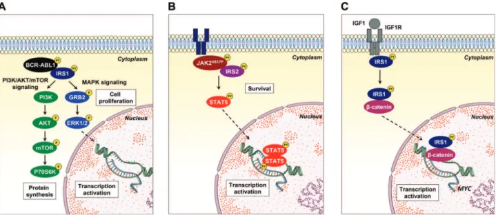
remains poorly understood. Recently, a great effort has been undertaken to develop and identify compounds capable of inhibiting signaling mediated by the IR/IRS and IGF1R/IRS axes. Reuveni et al. (109) identified that NT157, a compound that binds to IGF1R and induces a conformational change leading to the dissociation of IRS1/2 from the receptor and to the degradation of IRS1/2 by the proteasome, presented anti-neoplastic effects in solid tumors (109-113). The cancer cell panel in the initial study included K562 (CML) and Karpas (lymphoma) cell lines; thus, the results suggested that NT157 may exert antileukemic effects (109). Similarly, GZD824, a multikinase inhibitor, downregulated IRS1 signaling and reduced cell viability and tumor burden bothin vitro and in mice xenotransplanted with primary B-ALL cells (93). The participation of IRS1 and IRS2 in oncogenic pathways (namely, the BCR-ABL1 (68,69), JAK2V167F(76) and IRS1/ b-catenin (89) pathways) described by our research group corro-borates the participation of these proteins in the malignant phenotype of leukemias and suggests that these protein targets are druggable (Figure 3). Thus, a better understanding of the signaling pathway mediated by IRS proteins in hematopoietic-related processes, along with the increasing development of agonists and antagonists of this signaling axis, may generate new therapeutic approaches for hematological diseases.
In conclusion, the importance of IGF1R, EPOR and MPL signaling in cellular processes related to hematopoiesis has been recently consolidated; however, the mechanisms of intra-cellular regulation are continuously investigated. In this sense, the study of the participation of IRS proteins in hematopoietic processes still requires elucidation. The IRS proteins, particu-larly IRS1 and IRS2, play a relevant role in the signal transduc-tion of membrane receptors and the neoplastic phenotype induced by oncogenes. A summary of IRS signaling pathway alterations in hematological neoplasms is presented in Table 1. Future studies on the involvement of IRS proteins are neces-sary to open new avenues and augment the understanding of the complex signaling mediating normal hematopoiesis and malignant transformation.
Figure 3 -Noncanonical IRS1 signaling in hematological neoplasms.(A)IRS1 binds to and is activated by BCR-ABL1, inducing the activation of the PI3K/AKT/mTOR and MAPK signaling pathways, which contribute to cell proliferation.(B)IRS2 associates with JAK2 harboring the activating V617F mutation, which participates in STAT5 activation and cell survival.(C)Upon IGF1/IGF1R activation, IRS1 interacts with
Table 1-Alterations in the insulin receptor substrate (IRS) signaling pathway in hematological neoplasms.
Hematologic neoplasm Sample/cell line Notes Main approaches Publication
Chronic myeloid leukemia K562 IRS1 is constitutively phosphorylated on tyrosine residues and
associates with BCR-ABL1.
IP, WB Traina et al.(68)
Chronic myeloid leukemia K562 IRS1 silencing reduces cell proliferation and clonogenicity and
inhibits mTOR/Akt and MAPK activation.
shRNA-lentiviral delivery Machado-Neto et al. (69)
Chronic myeloid leukemia K562 and LAMA-84 IRS1 and IRS2 silencing reduces cell viability and metabolism. siRNA and transfection Zhao et al. (70)
Philadelphia-negative myeloproliferative neoplasm
HEL, U937 and primary samples
IRS2 is associated with the JAK2V617Fmutation and induces
survival in JAK2V617F-positive cells. NT157 reduces the viability
of primary cells from MPN patients.
IP, WB and
shRNA-lentiviral delivery
de Melo Campos et al. (76)
Acute myeloid leukemia Primary samples IRS1 mediates resistance to PI3K signaling inhibition. WB Bertacchini et al. (85)
Acute myeloid leukemia Primary samples IRS2 is upregulated by autocrine activation of IGF1/IGF1R
signaling upon Akt/mTOR inhibitor treatment.
WB Tamburini et al. (83)
Acute myeloid leukemia Primary samples IRS1 phosphorylation on serine 1101 is a biological marker
of p53 pathway deregulation.
Proteomics and network analyses
Quinta´s-Cardama et al. (87)
Myelodysplastic syndrome Primary samples IRS2 is downregulated and is associated with an increased
severity of cytopenia in MDS patients.
qPCR Machado-Neto et al. (40)
Myelodysplastic syndrome Primary samples IRS2 is downregulated in bone marrow mononuclear cells
from MDS patients compared with cells from healthy donors.
cDNA microarray Bar et al. (88)
Acute lymphoblastic leukemia Jurkat, MOLT4, Raji,
Namalwa and primary samples
IRS1 is highly expressed in ALL cell lines and primary samples.
Nuclear IRS1 associates withb-catenin and activatesb-catenin
signaling.
qPCR, WB and IP Fernandes et al. (89)
Acute lymphoblastic leukemia CCRF-CEM, NALM6
and REH
The activation of the IGF1R/IRS1 axis is a determinant of pro- or antiapoptotic responses to AMPK activators.
WB and cell viability assays
Leclerc et al. (91)
Acute lymphoblastic leukemia Primary samples IRS1 expression negatively correlates with survival,
independent of age and leukocyte count at diagnosis.
cDNA Microarray Juric et al. (92)
Acute lymphoblastic leukemia Primary samples IRS1 is a biomarker for the response to the multitarget
tyrosine kinase inhibitor GZD824.
WB and cell viability assays
Ye et al. (93)
Acute lymphoblastic leukemia Primary samples IRS4 is translocated, overexpressed and mutated in
ALL patients.
MC, FISH, WB and DNA sequencing
Karrman et al. (94) Kang et al. (95) Karrman et al. (96)
Chronic lymphocytic leukemia Primary samples IGF1R/IRS signaling is activated and promotes survival. WB, cell viability assays
and xenograft models
Yaktapour et al. (97)
Plasma cell neoplasms Murine primary tumors The activation of the IGF1R/IRS2/PI3K/p70S6K axis is important
in the development of plasma cell tumors.
Transfection and allograft models
Li et al. (98)
Multiple myeloma ANBL-6, Brown,
Delta-47, OPM-2, 8226, KMM1, H929, and MM-144
Activation of the IGF1R/IRS1 axis leads to the inhibition of apoptosis and the induction of cell proliferation.
WB, cell viability assays and xenograft models
Ge et al. (99)
Multiple myeloma OPM-2, 8226, MM1S and
HS-Sultan
IRS1 participates in a feedback loop that leads to mTOR inhibitor resistance.
WB Shi et al. (100)
Hairy cell leukemia Primary samples Gain-of-function mutations in IRS1 contribute to resistance
to vemurafenib (BRAFV600Einhibitor).
Deep targeted mutational and copy number analysis
Durham et al. (102)
Abbreviations: IP, immunoprecipitation; WB, western blotting; MPN, myeloproliferative neoplasm; MDS, myelodysplastic syndrome; qPCR, quantitative polymerase chain reaction; ALL, acute lymphoblastic leukemia; MC, metaphase cytogenetics; FISH, fluorescence in situ hybridization.
7
CLINICS
2018;73(
suppl
1):e566s
IRS
proteins
in
hematopoi
esis
Machado-Ne
to
JA
et
’ ABBREVIATIONS
AKT, AKT serine/threonine kinase; AML, acute myeloid leukemia; AMPK, AMP-activated protein kinase; ALL, acute lymphoblastic leukemia; B-ALL, B cell acute lymphoblastic leukemia; BAD, BCL2-associated death promoter; BAX, Bcl-2-associated X protein; BCL2, B cell lymphoma 2; BCL-XL, B cell lymphoma-extra large; BCR-ABL1, breakpoint cluster region-Abelson 1; BRAF, B-Raf proto-oncogene, serine/threonine kinase; CD, cluster of differentiation; CLL, chronic lympho-cytic leukemia; CML, chronic myeloid leukemia; CRK, CRK oncogene, adaptor protein; CRKL, CRK-like proto-oncogene, adaptor protein; DOK, docking protein; EPO, erythropoietin; EPOR, erythropoietin receptor; ERa, estro-gen receptor a; ERb, estrogen receptor b; ERK, extracel-lular signal-regulated kinase; FYN, FYN proto-oncogene, Src family tyrosine kinase; GAB2, GRB2-associated-binding protein 2; GRB2, growth factor receptor-bound protein 2; IFN-a, interferon-a; IGF1, insulin-like growth factor 1; IGF1R, insulin-like growth factor 1 receptor; IL, interleukin; IR, insulin receptor; IRS, insulin receptor substrate; JAK2, Janus kinase 2; MAPK, mitogen-activated protein kinase; MDS, myelodysplastic syndrome; miR, Micro RNA; MPL, MPL proto-oncogene, thrombopoietin receptor; MPN, myelo-proliferative neoplasms; mTOR, mammalian target of rapamycin; NCK, noncatalytic region of tyrosine kinase adaptor protein; NF-kB, nuclear factor-kappa B; NLS, nuclear localization signal; PDGFR, platelet-derived growth factor receptor; PH, pleckstrin homology; PI3K, phosphatidylinositol-4,5-bisphosphate 3-kinase; pre-B-ALL, B cell precursor acute lymphoblastic leukemia; PTB, phosphotyrosine binding; PTEN, phosphatase and tensin homolog; SH2, Src homo-logy; SHIP, SH2-containing inositol phosphatase; SHP2, Src homology 2 domain-containing protein-tyrosine phos-phatase 2; shRNA; short hairpin RNA; siRNA, small inter-fering RNA; SRC, SRC proto-oncogene, nonreceptor tyrosine kinase; STAT, signal transducer and activator of transcrip-tion; T-ALL, T cell acute lymphoblastic leukemia; TP53, tumor protein p53; TPO, thrombopoietin; TPOR, thrombo-poietin receptor
’ ACKNOWLEDGMENTS
The authors thank the Conselho Nacional de Desenvolvimento Científico e Tecnológico (CNPq), Coordenac¸ão de Aperfeic¸oamento de Pessoal de Nível Superior (CAPES) and Fundac¸ão de Amparo à Pesquisa do Estado de São Paulo (FAPESP) for financial support and Fernanda T. Udinal, from the Hemocentro Foundation of Ribeirão Preto, São Paulo, Brazil, for the English language review.
’ AUTHOR CONTRIBUTIONS
Machado-Neto JA, Fenerich BA, Rodrigues Alves APN, Fernandes JC, Scopim-Ribeiro R and Coelho-Silva JL participated in the preparation, completion andfinal approval of the manuscript. Traina F was the prin-cipal investigator and participated in the preparation, editing, completion andfinal approval of the manuscript. All authors read and approved the final version of the manuscript.
’ REFERENCES
1. Lee YH, White MF. Insulin receptor substrate proteins and diabetes. Arch Pharm Res. 2004;27(4):361-70, http://dx.doi.org/10.1007/BF029 80074.
2. Sun XJ, Rothenberg P, Kahn CR, Backer JM, Araki E, Wilden PA, et al. Structure of the insulin receptor substrate IRS-1 defines a unique signal transduction protein. Nature. 1991;352(6330):73-7, http://dx.doi.org/ 10.1038/352073a0.
3. Sun XJ, Wang LM, Zhang Y, Yenush L, Myers MG Jr, Glasheen E, et al. Role of IRS-2 in insulin and cytokine signalling. Nature. 1995;377(6545): 173-7, http://dx.doi.org/10.1038/377173a0.
4. Smith-Hall J, Pons S, Patti ME, Burks DJ, Yenush L, Sun XJ, et al. The 60 kDa insulin receptor substrate functions like an IRS protein (pp60IRS3) in adipose cells. Biochemistry. 1997;36(27):8304-10, http://dx.doi.org/ 10.1021/bi9630974.
5. Lavan BE, Fantin VR, Chang ET, Lane WS, Keller SR, Lienhard GE. A novel 160-kDa phosphotyrosine protein in insulin-treated embryonic kidney cells is a new member of the insulin receptor substrate family. J Biol Chem. 1997;272(34):21403-7, http://dx.doi.org/10.1074/jbc.272.34. 21403.
6. Cai D, Dhe-Paganon S, Melendez PA, Lee J, Shoelson SE. Two new substrates in insulin signaling, IRS5/DOK4 and IRS6/DOK5. J Biol Chem. 2003;278(28):25323-30, http://dx.doi.org/10.1074/jbc.M212430200. 7. Myers MG Jr, Sun XJ, Cheatham B, Jachna BR, Glasheen EM, Backer JM,
et al. IRS-1 is a common element in insulin and insulin-like growth factor-I signaling to the phosphatidylinositol 30-kinase. Endocrinology.
1993;132(4):1421-30, http://dx.doi.org/10.1210/endo.132.4.8384986. 8. Mardilovich K, Pankratz SL, Shaw LM. Expression and function of the
insulin receptor substrate proteins in cancer. Cell Commun Signal. 2009; 7:14, http://dx.doi.org/10.1186/1478-811X-7-14.
9. Shaw LM. The insulin receptor substrate (IRS) proteins: at the inter-section of metabolism and cancer. Cell Cycle. 2011;10(11):1750-6, http:// dx.doi.org/10.4161/cc.10.11.15824.
10. White MF, Maron R, Kahn CR. Insulin rapidly stimulates tyrosine phosphorylation of a Mr-185,000 protein in intact cells. Nature. 1985; 318(6042):183-6, http://dx.doi.org/10.1038/318183a0.
11. Patti ME, Sun XJ, Bruening JC, Araki E, Lipes MA, White MF, et al. 4PS/insulin receptor substrate (IRS)-2 is the alternative substrate of the insulin receptor in IRS-1-deficient mice. J Biol Chem. 1995;270(42):24670-3, http://dx.doi.org/10.1074/jbc.270.42.24670.
12. Bjornholm M, He AR, Attersand A, Lake S, Liu SC, Lienhard GE, et al. Absence of functional insulin receptor substrate-3 (IRS-3) gene in humans. Diabetologia. 2002;45(12):1697-702, http://dx.doi.org/10.1007/s00125-002-0945-z.
13. Grimm J, Sachs M, Britsch S, Di Cesare S, Schwarz-Romond T, Alitalo K, et al. Novel p62dok family members, dok-4 and dok-5, are substrates of the c-Ret receptor tyrosine kinase and mediate neuronal differentiation. J Cell Biol. 2001;154(2):345-54, http://dx.doi.org/10.1083/jcb.200102032. 14. Voliovitch H, Schindler DG, Hadari YR, Taylor SI, Accili D, Zick Y. Tyrosine phosphorylation of insulin receptor substrate-1 in vivo depends upon the presence of its pleckstrin homology region. J Biol Chem. 1995;270(30):18083-7, http://dx.doi.org/10.1074/jbc.270.30.18083. 15. Yenush L, Makati KJ, Smith-Hall J, Ishibashi O, Myers MG Jr, White MF.
The pleckstrin homology domain is the principal link between the insulin receptor and IRS-1. J Biol Chem. 1996;271(39):24300-6, http://dx.doi.org/ 10.1074/jbc.271.39.24300.
16. Burks DJ, Pons S, Towery H, Smith-Hall J, Myers MG Jr, Yenush L, et al. Heterologous pleckstrin homology domains do not couple IRS-1 to the insulin receptor. J Biol Chem. 1997;272(44):27716-21, http://dx.doi.org/ 10.1074/jbc.272.44.27716.
17. Sawka-Verhelle D, Tartare-Deckert S, White MF, Van Obberghen E. Insulin receptor substrate-2 binds to the insulin receptor through its phosphotyrosine-binding domain and through a newly identified domain comprising amino acids 591-786. J Biol Chem. 1996;271(11):5980-3, http:// dx.doi.org/10.1074/jbc.271.11.5980.
18. Backer JM, Wjasow C, Zhang Y. In vitro binding and phosphorylation of insulin receptor substrate 1 by the insulin receptor. Role of interactions mediated by the phosphotyrosine-binding domain and the pleckstrin-homology domain. Eur J Biochem. 1997;245(1):91-96, http://dx.doi.org/ 10.1111/j.1432-1033.1997.t01-1-00091.x.
19. Taniguchi CM, Emanuelli B, Kahn CR. Critical nodes in signalling pathways: insights into insulin action. Nat Rev Mol Cell Biol. 2006;7(2): 85-96, http://dx.doi.org/10.1038/nrm1837.
20. Lee CH, Li W, Nishimura R, Zhou M, Batzer AG, Myers MG Jr, et al. Nck associates with the SH2 domain-docking protein IRS-1 in insulin-stimulated cells. Proc Natl Acad Sci U S A. 1993;90(24):11713-7, http:// dx.doi.org/10.1073/pnas.90.24.11713.
21. Myers MG Jr, Grammer TC, Wang LM, Sun XJ, Pierce JH, Blenis J, et al. Insulin receptor substrate-1 mediates phosphatidylinositol 30-kinase
and p70S6k signaling during insulin, insulin-like growth factor-1, and interleukin-4 stimulation. J Biol Chem. 1994;269(46):28783-9. 22. Myers MG Jr, Wang LM, Sun XJ, Zhang Y, Yenush L, Schlessinger J,
et al. Role of IRS-1-GRB-2 complexes in insulin signaling. Mol Cell Biol. 1994;14(6):3577-87, http://dx.doi.org/10.1128/MCB.14.6.3577. 23. Myers MG Jr, Mendez R, Shi P, Pierce JH, Rhoads R, White MF.
The COOH-terminal tyrosine phosphorylation sites on IRS-1 bind SHP-2 and negatively regulate insulin signaling. J Biol Chem. 1998;273(41): 26908-14, http://dx.doi.org/10.1074/jbc.273.41.26908.
25. Shepherd PR. Mechanisms regulating phosphoinositide 3-kinase signal-ling in insulin-sensitive tissues. Acta Physiol Scand. 2005;183(1):3-12, http://dx.doi.org/10.1111/j.1365-201X.2004.01382.x.
26. Lietzke SE, Bose S, Cronin T, Klarlund J, Chawla A, Czech MP, et al. Structural basis of 3-phosphoinositide recognition by pleckstrin homology domains. Mol Cell. 2000;6(2):385-94, http://dx.doi.org/10.1016/S1097-2765 (00)00038-1.
27. Vivanco I, Sawyers CL. The phosphatidylinositol 3-Kinase AKT pathway in human cancer. Nat Rev Cancer. 2002;2(7):489-501, http://dx.doi.org/ 10.1038/nrc839.
28. Kelekar A, Chang BS, Harlan JE, Fesik SW, Thompson CB. Bad is a BH3 domain-containing protein that forms an inactivating dimer with Bcl-XL. Mol Cell Biol. 1997;17(12):7040-6, http://dx.doi.org/10.1128/MCB.17. 12.7040.
29. Beitner-Johnson D, Blakesley VA, Shen-Orr Z, Jimenez M, Stannard B, Wang LM, et al. The proto-oncogene product c-Crk associates with insulin receptor substrate-1 and 4PS. Modulation by insulin growth factor-I (IGF) and enhanced IGF-I signaling. J Biol Chem. 1996;271(16):9287-90, http://dx.doi.org/10.1074/jbc.271.16.9287.
30. Sun XJ, Pons S, Asano T, Myers MG Jr, Glasheen E, White MF. The Fyn tyrosine kinase binds Irs-1 and forms a distinct signaling complex during insulin stimulation. J Biol Chem. 1996;271(18):10583-7, http://dx. doi.org/10.1074/jbc.271.18.10583.
31. Lassak A, Del Valle L, Peruzzi F, Wang JY, Enam S, Croul S, et al. Insulin receptor substrate 1 translocation to the nucleus by the human JC virus T-antigen. J Biol Chem. 2002;277(19):17231-8, http://dx.doi.org/10.1074/ jbc.M110885200.
32. Prisco M, Santini F, Baffa R, Liu M, Drakas R, Wu A, et al. Nuclear translocation of insulin receptor substrate-1 by the simian virus 40 T antigen and the activated type 1 insulin-like growth factor receptor. J Biol Chem. 2002;277(35):32078-85, http://dx.doi.org/10.1074/jbc.M204658200. 33. Tu X, Batta P, Innocent N, Prisco M, Casaburi I, Belletti B, et al. Nuclear
translocation of insulin receptor substrate-1 by oncogenes and Igf-I. Effect on ribosomal RNA synthesis. J Biol Chem. 2002;277(46):44357-65, http://dx.doi.org/10.1074/jbc.M208001200.
34. Vuori K, Ruoslahti E. Association of insulin receptor substrate-1 with integrins. Science. 1994;266(5190):1576-8, http://dx.doi.org/10.1126/science. 7527156.
35. Morelli C, Garofalo C, Sisci D, del Rincon S, Cascio S, Tu X, et al. Nuclear insulin receptor substrate 1 interacts with estrogen receptor alpha at ERE promoters. Oncogene. 2004;23(45):7517-26, http://dx.doi.org/10.1038/ sj.onc.1208014.
36. Urbanska K, Pannizzo P, Lassak A, Gualco E, Surmacz E, Croul S, et al. Estrogen receptor beta-mediated nuclear interaction between IRS-1 and Rad51 inhibits homologous recombination directed DNA repair in medulloblastoma. J Cell Physiol. 2009;219(2):392-401, http://dx.doi.org/ 10.1002/jcp.21683.
37. Lavin DP, White MF, Brazil DP. IRS proteins and diabetic complications. Diabetologia. 2016;59(11):2280-91, http://dx.doi.org/10.1007/s00125-016-4072-7.
38. Kaushansky K. Lineage-specific hematopoietic growth factors. N Engl J Med. 2006;354(19):2034-45, http://dx.doi.org/10.1056/NEJMra052706. 39. Sun XJ, Pons S, Wang LM, Zhang Y, Yenush L, Burks D, et al. The IRS-2
gene on murine chromosome 8 encodes a unique signaling adapter for insulin and cytokine action. Mol Endocrinol. 1997;11(2):251-62, http:// dx.doi.org/10.1210/mend.11.2.9885.
40. Machado-Neto JA, Favaro P, Lazarini M, da Silva Santos Duarte A, Archangelo LF, Lorand-Metze I, et al. Downregulation of IRS2 in mye-lodysplastic syndrome: a possible role in impaired hematopoietic cell differentiation. Leuk Res. 2012;36(7):931-5, http://dx.doi.org/10.1016/ j.leukres.2012.03.002.
41. Schacher DH, VanHoy RW, Liu Q, Arkins S, Dantzer R, Freund GG, et al. Developmental expression of insulin receptor substrate-2 during dimethylsulfoxide-induced differentiation of human HL-60 cells. J Immunol. 2000;164(1):113-20, http://dx.doi.org/10.4049/jimmunol.164.1.113. 42. Underhill GH, George D, Bremer EG, Kansas GS. Gene expression
profiling reveals a highly specialized genetic program of plasma cells. Blood. 2003;101(10):4013-21, http://dx.doi.org/10.1182/blood-2002-08-2673.
43. Verdier F, Chretien S, Billat C, Gisselbrecht S, Lacombe C, Mayeux P. Erythropoietin induces the tyrosine phosphorylation of insulin receptor substrate-2. An alternate pathway for erythropoietin-induced phos-phatidylinositol 3-kinase activation. J Biol Chem. 1997;272(42):26173-8, http://dx.doi.org/10.1074/jbc.272.42.26173.
44. Miyakawa Y, Rojnuckarin P, Habib T, Kaushansky K. Thrombopoietin induces phosphoinositol 3-kinase activation through SHP2, Gab, and insulin receptor substrate proteins in BAF3 cells and primary murine megakaryocytes. J Biol Chem. 2001;276(4):2494-502, http://dx.doi.org/ 10.1074/jbc.M002633200.
45. Merchav S, Tatarsky I, Hochberg Z. Enhancement of human granulopoiesis in vitro by biosynthetic insulin-like growth factor I/somatomedin C and human growth hormone. J Clin Invest. 1988;81(3):791-7, http://dx.doi.org/ 10.1172/JCI113385.
46. Kurtz A, Zapf J, Eckardt KU, Clemons G, Froesch ER, Bauer C. Insulin-like growth factor I stimulates erythropoiesis in hypophysectomized rats. Proc Natl Acad Sci U S A. 1988;85(20):7825-9, http://dx.doi.org/ 10.1073/pnas.85.20.7825.
47. Ratajczak MZ, Kuczynski WI, Onodera K, Moore J, Ratajczak J, Kre-genow DA, et al. A reappraisal of the role of insulin-like growth factor I in the regulation of human hematopoiesis. J Clin Invest. 1994;94(1):320-7, http://dx.doi.org/10.1172/JCI117324.
48. Ratajczak J, Zhang Q, Pertusini E, Wojczyk BS, Wasik MA, Ratajczak MZ. The role of insulin (INS) and insulin-like growth factor-I (IGF-I) in regulating human erythropoiesis. Studies in vitro under serum-free conditions--comparison to other cytokines and growth factors. Leukemia. 1998;12(3):371-81, http://dx.doi.org/10.1038/sj.leu.2400927.
49. Klingmüller U. The role of tyrosine phosphorylation in proliferation and maturation of erythroid progenitor cells--signals emanating from the erythropoietin receptor. Eur J Biochem. 1997;249(3):637-47, http://dx. doi.org/10.1111/j.1432-1033.1997.t01-1-00637.x.
50. Remy I, Wilson IA, Michnick SW. Erythropoietin receptor activation by a ligand-induced conformation change. Science. 1999;283(5404):990-3, http://dx.doi.org/10.1126/science.283.5404.990.
51. Witthuhn BA, Quelle FW, Silvennoinen O, Yi T, Tang B, Miura O, et al. JAK2 associates with the erythropoietin receptor and is tyrosine phos-phorylated and activated following stimulation with erythropoietin. Cell. 1993;74(2):227-36, http://dx.doi.org/10.1016/0092-8674(93)90414-L. 52. Richmond TD, Chohan M, Barber DL. Turning cells red: signal
trans-duction mediated by erythropoietin. Trends Cell Biol. 2005;15(3):146-55, http://dx.doi.org/10.1016/j.tcb.2005.01.007.
53. Menon MP, Fang J, Wojchowski DM. Core erythropoietin receptor signals for late erythroblast development. Blood. 2006;107(7):2662-72, http://dx. doi.org/10.1182/blood-2005-02-0684.
54. Wojchowski DM, Menon MP, Sathyanarayana P, Fang J, Karur V, Houde E, et al. Erythropoietin-dependent erythropoiesis: New insights and ques-tions. Blood Cells Mol Dis. 2006;36(2):232-8, http://dx.doi.org/10.1016/ j.bcmd.2006.01.007.
55. Sathyanarayana P, Dev A, Fang J, Houde E, Bogacheva O, Bogachev O, et al. EPO receptor circuits for primary erythroblast survival. Blood. 2008;111(11):5390-9, http://dx.doi.org/10.1182/blood-2007-10-119743. 56. Verma R, Su S, McCrann DJ, Green JM, Leu K, Young PR, et al. RHEX,
a novel regulator of human erythroid progenitor cell expansion and erythroblast development. J Exp Med. 2014;211(9):1715-22, http://dx. doi.org/10.1084/jem.20130624.
57. Kaushansky K. The molecular mechanisms that control thrombopoiesis. J Clin Invest. 2005;115(12):3339-47, http://dx.doi.org/10.1172/JCI26674. 58. Johnston JA, Wang LM, Hanson EP, Sun XJ, White MF, Oakes SA, et al. Interleukins 2, 4, 7, and 15 stimulate tyrosine phosphorylation of insulin receptor substrates 1 and 2 in T cells. Potential role of JAK kinases. J Biol Chem. 1995;270(48):28527-30, http://dx.doi.org/10.1074/jbc.270.48.28527. 59. Yin T, Keller SR, Quelle FW, Witthuhn BA, Tsang ML, Lienhard GE, et al.
Interleukin-9 induces tyrosine phosphorylation of insulin receptor sub-strate-1 via JAK tyrosine kinases. J Biol Chem. 1995;270(35):20497-502, http://dx.doi.org/10.1074/jbc.270.35.20497.
60. Favre C, Gerard A, Clauzier E, Pontarotti P, Olive D, Nunes JA. DOK4 and DOK5: new Dok-related genes expressed in human T cells. Genes Immun. 2003;4(1):40-5, http://dx.doi.org/10.1038/sj.gene.6363891. 61. Gerard A, Ghiotto M, Fos C, Guittard G, Compagno D, Galy A, et al.
Dok-4 is a novel negative regulator of T cell activation. J Immunol. 2009;182(12):7681-9, http://dx.doi.org/10.4049/jimmunol.0802203. 62. Wang LM, Myers MG Jr, Sun XJ, Aaronson SA, White M, Pierce JH.
IRS-1: essential for insulin- and IL-4-stimulated mitogenesis in hematopoietic cells. Science. 1993;261(5128):1591-4, http://dx.doi.org/10.1126/science. 8372354.
63. Soon L, Flechner L, Gutkind JS, Wang LH, Baserga R, Pierce JH, et al. Insulin-like growth factor I synergizes with interleukin 4 for hema-topoietic cell proliferation independent of insulin receptor substrate expression. Mol Cell Biol. 1999;19(5):3816-28, http://dx.doi.org/10.1128/ MCB.19.5.3816.
64. Fantin VR, Keller SR, Lienhard GE, Wang LM. Insulin receptor substrate 4 supports insulin- and interleukin 4-stimulated proliferation of hema-topoietic cells. Biochem Biophys Res Commun. 1999;260(3):718-23, http:// dx.doi.org/10.1006/bbrc.1999.0967.
65. Uddin S, Fish EN, Sher D, Gardziola C, Colamonici OR, Kellum M, et al. The IRS-pathway operates distinctively from the Stat-pathway in hema-topoietic cells and transduces common and distinct signals during engage-ment of the insulin or interferon-alpha receptors. Blood. 1997;90(7):2574-82. 66. Uddin S, Yenush L, Sun XJ, Sweet ME, White MF, Platanias LC. Interferon-alpha engages the insulin receptor substrate-1 to associate with the phosphatidylinositol 30-kinase. J Biol Chem. 1995;270(27):15938-41,
http://dx.doi.org/10.1074/jbc.270.27.15938.
67. Platanias LC, Uddin S, Yetter A, Sun XJ, White MF. The type I interferon receptor mediates tyrosine phosphorylation of insulin receptor substrate 2. J Biol Chem. 1996;271(1):278-82, http://dx.doi.org/10.1074/jbc.271.1.278. 68. Traina F, Carvalheira JB, Saad MJ, Costa FF, Saad ST. BCR-ABL binds to
IRS-1 and IRS-1 phosphorylation is inhibited by imatinib in K562 cells.
9
FEBS Lett. 2003;535(1-3):17-22, http://dx.doi.org/10.1016/S0014-5793 (02)03845-0.
69. Machado-Neto JA, Favaro P, Lazarini M, Costa FF, Olalla Saad ST, Traina F. Knockdown of insulin receptor substrate 1 reduces proliferation and downregulates Akt/mTOR and MAPK pathways in K562 cells. Biochim Biophys Acta. 2011;1813(8):1404-11, http://dx.doi.org/10.1016/j.bbamcr. 2011.04.002.
70. Zhao H, Liu F, Jia R, Chang H, Li H, Miao M, et al. MiR-570 inhibits cell proliferation and glucose metabolism by targeting IRS1 and IRS2 in human chronic myelogenous leukemia. Iran J Basic Med Sci. 2017; 20(5):481-8.
71. Saad MJ, Carvalho CR, Thirone AC, Velloso LA. Insulin induces tyrosine phosphorylation of JAK2 in insulin-sensitive tissues of the intact rat. J Biol Chem. 1996;271(36):22100-4, http://dx.doi.org/10.1074/jbc.271.36. 22100.
72. Folli F, Kahn CR, Hansen H, Bouchie JL, Feener EP. Angiotensin II inhibits insulin signaling in aortic smooth muscle cells at multiple levels. A potential role for serine phosphorylation in insulin/angiotensin II crosstalk. J Clin Invest. 1997;100(9):2158-69, http://dx.doi.org/10.1172/ JCI119752.
73. Velloso LA, Folli F, Perego L, Saad MJ. The multi-faceted cross-talk between the insulin and angiotensin II signaling systems. Diabetes Metab Res Rev. 2006;22(2):98-107, http://dx.doi.org/10.1002/dmrr.611. 74. Velloso LA, Folli F, Sun XJ, White MF, Saad MJ, Kahn CR. Cross-talk between the insulin and angiotensin signaling systems. Proc Natl Acad Sci U S A. 1996;93(22):12490-5, http://dx.doi.org/10.1073/pnas.93.22.12490. 75. Carvalheira JB, Ribeiro EB, Folli F, Velloso LA, Saad MJ. Interaction between leptin and insulin signaling pathways differentially affects JAK-STAT and PI 3-kinase-mediated signaling in rat liver. Biol Chem. 2003;384(1):151-9, http://dx.doi.org/10.1515/BC.2003.016.
76. de Melo Campos P, Machado-Neto JA, Eide CA, Savage SL, Scopim-Ribeiro R, da Silva Souza Duarte A, et al. IRS2 silencing increases apoptosis and potentiates the effects of ruxolitinib in JAK2V617F-positive myeloproliferative neoplasms. Oncotarget. 2016;7(6):6948-59. 77. Chang YC, Lin HC, Chiang YH, Chen CG, Huang L, Wang WT, et al.
Targeted next-generation sequencing identified novel mutations in triple-negative myeloproliferative neoplasms. Med Oncol. 2017;34(5):83, http:// dx.doi.org/10.1007/s12032-017-0944-z.
78. Doepfner KT, Spertini O, Arcaro A. Autocrine insulin-like growth factor-I signaling promotes growth and survival of human acute myeloid leukemia cells via the phosphoinositide 3-kinase/Akt pathway. Leukemia. 2007;21(9):1921-30, http://dx.doi.org/10.1038/sj.leu.2404813.
79. Zhou HS, Carter BZ, Andreeff M. Bone marrow niche-mediated survival of leukemia stem cells in acute myeloid leukemia: Yin and Yang. Cancer Biol Med. 2016;13(2):248-59, http://dx.doi.org/10.20892/j.issn.2095-3941. 2016.0023.
80. Malaguarnera R, Belfiore A. The emerging role of insulin and insulin-like growth factor signaling in cancer stem cells. Front Endocrinol (Lausanne). 2014;5:10.
81. Doepfner KT, Boller D, Arcaro A. Targeting receptor tyrosine kinase signaling in acute myeloid leukemia. Crit Rev Oncol Hematol. 2007;63(3): 215-30, http://dx.doi.org/10.1016/j.critrevonc.2007.05.005.
82. Chapuis N, Tamburini J, Cornillet-Lefebvre P, Gillot L, Bardet V, Willems L, et al. Autocrine IGF-1/IGF-1R signaling is responsible for constitutive PI3K/Akt activation in acute myeloid leukemia: therapeutic value of neutralizing anti-IGF-1R antibody. Haematologica. 2010;95(3):415-23, http://dx.doi.org/10.3324/haematol.2009.010785.
83. Tamburini J, Chapuis N, Bardet V, Park S, Sujobert P, Willems L, et al. Mammalian target of rapamycin (mTOR) inhibition activates phospha-tidylinositol 3-kinase/Akt by up-regulating insulin-like growth factor-1 receptor signaling in acute myeloid leukemia: rationale for therapeutic inhibition of both pathways. Blood. 2008;111(1):379-82, http://dx.doi. org/10.1182/blood-2007-03-080796.
84. Abe S, Funato T, Takahashi S, Yokoyama H, Yamamoto J, Tomiya Y, et al. Increased expression of insulin-like growth factor i is associated with Ara-C resistance in leukemia. Tohoku J Exp Med. 2006;209(3):217-28, http://dx.doi.org/10.1620/tjem.209.217.
85. Bertacchini J, Guida M, Accordi B, Mediani L, Martelli AM, Barozzi P, et al. Feedbacks and adaptive capabilities of the PI3K/Akt/mTOR axis in acute myeloid leukemia revealed by pathway selective inhibition and phosphoproteome analysis. Leukemia. 2014;28(11):2197-205, http://dx. doi.org/10.1038/leu.2014.123.
86. Bullinger L, Dohner K, Dohner H. Genomics of Acute Myeloid Leukemia Diagnosis and Pathways. J Clin Oncol. 2017;35(9):934-46, http://dx.doi. org/10.1200/JCO.2016.71.2208.
87. Quintas-Cardama A, Hu C, Qutub A, Qiu YH, Zhang X, Post SM, et al. p53 pathway dysfunction is highly prevalent in acute myeloid leukemia independent of TP53 mutational status. Leukemia. 2017;31(6):1296-305, http://dx.doi.org/10.1038/leu.2016.350.
88. Bar M, Stirewalt D, Pogosova-Agadjanyan E, Wagner V, Gooley T, Abbasi N, et al. Gene expression patterns in myelodyplasia underline the role of apoptosis and differentiation in disease initiation and progression. Transl Oncogenomics. 2008;3:137-49.
89. Fernandes JC, Rodrigues Alves AP, Machado-Neto JA, Scopim-Ribeiro R, Fenerich BA, da Silva FB, et al. IRS1/beta-Catenin Axis Is Activated and Induces MYC Expression in Acute Lymphoblastic Leukemia Cells. J Cell Biochem. 2017;118(7):1774-81, http://dx.doi.org/10.1002/jcb.25845. 90. Chen J, Wu A, Sun H, Drakas R, Garofalo C, Cascio S, et al. Functional
significance of type 1 insulin-like growth factor-mediated nuclear trans-location of the insulin receptor substrate-1 and beta-catenin. J Biol Chem. 2005;280(33):29912-20, http://dx.doi.org/10.1074/jbc.M504516200. 91. Leclerc GM, Leclerc GJ, Fu G, Barredo JC. AMPK-induced activation of
Akt by AICAR is mediated by IGF-1R dependent and independent mechanisms in acute lymphoblastic leukemia. J Mol Signal. 2010;5:15, http://dx.doi.org/10.1186/1750-2187-5-15.
92. Juric D, Lacayo NJ, Ramsey MC, Racevskis J, Wiernik PH, Rowe JM, et al. Differential gene expression patterns and interaction networks in BCR-ABL-positive and -negative adult acute lymphoblastic leukemias. J Clin Oncol. 2007;25(11):1341-9, http://dx.doi.org/10.1200/JCO.2006.09.3534. 93. Ye W, Jiang Z, Lu X, Ren X, Deng M, Lin S, et al. GZD824 suppresses the growth of human B cell precursor acute lymphoblastic leukemia cells by inhibiting the SRC kinase and PI3K/AKT pathways. Oncotarget. 2017; 8(50):87002-15.
94. Karrman K, Kjeldsen E, Lassen C, Isaksson M, Davidsson J, Andersson A, et al. The t(X;7)(q22;q34) in paediatric T-cell acute lymphoblastic leukaemia results in overexpression of the insulin receptor substrate 4 gene through illegitimate recombination with the T-cell receptor beta locus. Br J Haematol. 2009;144(4):546-51, http://dx.doi.org/10.1111/ j.1365-2141.2008.07453.x.
95. Kang DH, Kim SH, Jun JW, Lee YW, Shin HB, Ahn JY, et al. Simultaneous translocation of both TCR Loci (14q11) with rare partner loci (Xq22 and 12p13) in a case of T-lymphoblastic leukemia. Ann Lab Med. 2012; 32(3):220-4, http://dx.doi.org/10.3343/alm.2012.32.3.220.
96. Karrman K, Isaksson M, Paulsson K, Johansson B. The insulin receptor substrate 4 gene (IRS4) is mutated in paediatric T-cell acute lympho-blastic leukaemia. Br J Haematol. 2011;155(4):516-9, http://dx.doi.org/ 10.1111/j.1365-2141.2011.08709.x.
97. Yaktapour N, Ubelhart R, Schuler J, Aumann K, Dierks C, Burger M, et al. Insulin-like growth factor-1 receptor (IGF1R) as a novel target in chronic lymphocytic leukemia. Blood. 2013;122(9):1621-33, http://dx. doi.org/10.1182/blood-2013-02-484386.
98. Li W, Hyun T, Heller M, Yam A, Flechner L, Pierce JH, et al. Activation of insulin-like growth factor I receptor signaling pathway is critical for mouse plasma cell tumor growth. Cancer Res. 2000;60(14):3909-15. 99. Ge NL, Rudikoff S. Insulin-like growth factor I is a dual effector of
multiple myeloma cell growth. Blood. 2000;96(8):2856-61.
100. Shi Y, Yan H, Frost P, Gera J, Lichtenstein A. Mammalian target of rapamycin inhibitors activate the AKT kinase in multiple myeloma cells by up-regulating the insulin-like growth factor receptor/insulin receptor substrate-1/phosphatidylinositol 3-kinase cascade. Mol Cancer Ther. 2005;4(10):1533-40, http://dx.doi.org/10.1158/1535-7163.MCT-05-0068. 101. Mitsiades CS, Mitsiades N, Poulaki V, Schlossman R, Akiyama M,
Chauhan D, et al. Activation of NF-kappaB and upregulation of intra-cellular anti-apoptotic proteins via the IGF-1/Akt signaling in human multiple myeloma cells: therapeutic implications. Oncogene. 2002;21(37): 5673-83, http://dx.doi.org/10.1038/sj.onc.1205664.
102. Durham BH, Getta B, Dietrich S, Taylor J, Won H, Bogenberger JM, et al. Genomic analysis of hairy cell leukemia identifies novel recurrent genetic alterations. Blood. 2017;130(14):1644-8.
103. Tamemoto H, Kadowaki T, Tobe K, Yagi T, Sakura H, Hayakawa T, et al. Insulin resistance and growth retardation in mice lacking insulin receptor substrate-1. Nature. 1994;372(6502):182-6, http://dx.doi.org/10.1038/ 372182a0.
104. Burks DJ, Font de Mora J, Schubert M, Withers DJ, Myers MG, Towery HH, et al. IRS-2 pathways integrate female reproduction and energy homeostasis. Nature. 2000;407(6802):377-82, http://dx.doi.org/10.1038/ 35030105.
105. Fantin VR, Wang Q, Lienhard GE, Keller SR. Mice lacking insulin receptor substrate 4 exhibit mild defects in growth, reproduction, and glucose homeostasis. Am J Physiol Endocrinol Metab. 2000;278(1):E127-33, http://dx.doi.org/10.1152/ajpendo.2000.278.1.E127.
106. Ma Z, Gibson SL, Byrne MA, Zhang J, White MF, Shaw LM. Suppression of insulin receptor substrate 1 (IRS-1) promotes mammary tumor meta-stasis. Mol Cell Biol. 2006;26(24):9338-51, http://dx.doi.org/10.1128/ MCB.01032-06.
107. Gibson SL, Ma Z, Shaw LM. Divergent roles for IRS-1 and IRS-2 in breast cancer metastasis. Cell Cycle. 2007;6(6):631-7, http://dx.doi.org/10.4161/ cc.6.6.3987.
108. Sakurai Y, Kubota N, Takamoto I, Obata A, Iwamoto M, Hayashi T, et al. Role of insulin receptor substrates in the progression of hepatocellular carcinoma. Sci Rep. 2017;7(1):5387, http://dx.doi.org/10.1038/s41598-017-03299-3.
110. Ibuki N, Ghaffari M, Reuveni H, Pandey M, Fazli L, Azuma H, et al. The tyrphostin NT157 suppresses insulin receptor substrates and augments therapeutic response of prostate cancer. Mol Cancer Ther. 2014;13(12): 2827-39, http://dx.doi.org/10.1158/1535-7163.MCT-13-0842.
111. Flashner-Abramson E, Klein S, Mullin G, Shoshan E, Song R, Shir A, et al. Targeting melanoma with NT157 by blocking Stat3 and IGF1R signaling. Oncogene. 2016;35(20):2675-80, http://dx.doi.org/10.1038/onc.2015.229.
112. Sanchez-Lopez E, Flashner-Abramson E, Shalapour S, Zhong Z, Taniguchi K, Levitzki A, et al. Targeting colorectal cancer via its microenvironment by inhibiting IGF-1 receptor-insulin receptor substrate and STAT3 signaling. Oncogene. 2016;35(20):2634-44, http://dx.doi.org/10.1038/onc.2015.326. 113. Garofalo C, Capristo M, Mancarella C, Reunevi H, Picci P, Scotlandi K.
Preclinical Effectiveness of Selective Inhibitor of IRS-1/2 NT157 in Osteosarcoma Cell Lines. Front Endocrinol (Lausanne). 2015;6:74.
11