ABSTRACT
ORIGINAL AR
Department of Clinical Genetics, National Institute of Genetic
Engineering and Biotechnology (NIGEB), Tehran, Iran, and Hematology,
Oncology and Stem Cell Research Center, Medical Sciences, University of
Tehran, Tehran, Iran
CONTEXT AND OBJECTIVE: Overexpression of the multidrug resistance-associated protein 1 (MRP1) gene has been linked with resistance to chemotherapy in vitro, but little is known about its clinical impact on acute leukemia patients. Our aim was to investigate the possible association between MRP1 gene expression level and clinical outcomes among Iranian leukemia patients. DESIGN AND SETTING: This was an analytical cross-sectional study on patients referred to the Hematology, Oncology and Stem Cell Research Center, Sharyatee Public Hospital, whose diagnosis was acute myelogenous leukemia (AML) or acute lymphoblastic leukemia (ALL). All molecular work was performed at NIGEB (public institution).
METHODS: To correlate with prognostic markers and the clinical outcome of acute leukemia, MRP1 gene expression was assessed in 35 AML cases and 17 ALL cases, using the quantitative real-time polymerase chain reaction and comparing this to the chemotherapy response type.
RESULTS: Mean expression in AML patients in complete remission (0.032 ± 0.031) was significantly lower than in relapsed cases (0.422 ± 0.297). In contrast, no signifi cant difference in MRP1 mRNA level was observed between complete remission and relapsed ALL patients. There was a difference in MRP1 expres-sion between patients with unfavorable and favor-able cytogenetic prognosis (0.670 ± 0.074 and 0.028 ± 0.013, respectively). MRP1 expression in M5 was signifi cantly higher (p-value = 0.001) than in other subtypes.
CONCLUSIONS: The fi ndings suggest that high MRP1 expression was associated with poor clinical outcome and was correlated with the M5 subtype and poor cytogenetic subgroups among AML patients but not among ALL patients. KEY WORDS: Multidrug resistance. Chemo-therapy. Polymerase chain reaction. Leukemia, myeloid. Leukemia, lymphoblastic, acute.
INTRODUCTION
The multidrug resistance (MDR) phe-notype in some cancers, especially leukemia, remains a major problem in chemotherapy treatment.1 Although new drugs and
treat-ment protocols have improved the disease prognosis among leukemia patients, initially responsive tumors ultimately relapse and de-velop resistance to the drugs.2 When leukemic
cells become resistant to an antineoplastic agent, treatment becomes very diffi cult be-cause the range of resistance generally extends even to drugs to which leukemic cells have never been exposed.1
Several mechanisms for MDR have been identifi ed. One of the major mechanisms for drug resistance is associated with altered anti-cancer drug transport, mediated by members of the human-adenosine triphosphate bind-ing cassette (ABC) transporter superfamily proteins.3,4 These transporters are capable of
decreasing the intracellular drug concentration
in vitro.5 One of the best known multidrug
resistance genes is the multidrug resistance gene, multidrug resistance-associated protein 1 (MDR1), which is located on chromosome 7 and codes a p-glycoprotein (Pgp). The role of Pgp in inducing MDR has been confi rmed by in vitro studies.6 However, trial usage of
Pgp modulators in leukemia has produced controversial results7 and it seems that MDR1
function in acute myelogenous leukemia (AML) patients does not correspond to in vitro drug resistance.8 In several studies on
drug-resistant cell lines with increased drug effl ux, no signifi cant MDR1 expression was observed.9 In addition, the kinetics of
anthra-cycline transport by MDR1 and MRP1 are very similar.10 This suggests that alternative
proteins, such as MRP1, may play a role in the MDR phenotype.11
The MRP1 gene is a member of the ABC-transporter superfamily of membrane
drug transporter genes, located on chromo-some 16p13. Its protein product has been shown to transport chemicals conjugated with sulfate, glutathione or glucuronate and various other organic anions.12 Several
studies have shown that MRP1 expression confers in vitro resistance to a wide range of anticancer drugs, such as anthracyclines, vincristine and epipodophyllotoxins.13
However, the role of MRP1 in inducing the MDR phenotype in cancer patients is still controversial.14 These differences are
partly due to the variety of detection meth-ods employed, as well as the defi nition of overexpression used.15 Nevertheless, from a
pharmacological viewpoint, it is important to determine whether or not the expression of MRP1 may change with the disease state and hence whether the expression of Pgp affects the clinical outcome. Relevant investi-gations published over the last few years have used different methods such as Northern blot,16 dye exclusion,14 fl ow cytometry9,17
and reverse-transcriptase polymerase chain reaction (PCR)17 to detect MRP1 expression
in cell line samples.14 However, these assays
have either proven diffi cult to standardize or tedious to perform. Very importantly, all of them are semiquantitative and therefore the amount of expression cannot be expressed as defi nite values.
OBJECTIVE
In this study, we aimed to investigate the expression of MRP1 in leukemia patients at transcription level and examined whether the messenger RNA (mRNA) level of MRP1
MATERIALS AND METHODS
Patients and samples
Peripheral blood (PB) and bone marrow (BM) aspirates from 52 patients, including 35 patients with AML and 17 patients with ALL, were collected. The samples were col-lected from patients who were at the diagnosis, remission or relapse stage. For some of the patients, MRP1 expression evaluations could be followed through the course of the treat-ment or at regular check-ups. The patients’ ages were from 15 to 50+ years (mean = 35). Two patients were classified with M0, four patients with M1, seven patients with M2, six patients with M3, seven patients with M4 and eleven patients with M5. Five patients were classified with L1 and twelve patients with L2. The white blood cell (WBC) count ranged from 3,500 to 200,000. The majority of the patients were CD34 negative (n = 35).
For each patient, several clinical and pathological characteristics including age, sex, white and red blood cell counts at diagnosis, leukemia FAB (French-American-British) subtype, CD34 expression and karyotype were considered. The patients were divided into three groups: CR (complete remission), Relapsed and NR (no response). The response to treatment was classified as described previ-ously.18 Patients were considered to be in
the CR group if the criteria established were met, including cellular marrow with < 5% blast cells, neutrophil count > 1.5 x 10⁹/l,
platelet count ≥ 100 x 109/l and no evidence
of leukemia at other sites observed within six months. The NR group included patients with cellular marrow > 5% blast cells, or evidence of leukemia at other sites. Finally, the Relapsed group consisted of patients who presented a relapse within six months after remission. The resistant HL-60 cell line, which is known to overexpress MRP1 (generously provided by Dr. Dizage), and peripheral blood from 10 healthy individuals were used, respectively, as overexpression and normal controls.
Total RNA isolation and complementary DNA (cDNA) synthesis
Leukemic blasts from PB and BM samples were separated by Ficoll-Paque Plus® density
gradient centrifugation, in accordance with the manufacturer’s instruction (Amersham Biosciences), and then suspended in phos-phate-buffer saline (PBS). Total RNA was isolated from lymphocytes using the TRIZOL reagent (Gibco BRL), in accordance with the manufacturer’s protocol. Pelleted RNA was
resuspended in extragene E solution. Its con-centration was determined by spectrophotom-etry and its purity was assessed in relation to an OD260/OD280 (optical density) absorption ratio greater than 1.7. RNA was stored at -80 °C until use. One µg of total RNA from each sample was used to synthesize first-strand cDNA. The RNA was incubated for one hour at 42 °C in a 20 µl of RT buffer containing 100 units of Moloney Murine Leukemia Virus (MMLV) reverse transcriptase, 20 units of RNasin, 1 mm of each dNTPand random hexamer primer (all from Promega). The resulting cDNA was diluted in diethylpyro-carbonate (DEPC) treated pure water and was used in the real-time PCR reaction.
Real-time polymerase chain reaction
The sequences of primers for assessing
MRP1 expression were as follows: forward 5´ - CGG AAA CCA TCC ACG ACC CTA ATC - 3´ and reverse 5´ - ACC TCC TCA TTC GCA TCC ACC TGG - 3´. The sequences of primers for assessing β2M (β -2-microglobu-lin) expression were: forward 5´ - CTA TCC AGC GTA CTC CAA AG - 3´ and reverse 5´ - GAC AAG TCT GAA TGC TCC AC - 3´. The primers were designed using Primer Premier 5.0 software and synthesized by MWG Biotech AG. All primer sequences were checked by means of the alternative splicing electronic real-time PCR (ASePCR) program (http://genome.ewha.ac.kr/ASePCR) for absence of any false priming sites. The length of the amplicon was 294 bp for MRP1 and 147 bp for β2M.
To quantify the gene expression, we used the LightcyclerTM system (Roche Applied
Sciences) and the Fast-Start DNA Master SYBR-Green I kit (Roche Applied Sciences). All reactions were carried out in a total volume of 20 µl in capillary tubes. Each reaction mix contained 0.6 µm of each primer, 2.5 mM MgCl2 and 2 µl of Fast-Start Master solu-tion. A total of 18 µl of this reaction mix was placed into glass capillaries and 2 µl of cDNA (based on 1 µg total RNA) was added as the template. The capillary tubes were capped, centrifuged (2500 rpm, 1 second) and placed in the carousel under reduced light conditions. The PCR conditions were optimized with regard to primer and MgCl2 concentrations and annealing temperatures.
A standard Lightcycler PCR program was established for each gene. The thermal cycling consisted of an initial denaturation step at 95 °C for 10 minutes followed by a three-step (primer annealing,
amplifica-tion and quantificaamplifica-tion) program repeated for 50 cycles with temperature ramp rate of 20 °C/second. The program consisted of 95 °C for 1 second, 64 °C for 10 seconds and 72 °C for 40 seconds with single fluorescence acquisition at the end of the elongation step. The third segment consisted of a melting curve program at 95 °C for 0 seconds, 72 °C for 10 seconds and 95 °C for 0 seconds with a liner temperature transition rate of 0.1 °C/seconds with continuous fluorescence acquisition. Finally, a cooling program cooled the reaction mixture to 40 °C. The β-2-microglobulin PCR program was the same except that the annealing temperature in the second segment was 50 °C for 10 seconds. To ascertain that the fluorescence signals were associated with specific products, melting curves for each reaction analyzed and the PCR products were checked on 1.5% agarose gel electrophoresis for the absence of nonspecific bands.
Cytogenetic analysis
Cytogenetic studies on nonstimulated bone marrow samples were performed using a standard protocol. Bone marrow cells were cultured for 24 and 48 hours in RPMI 1640 (Roswell Park Memorial Institute) culturing medium with 10% fetal calf serum (FCS). The cultures were harvested and 20 banded metaphases were analyzed. The results were described in accordance with the Inter-national System for Human Cytogenetic Nomenclature.19
Data analysis
The raw data were analyzed using version 3.03 of the Lightcycler software. The cross-ing point was defined as the cycle number at which the fit line in the log-linear portion of the plot intersected the threshold level. An external standard curve for MRP1 and β2M
was generated from serial dilution of mRNA of each gene. The standard curve was constructed from the plot of crossing points against the copy number of serially diluted standard samples. For each sample, the amounts of
MRP1 and the housekeeping gene were mea-sured. Finally, the relative copy number was calculated as the ratio of MRP1 to β2M copy number in each sample.
between clinical characteristics and expres-sion levels were determined using Pearson’s chi-squared test. The statistical significance limit was defined as p ≤ 0.05.
RESULTS
Real-time polymerase chain reaction validation
The real-time PCR products showed only one band of the expected size upon electro-phoresis and only a single melting temperature peak was observed for each reaction, thus suggesting that nonspecific amplification did not occur. To establish optimal conditions for quantitative analysis, a calibration curve was prepared using serial dilutions of MRP1
RNA (Figure 1A). The calibration curve showed a good correlation between transcript
A
B
Figure 1. Amplification (A) and calibration curve (B) for serial dilutions of multidrug resistance-associated protein 1 (MRP1). The calibration curve shows a good correla-tion between transcript copy number and threshold cycle (r = -1.00). The calibracorrela-tion curve slope (-3.336) is close to the optimal curve slope and represents high polymerase chain reaction (PCR) efficiency (0.98).
Cycle number
Fluorescence (F1)
x x x x x x
x x x x x x x x x
x x x x x x x x x x x x x x x x x x x x x x x x x x
Log concentration
Slope: -3.336 r: -1.00
Cycle number
x x x x x x x x x x x x x x x x x
x x x x x x x x x x x
copy number and threshold cycle (r = -1.00). To ensure high PCR efficiency, we tried to reach a calibration curve slope near to -3.322 (optimum curve slope) and y-intercepts close to the Ct value of the negative control. Our assay was linear from 6 x 10⁵ copies to 6 x 10¹⁰
copies (Figure 1B).
Determination of cutoff values
To obtain a cutoff value to discriminate between normal and upregulated states of samples, we examined the expression of MRP1
mRNA in the cell line and healthy blood by means of real-time PCR. The final results were expressed as the ratio of MRP1 to β2M copy number in each sample. The mean MRP1 ex-pression was 0.235 in resistant HL-60 but was 0.0219 in healthy samples (Figure 2). On the
basis of the MRP1 expression value in healthy samples, we defined the cutoff for MRP1 as 0.0575 (mean ± 2 standard deviations, SD). Therefore, all values above this cutoff were assumed to represent overexpression.
As a second approach, we defined an alternative cutoff value based on the MRP1
expression level in newly diagnosed cases. Ac-cording to the mean expression level in newly diagnosed patients, we assumed 0.0636 and 0.1262 as alternative cutoff values for AML and ALL, respectively.
Expression of MRP1 in relapsed and non-response patients
A statistically significant (p < 0.05) increase in MRP1 expression was observed when samples from the Relapsed and NR groups of AML patients were compared with samples from patients with CR (Table 1).
MRP1 overexpression was observed in all AML cases at relapse and in all but one NR patients (Figure 3A). The mean expression in the CR group (0.032 ± 0.031) was significantly lower than the mean expression in the Relapsed group (0.422 ± 0.297) and NR group (0.619 ± 0.284). Table 2 shows the mean difference between normal and AML samples and their p-values. Although no significant difference was observed between NR and Relapsed pa-tients (p = 0.171), the MRP1 expression level in both groups was meaningfully (p < 0.005) different from the Healthy or CR groups and there appeared to be a tendency towards higher
MRP1 expression at the time of relapse. Among the 17 ALL cases studied, eight cases showed overexpression of MRP1, includ-ing five CR, two Relapsed and one NR patient (Figure 3B). In contrast, when ALL cases were analyzed according to the second cutoff value, only four cases showed overexpression (one CR, one NR and two Relapsed). The differ-ence in mean expression was not significant between CR and Relapsed (0.169 ± 0.275 compared with 0.055 ± 0.079; p = 0.608) or CR and NR (0.169 ± 0.275 compared with 0.053 ± 0.095; p = 0.680)
Correlation of MRP1 expression with other clinical characteristics
The correlations of other well-known variables such as gender, age, WBC count at diagnosis, CD34expression, FAB subtype and cytogenetic findingswith MRP1 mRNA copy number was also analyzed. There was no clear
expres-sion (Table 1). However, cytogenetic group and FAB subtype were two main factors that were shown to correlate with MRP1 transcript level in both ALL and AML patients.
The patients were assigned to different ge-netic subgroups according to the chromosomal abnormalities identified in the leukemic cells and the findings were described in accordance with the international nomenclature.20 We
defined abnormalities such as inv16, t(15;17), t(8;21) and deletion 16 as favorable normal cytogenetic prognostic categories, +8 as in-termediate and -5/del, -7/del and 11q2.3 as unfavorable. Abnormalities associated with AML were found in 11 cases including t(8;21), inv(16), t(15;17), del 7, del 11q2.3, t(12;22), deletion 16 and trisomy 8. According to this scheme, we found that there was a difference in MRP1 expression between patients with un-favorable and un-favorable cytogenetic prognoses (0.670 ± 0.074 and 0.028 ± 0.013, respec-tively), and the mean difference between two groups was 0.642 ± 0.183 (p-value = 0.006). Interestingly, we found one patient in the NR group with no apparently chromosomal ab-normality whose MRP1 expression was lower than the cutoff. Four patients with favorable cytogenetic results had no MRP1 expression. Three of those cases had inv16 and one case had del 16, and all these four cases were in the CR group.
There was also heterogeneity of MRP1
expression among AML patients with different FAB subtypes. The AML patients presented six subtypes, including M0 to M5. According to our data, the mean expression of MRP1
in the M5 subtype (0.6415 ± 0.071) was significantly higher (p-value = 0.001) than in the other subtypes. In contrast, normal
MRP1 expression was relatively more frequent in M0, M1 and M3 cases (100%, 50% and 66%, respectively) and less frequent in M2, M4 and M5 (28%, 0% and 0%).
According to the alternative cutoff, the p-values in ALL and AML cases underwent
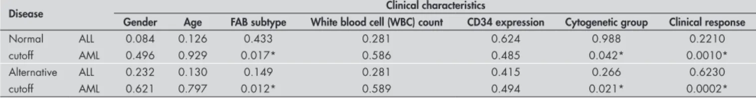
Table 1. Correlation between multidrug resistance-associated protein 1 (MRP1) expression and clinical characteristics of acute myelogenous leukemia (AML) and acute lymphoblastic leukemia (ALL) patients according to different cutoffs. The p-values for AML patients suggest that the French-American-British (FAB) subtype and cytogenetic risk group are correlated with MRP1 expression level (0.017 and 0.042, respectively). In contrast, the p-values for ALL patients show no correlation between MRP1 expression and clinical findings. The alternative cutoff makes the p-values different but no significant changes are observed in the final results
Disease Clinical characteristics
Gender Age FAB subtype White blood cell (WBC) count CD34 expression Cytogenetic group Clinical response
Normal ALL 0.084 0.126 0.433 0.281 0.624 0.988 0.2210
cutoff AML 0.496 0.929 0.017* 0.586 0.485 0.042* 0.0010*
Alternative ALL 0.232 0.130 0.149 0.281 0.415 0.266 0.6230
cutoff AML 0.621 0.797 0.012* 0.589 0.494 0.021* 0.0002*
*The mean difference is significant at the 0.05 level.
Figure 2. Expression of multidrug resistance-associated protein 1 (MRP1) messenger RNA (mRNA) in peripheral blood of healthy individuals and the HL-60 cell line. The copy number of MRP1 is expressed as the ratio of MRP1 to β2M copy number (each circle represents a sample). In accordance with MRP1 expression in healthy individu-als, the cutoff is defined as mean ± 2 standard deviations (SD). The HL-60 resistant cell line showed MRP1 expression levels higher than the cutoff.
0.2000
MRP1
copy
0.2350
Cell line Normal
Mean: 0.00219 Cutoff 0.1000
0.0000
little modification. The overall result did not change, although greater significance was ob-served in correlations between mRNA levels and cytogenetic risk group or FAB subtype (Table 1).
DISCUSSION
Although the antineoplastic drugs cur-rently available are effective for treating vari-ous cancers, they may prove to be ineffective in treating some primary or relapsed types of neoplasia. Multidrug resistance is seen to be the most significant barrier to effective
treatment of malignant tumors in general. Identification of variables that might be used to predict the response of tumors to treatment is constantly under discussion in oncology. Several genes are thought to participate in the MDR phenotype. Because many kinds of antileukemic agents can be substrates for the efflux pumps, upregulation of these pumpsleads to insufficient concentrations of agents in cancer cells, even when used at maximum dosages.
be correlated to poor outcomes is medi-ated by the MDR1 gene.3,6 However, clinical
investigations into the role of MDR1 have yielded inconsistent results that are diffi cult to interpret.14 In several papers, the expression
of Pgp did not correlate with the multidrug resistance phenotype.9,21 Thus, alternative
proteins such as MRP1 may also contrib-ute towards resistance in accontrib-ute leukemia cases. Although in several studies MRP1
has been shown to be involved in multidrug resistance,22,23 in other investigations MRP1
expression appeared to have no impact on treatment outcome in AML cases.24-26MRP1
has been detected in a wide variety of solid and hematological tumors, but evaluation of the presence of MRP1 protein or its cognate
Table 2. Multidrug resistance-associated protein 1 (MRP1) expression level in acute myelogenous leukemia (AML) and acute lymphoblastic leukemia (ALL) samples. Ex-pression of MRP1 in the nonresponse (NR) group (phase i) compared with normal, complete remission (CR) and early relapsed groups (phase j). In ALL samples, no signifi cant difference was observed. In contrast, in AML samples from relapsed and NR groups, the MRP1 level is signifi cantly different from the healthy and CR groups (p-value = 0.001), but the mean difference between the NR and Relapsed patients is nonsignifi cant (p-value = 0.171)
Phase (j) Phase (i) Mean difference (i-j) Standard error p-value
ALL Normal 0.0317 0.0885 0.988
CR 0.1160 0.0938 0.680
NR Relapsed 0.0022 0.0966 1.000
AML Normal 0.6003* 0.0817 0.001
CR 0.5874* 0.0728 0.001
NR Relapsed 0.1969 0.0858 0.171
*Signifi cant at the 0.01 level.
Figure 3. The expression of multidrug resistance-associated protein 1 (MRP1) messenger RNA (mRNA) at onset and relapse in cases of acute myelogenous leukemia (AML) (A) and acute lymphoblastic leukemia (ALL) (B) is shown (each circle represents a sample). In AML, the distribution of copy numbers in CR and healthy individuals is different from the distribution in relapsed patients. Patients in the NR group (except one) show higher messenger RNA (mRNA) levels than do relapsed patients. In contrast, mRNA levels in ALL patients have similar distributions in the different groups.
1.00000
0.80000
0.60000
0.40000
0.20000
0.00000
Normal CR Relapse NR
MRP1
copy
Cutoff Cutoff
1.00000
0.80000
0.60000
0.40000
0.20000
0.00000
Normal CR Relapse NR
MRP1
copy
A B
mRNA in a tumor sample is complicated by the fact that the MRP1 gene is expressed in the tissue from which these tumors originate and the protein is also expressed in all lineages of normal hematopoietic cells. Thus, the clinical impact of MRP1 expression remains contro-versial. In addition, one of the main reasons for the contradictory results from studies on drug resistance relates to methodology, in addition to differences in the defi nition of overexpression.15
Lack of adequate quantitative methods for assessing the amount of transcription is the most important problem making the data hard to interpret. 3-(4,5-dimethylthia-zol-2-Yl)-2,5-diphenyltetrazolium bromide (MTT) assaying is a widely used and validated
method for analyzing in vitro drug resis-tance, but it may not be useful for detecting resistance induced by ABC transporters. This is partly because the leukemia cells are heterogeneous.27 The fl ow cytometry method
can overcome the heterogeneity of leukemic cells by targeting leukemic cells with specifi c antibodies.9,17 However, even with
second-ary antibodies, fl ow cytometry sensitivity is very low. Northern blot assaying is simple to perform, but the sensitivity of this technique is the lowest of all the methods described for mRNA quantification.16 Competitive
reverse-transcriptase PCR assaying, which is widely used for mRNA quantifi cation, is correlated with a high risk of contamination because it needs post-PCR treatments.17 In
addition, this method is semiquantitative and only yields data discontinuously, such that the cutoff cannot be determined. All of the methods described are diffi cult to standardize and hard to perform. In contrast, real-time PCR is simple to use, and requires only minimal experience and skills. It provides maximum sensitivity (e.g. 10,000 times more sensitive than northern blot) and is the only method that allows quantifi cation.
In many studies, real-time PCR has been widely used to quantify ABC transporter mRNA levels in different cancer cells. In these studies, MRP1 expression did not correlate strongly with clinical resistance in leukemia patients. Fujimaki et al. used real-time PCR to determine MDR1 and MRP1 transcript level impact in AML patients. They found that increased MDR1 butnot MRP1 expression at diagnosis correlated with the multidrug resis-tance phenotype.23 Considering the molecular
aspect of gene expression regulation, the
MDR1 expression in leukemic cells exposed to antineoplastic agents is regulated by two distinct processes: stabilization of messenger RNA and initiation of the translation process.
In vitro study of these phenomena has revealed that MDR1 overexpression does not always occur via the activation of transcription.28
In contrast, MRP1 protein levels increase ac-cording to MRP1 mRNA levels in vivo.29 The
short-lived MDR1 mRNA of naive cells (not exposed to drugs) is stabilized (half-life greater than 10 hours) following short-term drug exposure. However, this stabilized mRNA has not been associated with polyribosome translation and does not direct Pgp synthesis.28
Furthermore, identification of a valid refer-ence gene for data normalization remains the most stubborn of the problems in quantitative PCR. GAPDH and β-Actin genes continue to be utilized as normalizers despite continuing reports that emphasize the problems associated with their use.30 It is now well documented that GAPDH and β-Actin mRNA levels are not constant, even in cellular subpopulations of the same pathological origin.31
To overcome this problem, we have de-signed and validated a real-time PCR assay to monitor MRP1 transcript levels in acute leukemia at different stages of the disease. We used β2M because it has no pseudogene and the use of β2M as a housekeeping gene has a critical impact on the interpretation of PCR data.32 In our assay, the dynamic
quantifica-tion range (from 6 x 10⁵ copies to 6 x 10¹⁰
copies) was satisfactory for clinical use. When this system was applied to clinical samples, we found that there was a statistically significant increase (p < 0.01) in the average MRP1
expression level at the time of nonresponse or early relapse, compared with patients in remission. The MRP1 expression level could be correlated with clinical response. This result is in agreement with observations by other authors who also found statistically significant increases in MRP1 expression in relapsed leukemia patients.5,16,22,33 In addition,
the mean expression level in relapsed samples is somewhat lower than in NR cases. This may be due to the nature of cancer cells and chemotherapy-induced events. Treatment induces rapid apoptosis of the sensitive cell fraction, leaving a small but substantial num-ber of resistant cells.
As mentioned earlier, the relationship between MRP1 expression and response to treatment in patients affected by leukemia is still controversial. We addressed the question of whether or not MRP1 expression would be elevated in Relapsed and NR patients, thus offering a possible explanation for their relapse phenotype. Healthy individuals served as controls, such that their MRP1 levels would be the base levels to which the MRP1 levels of the patients would be compared. As we have
reported, the MRP1 levels in Relapsed and NR patients were significantly higher than those in the controls, thus suggesting that the lack of optimal response to drug treatment may be due to this elevated expression.
The patients in the CR group were also tested because the results from that group might have been revealing and interesting, if the MRP1 levels in this group were also found to be high. This would have made the hypothesis that MRP1 levels had a role in drug response at least less likely.
However, in all the AML patients in CR, the MRP1 levels were similar to those in healthy controls. Therefore, it is possible that the observed CR phenotype is like this precisely because these patients’ MRP1 levels never increased in response to drug treatment, and that this contributed towards the effective-ness of the treatment.
Although this correlation was obvious in AML patients, no meaningful correlation was observed between MRP1 mRNA levels and response to chemotherapy in ALL samples. Af-ter we redefined the cutoff value according to the mRNA level in newly diagnosed patients, some of the samples that had previously been assumed to present overexpression rolled back to the normal group, but no change in the overall results was observed either in AML or in ALL patients. When we used the first cutoff value, MRP1 overexpression was observed in the relapsed ALL patients but, according to the alternative cutoff, no cases of ALL in the relapsed or NR group showed overexpression. It seems that the basic amounts of MRP1
mRNA in various types of leukemia are dif-ferent and that attention must be paid to this phenomenon when defining the cutoff value. Hence, defining the cutoff according to the mRNA level in newly diagnosed cases in each disease could solve the paradox of meaning-less overexpression of ABC transporter genes observed in cancer cells.23
According to our data, there was no clear relationship between MRP1 expression and the patients’ gender, age, WBC count and CD34 expression. However, clinical parameters such as FAB subtype and
cytoge-netic risk group were found to be significantly related to the CR rate among AML patients. Genetic alterations could be detected in approximately 80% of de novo AML cases. These alterations are not only recognized as important initiating events in the process of leukemogenesis but also as indicators of clini-cal outcome. Deletion of the MRP1 gene in AML patients with the 46, inv(16) karyotype was associated with a favorable effect on dis-ease outcome.33 Similarly, in this study, we
found four cases of AML with no MRP1 ex-pression, in which the cytogenetic group was favorable, including three cases with inv16 and one case with del 16. Our data suggest that changes at DNA level could contribute towards remission in AML and emphasize the role of MRP1 in chemotherapy resis-tance. FAB subtype represents an additional relevant prognostic factor in relapsed AML cases. In our study, heterogeneity of MRP1
expression among patients with different FAB subtypes was observed. Among the subtypes in AML cases, the mean MRP1 expression in the M5 subtype (0.6415 ± 0.071) was significantly higher than in other subtypes (p-value = 0.001). The M5 subtype seemed to correlate negatively with Pgp function, without a better prognosis,34 so it is possible
that MRP1 overexpression is responsible for the MDR phenotype in the M5 subtype of AML patients.
CONCLUSIONS
In summary, the mRNA level of MRP1
1. Sonneveld P. Multidrug resistance in haematological malignan-cies. J Intern Med. 2000;247(5):521-34.
2. Büchner T. Treatment of adult acute leukemia. Curr Opin Oncol. 1997;9(1):18-25.
3. Marie JP, Zhou DC, Gurbuxani S, Legrand O, Zittoun R. MDR1/P-glycoprotein in haematological neoplasms. Eur J Cancer. 1996;32A(6):1034-8.
4. Cole SP, Bhardwaj G, Gerlach JH, et al. Overexpression of a transporter gene in a multidrug-resistant human lung cancer cell line. Science. 1992;258(5088):1650-4.
5. Grant CE, Valdimarsson G, Hipfner DR, Almquist KC, Cole SP, Deeley RG. Overexpression of multidrug resistance-associated protein (MRP) increases resistance to natural product drugs. Cancer Res. 1994;54(2):357-61.
6. Legrand O, Perrot JY, Simonin G, Baudard M, Marie JP. JC-1: a very sensitive fluorescent probe to test Pgp activity in adult acute myeloid leukemia. Blood. 2001;97(2):502-8. 7. Solary E, Drenou B, Campos L, et al. Quinine as a
multi-drug resistance inhibitor: a phase 3 multicentric randomized study in adult de novo acute myelogenous leukemia. Blood. 2003;102(4):1202-10.
8. Broxterman HJ, Sonneveld P, Pieters R, et al. Do P-glycoprotein and major vault protein (MVP/LRP) expression correlate with in vitro daunorubicin resistance in acute myeloid leukemia? Leukemia. 1999;13(2):258-65.
9. Wuchter C, Leonid K, Ruppert V, et al. Clinical significance of P-glycoprotein expression and function for response to induction chemotherapy, relapse rate and overall survival in acute leukemia. Haematologica. 2000;85(7):711-21.
10. Marbeuf-GueyeC, Broxterman HJ, Dubru F, Priebe W,
Garnier-Suillerot A. Kinetics of anthracycline efflux from multidrug resistance protein-expressing cancer cells compared with P-glycoprotein-expressing cancer cells. Mol Pharmacol. 1998;53(1):141-7.
11. Legrand O, Zittoun R, Marie JP. Role of MRP1 in mul-tidrug resistance in acute myeloid leukemia. Leukemia. 1999;13(4):578-84.
12. Cole SP, Deeley RG. Multidrug resistance mediated by the ATP-binding cassette transporter protein MRP. Bioessays. 1998;20(11):931-40.
13. Lockhart AC, Tirona RG, Kim RB. Pharmacogenetics of ATP-binding cassette transporters in cancer and chemotherapy. Mol Cancer Ther. 2003;2(7):685-98.
14. Beck WT, Grogan TM. Methods to detect P-glycoprotein and implications for other drug resistance-associated proteins. Leukemia. 1997;11(7):1107-9.
15. Beck WT, Grogan TM, Willman CL, et al. Methods to detect P-glycoprotein-associated multidrug resistance in patients’ tumors: consensus recommendations. Cancer Res. 1996;56(13):3010-20.
16. Ohno N, Tani A, Chen ZS, et al. Prognostic significance of multidrug resistance protein in adult T-cell leukemia. Clin Cancer Res. 2001;7(10):3120-6.
17. Plasschaert SL, Vellenga E, de Bont ES, et al. High functional P-glycoprotein activity is more often present in T-cell acute lymphoblastic leukaemic cells in adults than in children. Leuk Lymphoma. 2003;44(1):85-95.
18. Michieli M, Damiani D, Ermacora A, et al. P-glycoprotein, lung resistance-related protein and multidrug resistance associated protein in de novo acute non-lymphocytic leukaemias: biological and clinical implications. Br J Haematol. 1999;104(2):328-35. 19. Mitelman F. ISCN 1995. An international system for human
cytogenetic nomenclature. Basel: Karger; 1995.
20. Mitelman F. ISCN 1991. Guidelines for cancer cytogenetics: supplement to an international system for human cytogenetic nomenclature. Basel: Karger; 1991.
21. Bailly JD, Muller C, Jaffrézou JP, et al. Lack of correlation between expression and function of P-glycoprotein in acute myeloid leukemia cells lines. Leukemia. 1995;9(5):799-807. 22. Zhou DC, Zittoun R, Marie JP. Expression of multidrug
resistance-associated protein (MRP) and multidrug resis-tance (MDR1) genes in acute myeloid leukemia. Leukemia. 1995;9(10):1661-6.
23. Fujimaki S, Funato T, Harigae H, et al. Quantitative analysis of a MDR1 transcript for prediction of drug resistance in acute leukemia. Clin Chem. 2002;48(6 Pt 1):811-7.
24. Filipits M, Suchomel RW, Zöchbauer S, Brunner R, Lechner K, Pirker R. Multidrug resistance-associated protein in acute myeloid leukemia: No impact on treatment outcome. Clin Cancer Res. 1997;3(8):1419-25.
25. den Boer ML, Pieters R, Kazemier KM, et al. Relationship between major vault protein/lung resistance protein, multidrug resistance-associated protein, P-glycoprotein expression, and drug resistance in childhood leukemia. Blood. 1998:91(6):2092-8. 26. van der Kolk DM, de Vries EG, Noordhoek L, et al. Activity and
expression of the multidrug resistance proteins P-glycoprotein, MRP1, MRP2, MRP3 and MRP5 in de novo and relapsed acute myeloid leukemia. Leukemia. 2001;15(10):1544-53. 27. Steinbach D, Friedrich J, Dawczynski K, et al. Are MTT
assays the right tool to analyze drug resistance caused by ABC-transporters in patient samples? Leuk Lymphoma. 2005;46(9):1357-63.
28. Yague E, Armesilla AL, Harrison G, et al. P-glycoprotein (MDR1) expression in leukemic cells is regulated at two distinct steps, mRNA stabilization and translational initiation. J Biol Chem. 2003;278(12):10344-52.
29. Manohar CF, Bray JA, Salwen HR, et al. MYCN-mediated regulation of the MRP1 promoter in human neuroblastoma. Oncogene. 2004;23(3):753-62.
30. Zhu G, Chang Y, Zuo J, et al. Fudenine, a C-terminal truncated rat homologue of mouse prominin, is blood glucose-regulated
and can up-regulate the expression of GAPDH. Biochem
Bio-phys Res Commun. 2001;281(4):951-6.
31. Goidin D, Mamessier A, Staquet MJ, Schmitt D, Berthier-Vergnes O. Ribosomal 18S RNA prevails over glyceraldehyde-3-phosphate dehydrogenase and beta-actin genes as internal standard for quantitative comparison of mRNA levels in invasive and noninvasive human melanoma cell subpopulations. Anal Biochem. 2001;295(1):17-21.
32. Oselin K, Mrozikiewicz PM, Pähkla R, Roots I. Quantitative determination of the human MRP1 and MRP2 mRNA expres-sion in FACS-sorted peripheral blood CD4+, CD8+, CD19+, and CD56+ cells. Eur J Haematol. 2003;71(2):119-23. 33. Kuss BJ, Deeley RG, Cole SP, et al. Deletion of gene for
multidrug resistance in acute myeloid leukemia with inver-sion in chromosome 16: prognostic implications. Lancet. 1994;343(8912):1531-4.
34. Leith CP, Kopecky KJ, Chen IM, et al. Frequency and clini-cal significance of the expression of the multidrug resistance proteins MDR1/P-glycoprotein, MRP1, and LRP in acute myeloid leukemia: a Southwest Oncology Group Study. Blood. 1999;94(3):1086-99.
Acknowledgements: The authors are grateful to Dr. Dizage for donating resistant HL-60 cell line.
Sources of funding: This work was supported by a grant provided by the National Institute of Genetic Engineering and Biotechnology (NIGEB) and Hematology, Oncology, Stem Cell Research Center, Medical Sciences, University of Tehran, Tehran, Iran. Grant number 220.
Conflict of interest: Not declared Date of first submission: June 25, 2007 Last received: May 10, 2008 Accepted: May 27, 2008
AUTHOR INFORMATION Frouzandeh Mahjoubi, PhD. Project leader and supervisor
of Masoud Golalipour, Department of Clinical Genetics, National Institute of Genetic Engineering and Biotechnology (NIGEB), Tehran, Iran.
Masoud Golalipour, PhD. PhD student who performed all the laboratory work, Department of Clinical Genetics, National Institute of Genetic Engineering and Biotechnology (NIGEB), Tehran, Iran.
Ardeshir Ghavamzadeh, MD. Hematology, Oncology and Stem Cell Research Center, Medical Sciences, University of Tehran, Tehran, Iran.
Kamran Alimoghaddam, MD. Hematology, Oncology and Stem Cell Research Center, Medical Sciences, University of Tehran, Tehran, Iran.
Address for correspondence:
Dr. Frouzandeh Mahjoubi
Department of Clinical Genetics, National Institute of Genetic Engineering and Biotechnology (NIGEB) Pajoohesh Blvd.,
Karaj Highway, 15th Km, Tehran, Iran Tel. 9821-44580389 — Fax. 9821-44580399 E-mail: [email protected] or [email protected]
Copyright © 2008, Associação Paulista de Medicina
RESUMO
Expressão do gene MRP1 em leucemias agudas
CONTEXTO E OBJETIVO: A superexpressão do gene de resistência a múltiplas drogas associado à proteína 1 (MRP1) tem sido ligada à resistência à quimioterapia in vitro, porém pouco é conhecido sobre seu impacto clínico nos pacientes com leucemia aguda. Nosso objetivo foi investigar a possível associação entre a expressão do gene MRP1 e os desfechos clínicos em pacientes iranianos com leucemia.
DESENHO E LOCAL: Este foi um estudo analítico transversal em pacientes encaminhados ao Centro de Pesquisa em Hematologia, Oncologia e Células Tronco do Hospital Público de Sharyatee, com diagnóstico de leucemia mielóide aguda (LMA) ou leucemia linfoblástica aguda (LLA). Todo trabalho molecular foi realizado no NIGEB (instituição pública).
MÉTODOS: Para correlação de marcadores prognósticos e desfechos clínicos da leucemia aguda, a expres-são do MRP1 foi avaliada em 35 casos de LMA e 17 de LLA, usando a reação da cadeia de polimerase quantitativa em tempo real, e comparando este dado ao tipo de resposta à quimioterapia.
RESULTADOS: A média da expressão em pacientes com LMA em remissão completa (0,032 ± 0,031) foi significativamente menor que aquela dos casos recidivantes (0,422 ± 0,297). Por outro lado, não foram observadas diferenças significativas nos níveis de mRNA para MRP1 entre os casos de LLA com remissão completa e os casos recidivantes. Houve uma diferença na expressão de MRP1 entre pacientes com prognós-tico citogenéprognós-tico não-favorável e favorável (0,670 ± 0,074 e 0,028 ± 0,013, respectivamente). A expressão de MRP1 em M5 foi significativamente maior (valor de p = 0,001) do que em outros subtipos. CONCLUSÕES: Os achados sugerem que a alta expressão de MRP1 se associou com o pior desfecho clínico, estando correlacionada com o subtipo M5 e os subgrupos citogenéticos menos favoráveis para os pacientes com LMA, mas não para pacientes com LLA.