Luís Filipe Madureira Fonseca
Licenciado em Bioquímica
Bioremediation and CO
2scavenging using
molybdenum-containing enzymes
Dissertação para obtenção do Grau de Mestre em
Bioquímica
Orientadores: José João Galhardas de Moura, Professor Catedrático,
Faculdade de Ciências e Tecnologia da Universidade Nova de
Lisboa
Isabel Maria Andrade Martins Galhardas de Moura, Professora
Catedrática, Faculdade de Ciências e Tecnologia da
Universidade Nova de Lisboa
Co-orientadora: Luísa Bernardina Lopes Maia, Investigadora Pós-Doc,
Faculdade de Ciências e Tecnologia da Universidade Nova de
Lisboa
Júri:
Presidente: José Ricardo Ramos Franco Tavares
Arguente: Stéphane Pierre Bensson
Luís Filipe Madureira Fonseca
Licenciado em Bioquímica
Bioremediation and CO
2scavenging using
molybdenum-containing enzymes
Dissertação para obtenção do Grau de Mestre em
Bioquímica
Orientadores: José João Galhardas de Moura, Professor Catedrático,
Faculdade de Ciências e Tecnologia da Universidade Nova de
Lisboa
Isabel Maria Andrade Martins Galhardas de Moura, Professora
Catedrática, Faculdade de Ciências e Tecnologia da
Universidade Nova de Lisboa
Co-orientadora: Luísa Bernardina Lopes Maia, Investigadora Pós-Doc,
Faculdade de Ciências e Tecnologia da Universidade Nova de
Lisboa
Júri:
Presidente: José Ricardo Ramos Franco Tavares Arguentes: Stéphane Pierre Bensson
Bioremediation and CO2 scavenging using molybdenum-containing enzymes.
Copyright © Luís Filipe Madureira Fonseca, Faculdade de Ciências e Tecnologia, Universidade Nova de Lisboa.
A
GRADECIMENTOS
O trabalho desenvolvido no âmbito desta dissertação, apenas foi possível devido à contribuição de diversas pessoas a quem gostaria de expressar os meus agradecimentos.
Em primeiro lugar, gostaria de agradecer aos meus orientadores Prof. José J. G. Moura e Prof.ª Isabel Moura, por me terem aceite nos seus grupos de investigação e por me proporcionarem todas as condições necessárias para a realização deste trabalho.
Um agradecimento à Doutora Luísa Maia, por ter orientado de perto esta dissertação, pela sua paciência e por me ter transmitido conhecimentos que em muito contribuíram para ampliar a minha formação científica.
Aos meus colegas, Francisco, Joana e Lara um agradecimento pela amizade, companheirismo e entreajuda que sempre existiu ao longo da preparação desta dissertação e, pelas animadas conversas que sempre ajudaram a desanuviar nos dias em que “a ciência” corria menos bem.
Uma palavra de agradecimento também à Célia Silveira, pelas frutuosas conversas e discussões científicas e por toda a ajuda, quer com os crescimentos bacterianos, quer na preparação dos ensaios cinéticos.
Gostaria também de demonstrar a minha gratidão a todos os membros dos grupos
BioIn e Bioprot pelo excelente acolhimento. Em particular, à Cíntia Carreira, Cláudia Nóbrega e Rute Nunes pela ajuda, disponibilidade e conselhos.
Não posso deixar de agradecer à técnica dos laboratórios 407 e 617, Ana Teresa Lopes, pela constante disponibilidade e ajuda; nem olvidar um agradecimento especial às técnicas do 4º piso: Idalina Martins e Maria da Conceição Luís por tornarem o meu trabalho no laboratório mais fácil.
agradeço o facto de, independentemente da distância, terem estado sempre presentes, inclusivamente quando eu não podia estar!…
Rute, obrigado por teres estado presente nos momentos menos bons, por teres tornado menos trabalhosa a escrita desta tese e por me teres mostrado que com paciência e dedicação tudo é possível.
A
BSTRACT
Carbon dioxide valorization, will not only help to relieve the greenhouse effect but might also allow us to transform it in value-added chemicals that will help overcoming the energy crisis. To accomplish this goal, more research that focus on sequestering CO2 and endeavors through a carbon-neutral or carbon-negative strategy is
needed in order to handle with the dwindling fossil fuel supplies and their environmental impact. Formate dehydrogenases are a promising means of turning CO2
into a biofuel that will allow for a reduction of greenhouse gas emissions and for a significant change to the economic paramount. The main objective of this work was to assess whether a NAD+-independent molybdenum-containing formate dehydrogenase is able to catalyze the reduction of CO2 to formate. To achieve this, a
molybdenum-containing formate dehydrogenase was isolated from the sulfate reducing bacteria Desulfovibrio desulfuricans ATCC 27774. Growth conditions were found that allowed for a greater cellular mass recovery and formate dehydrogenase expression. After growth trials, kinetic assays for formate oxidation and CO2 reduction were
performed and kinetic parameters determined. For the formate oxidation reaction, a KM
of 49 μM and a turnover constant of 146 s-1 were determined. These kinetic parameters
are in agreement with those determined by Mota, et al. (2011). Finally, we found that this molybdenum-containing enzyme was able to catalyze the reduction of CO2 to
formate with a turnover constant of 4.6 s-1 and a K
M of 13 μM. For the first time a
NAD+-independent molybdenum-containing formate dehydrogenase was found to
catalyze CO2 reduction, allowing its use as a biocatalyst in energetically efficient CO2
fixation processes that can be directed towards bioremediation or as an alternative and renewable energy source. Characterizing these enzymes may lead to the development of more efficient synthetic catalysts, make them readily available and more suited for practical applications.
Keywords: formate dehydrogenase; molybdoenzymes; CO2 reduction;
R
ESUMO
A valorização do CO2, através da sua conversão em produtos de valor
acrescentado, não só é importante para diminuir o efeito de estufa mas também para obter biocombustíveis que nos podem auxiliar a ultrapassar a crise energética. Para atingir este objectivo, é necessário focar a investigação no sequestro de CO2 e na sua
valorização, de modo a lidar com a crescente escassez de combustíveis fósseis e com o impacto ambiental que a sua utilização acarreta. As formato desidrogenases são um meio promissor para transformar CO2 num biocombustível que permitirá, além da
redução das emissões de gases com efeito de estufa, operar uma alteração significativa no contexto económico. O principal objectivo deste trabalho foi determinar se uma formato desidrogenase contendo molibdénio e independente de NAD+, é capaz de
catalisar a redução de CO2 a formato. Para tal, foi isolada uma formato desidrogenase da
bactéria redutora de sulfato Desulfovibrio desulfuricans ATCC 27774. Foram encontradas as condições de crescimento bacteriano que permitiam, simultaneamente, uma maior recuperação de massa celular e de expressão da enzima. Seguidamente, caracterizou-se cineticamente a reacção de oxidação de formato a CO2 tendo-se
determinado um KM para o formato de 49 μM e uma constante catalítica de 146 s -1. Os
parâmetros cinéticos para esta reacção, estão de acordo com os que foram obtidos por Mota, et al. (2011). Finalmente, verificou-se que esta enzima é capaz de catalisar a redução de CO2 com uma constante catalítica de 4.6 s
-1 e um K
M de 13 μM. Permitindo,
pela primeira vez, demonstrar que uma formato desidrogenase independente de NAD+ e
contendo molibdénio, é capaz de catalisar a redução de dióxido de carbono a formato possibilitando a sua utilização como biocatalisador energicamente eficiente na fixação de CO2, a sua aplicação em processos de bioremediação ou como uma fonte de energia
alternativa e renovável. Caracterizar estas enzimas torna possível o desenvolvimento de catalisadores sintéticos mais eficientes, facilmente disponíveis e mais adequados a aplicações práticas.
T
ABLE OF
C
ONTENTS
AGRADECIMENTOS ... VII ABSTRACT ... IX RESUMO ... XI TABLE OF CONTENTS ... XIII FIGURES INDEX ... XV TABLE INDEX ... XIX ABBREVIATIONS ... XXI
I. INTRODUCTION ... 1
I.1. CARBON DIOXIDE ... 4
I.2. REDUCED CARBON DIOXIDE AS A NOVEL SOURCE OF ENERGY ... 7
I.3. ENZYMES: FROM BIOREMEDIATION AND CHEMICALS TO BIOFUELS ... 8
I.4. THE MOLYBDENUM AND TUNGSTEN CONTAINING ENZYMES ... 10
I.4.1. Formate dehydrogenase ... 16
I.4.1.1. Formate dehydrogenase – Structural Studies ... 17
I.4.1.2. Formate dehydrogenase – Mechanistic Studies ... 23
I.5. SUBJECT AND OBJECTIVE OF THIS WORK ... 29
II. MATERIALS AND METHODS ... 31
II.1. BACTERIAL STRAIN, CULTURE MEDIA AND GROWTH CONDITIONS ... 33
II.2. SOLUBLE EXTRACT PREPARATION ... 37
II.3. IN GEL ACTIVITY ASSAYS ... 38
II.4. FDH PURIFICATION ... 39
II.5. PROTEIN CONTENT QUANTIFICATION ... 39
II.6. PRELIMINARY FDH REDUCTION STUDIES ... 39
II.7. STEADY-STATE KINETIC ASSAYS ... 40
II.7.1. CO2 solutions preparation ... 41
II.7.2. Analysis of initial rate data ... 41
III. RESULTS AND DISCUSSION ... 43
III.1.2. Gaseous phase ... 45
III.1.2.1. Gaseous phase influence in the pH of the media ... 48
III.1.3. Inoculum volume and growth stage harvest ... 50
III.2. FDH PURIFICATION ... 57
III.3. PRELIMINARY KINETIC STUDIES ... 61
III.3.1. Triggering the enzymatic reaction ... 61
III.3.2. The role of the sulfhydryl reducing agent ... 66
III.3.3. Atmospheric O2 interference in the kinetic assays ... 67
III.4. STEADY-STATE KINETIC STUDIES ... 69
III.4.1. Formate oxidation studies ... 69
III.4.2. Carbon dioxide reduction studies ... 71
IV. CONCLUSIONS, FINAL REMARKS AND FUTURE WORK ... 79
V. BIBLIOGRAPHY ... 85
VI. APPENDIXES ... 95
VI.1. GROWTH MEDIA ... 97
VI.2. GEL ELECTROPHORESIS ... 103
VI.3. PERIPLASMATIC SOLUBLE EXTRACT PREPARATION FLOWCHART ... 106
VI.4. CELL SOLUBLE EXTRACT PREPARATION FLOWCHART ... 107
VI.5. PURIFICATION FLOWCHART ... 108
VI.6. CARBONATE SPECIES AND PH DEPENDENCY ... 109
F
IGURES
I
NDEX
FIGURE I.1–GLOBAL GHG EMISSIONS FOR 2010. ... 3
FIGURE I.2–ATMOSPHERIC CARBON DIOXIDE CONCENTRATIONS, IN PPM, SINCE RECORD BEGAN AT MAUNA LOA OBSERVATORY IN 1958. ... 4
FIGURE I.3–GLOBAL GHG EMISSIONS FOR 2013 BY SOURCE AND TYPE. ... 5
FIGURE I.4–POSSIBLE CHEMICAL TRANSFORMATIONS OF CO2. ... 6
FIGURE I.5–CARBON CAPTURE AND STORAGE FACILITIES IMPLEMENTED THROUGHOUT THE WORLD. ... 6
FIGURE I.6 – PYRANOPTERIN COFACTOR PRESENT IN MONONUCLEAR MO/W-CONTAINING ENZYMES [38,39]. TOP: STRUCTURE OF THE PYRANOPTERIN COFACTOR. BOTTOM: THE COFACTOR CAN BE FOUND IN THE SIMPLEST MONOPHOSPHATE FORM (R IS A HYDROGEN ATOM), OR ESTERIFICATED WITH DIFFERENT NUCLEOTIDES (R CAN BE ONE CYTOSINE MONOPHOSPHATE OR GUANOSINE MONOPHOSPHATE). ... 11
FIGURE I.7 – ACTIVE SITE STRUCTURES AMONGST THE DIFFERENT FAMILIES OF MO AND W PYRANOPTERIN-DEPENDENT ENZYMES. ... 14
FIGURE I.8–DIFFERENT MOLYBDENUM COORDINATION IN THE THREE SUBFAMILIES WITHIN THE DMSOR FAMILY OF MO/W-ENZYMES. ... 15
FIGURE I.9–FDH-H FROM E. COLI.LEFT:THREE-DIMENSIONAL VIEW OF FDH-H.RIGHT:ARRANGEMENT OF THE REDOX CENTERS SHOWN IN THE SAME ORIENTATION. ... 18
FIGURE I.10-THE MO ACTIVE SITE OF E. COLI FDH-H, AND CONSERVED RESIDUES SECYS140,HIS141 AND ARG333. ... 18
FIGURE I.11 – FDH-N STRUCTURE FROM E. COLI. LEFT: THREE-DIMENSIONAL VIEW OF FDH-N.RIGHT: ARRANGEMENT OF THE REDOX CENTERS THAT COMPOSE THE ELECTRON TRANSFER PATHWAY. ... 19
FIGURE I.12–THE MOLYBDENUM-CONTAINING ACTIVE SITE OF FDH-H, ISOLATED FROM E. COLI, AND THE CONSERVED RESIDUES SECYS196,HIS197 AND ARG446. ... 20
FIGURE I.13– FDH STRUCTURE FROM D. GIGAS. LEFT: THREE-DIMENSIONAL VIEW OF W-FDH FROM D. GIGAS.RIGHT:ARRANGEMENT OF THE REDOX CENTERS THAT COMPOSE THE ELECTRON TRANSFER PATHWAY IN THE SAME ORIENTATION. ... 21
FIGURE I.14–THE W ACTIVE SITE OF D. GIGASFDH AND THE CONSERVED RESIDUES SECYS157,HIS158 AND ARG407. ... 21
FIGURE I.15–ALIGNMENT OF THE Α SUBUNITS OF FDH-H(PURPLE) AND FDH-N(BEIGE) FROM E. COLI WITH THE Α SUBUNIT OF FDH FROM D. GIGAS (BLUE) EXHIBITING AN RMSD OF 1.8 Å. LEFT: OVERALL SUPERIMPOSITION OF THE THREE Α SUBUNITS.RIGHT:DETAIL OF THE ACTIVE CENTERS AND OF THE CONSERVED RESIDUES. ... 22
FIGURE I.17-REACTION MECHANISM FOR FORMATE OXIDATION BY FDH, PROPOSED BY RAAIJMAKERS ET
AL. [60]. ... 25
FIGURE I.18 – REACTION MECHANISM PROPOSED FOR FDH ACTIVATION AND FORMATE OXIDATION AS
PROPOSED BY MOTA, ET AL. (2011). TOP: ACTIVATION OF THE METALLIC CENTER OF FDH VIA
SULFUR-SHIFT.BOTTOM:CATALYTIC CYCLE FOR FORMATE OXIDATION BY FDH. ... 26
FIGURE II.1–SCHEMATIC REPRESENTATION OF THE ACCLIMATIZATION PROCEDURE OF D. DESULFURICANS
CELLS TO THE DIFFERENT MEDIA TESTED. ... 34
FIGURE II.2–SCHEMATIC REPRESENTATION OF THE SCREENING DONE TO DETERMINE WHICH OF THE TEST
MEDIA WOULD ALLOW FOR A HIGHER CELL MASS TO BE HARVESTED. ... 34
FIGURE II.3–D. DESULFURICANS CELLS, MAGNIFIED 1000X. ... 37
FIGURE III.1–IN GEL FDH ACTIVITY ASSAY FOR D. DESULFURICANS SOLUBLE EXTRACTS GROWN IN VMN
MEDIUM UNDER DIFFERENT GAS PHASES. ... 46
FIGURE III.2–IN GEL FDH ACTIVITY ASSAYS FOR D. DESULFURICANS CELLS DISRUPTED WITH A FRENCH
PRESS AFTER GROWING IN DIFFERENT MEDIA AND UNDER DIFFERENT GAS PHASES. ... 48
FIGURE III.3–D. DESULFURICANS GROWTH CURVE IN VMN MEDIUM WITH A 2% INOCULUM. ... 50
FIGURE III.4– EFFECT OF DIFFERENT INOCULUM VOLUMES ON D. DESULFURICANS GROWTH CURVES IN
VMN MEDIUM AND RESPECTIVE SIGMOIDAL FITS. ... 51
FIGURE III.5 – EFFECT OF INOCULUM VOLUME AND DIFFERENT GROWTH STAGE HARVESTING IN D.
DESULFURICANSFDH EXPRESSION. ... 53
FIGURE III.6 – EFFECT OF INOCULUM VOLUME, DIFFERENT GROWTH STAGE HARVESTING AND THE
PRESENCE OR ABSENCE OF PMSF AND DTT IN THE EXPRESSION OF D. DESULFURICANSFDH. ... 54
FIGURE III.7–CRUDE EXTRACT FRACTIONATION ON AN ANION EXCHANGE CHROMATOGRAPHY,DEAEBIO
GEL EQUILIBRATED WITH 10 MMTRIS-HCL. ... 57
FIGURE III.8–SDS-PAGE FROM THE FRACTIONS COLLECTED AFTER THE FIRST CHROMATOGRAPHIC STEP.
... 58
FIGURE III.9–SCHEMATIC REPRESENTATION OF THE FORMATE OXIDATION REACTION, CATALYZED BY FDH
IN THE PRESENT OF A MEDIATOR. ... 61
FIGURE III.10–D. DESULFURICANS FDH TIMECOURSES WITH OR WITHOUT THE ACTIVATION PROCEDURE. 62
FIGURE III.11–FDH SPECTRA, AS PURIFIED AND EVOLUTION AFTER DTT ADDITION. ... 64
FIGURE III.12– DTT-TREATED FDH SPECTRA AND AFTER THE ADDITIONS OF 83 AND 230 ΜM SODIUM
FORMATE. ... 65
FIGURE III.13–KINETICS OF FORMATE OXIDATION CATALYZED BY D. DESULFURICANSFDH (w). ... 69
FIGURE III.14 – SCHEMATIC REPRESENTATION OF THE CARBON DIOXIDE REDUCTION REACTION,
CATALYZED BY FDH IN THE PRESENT OF A MEDIATOR. ... 71
FIGURE III.15 – MEDIATOR REDUCTION TIMECOURSE IN THE PRESENCE OF FORMATE AND ITS
RE-OXIDATION AFTER SODIUM CARBONATE ADDITION. ... 72
FIGURE III.18–KINETICS OF CARBON DIOXIDE REDUCTION CATALYZED BY D. DESULFURICANSFDH(n). ... 75
FIGURE VI.1 – ELECTROPHORETIC PROFILE OF FERMENTAS UNSTAINED PROTEIN MARKER IN A 12%
FIGURE VI.2–PERIPLASMATIC SOLUBLE EXTRACT PREPARATION FLOWCHART. ... 106
FIGURE VI.3–CELL SOLUBLE EXTRACT PREPARATION FLOWCHART. ... 107
FIGURE VI.4–D. DESULFURICANS ATCC27774 PURIFICATION FLOWCHART. ... 108
FIGURE VI.5–CARBONATESPECIES PRESENT IN SOLUTION AND THEIR DEPENDENCY WITH THE PH VALUE.
T
ABLE
I
NDEX
TABLE II-1–CULTURE MEDIA COMPOSITIONS, PER LITER. ... 36
TABLE III-1–EVALUATION OF CELL GROWTH IN THE VARIOUS MEDIA USED. ... 47
TABLE III-2–MEDIA PH VALUE VARIATIONS FOLLOWING CELL HARVEST. ... 49
TABLE III-3 – GROWTH PARAMETERS AND MODEL FIT CONVERGENCE QUALITY DESCRIPTORS FOR D. DESULFURICANS IN VMN MEDIUM, USING DIFFERENT INOCULUM VOLUMES. ... 52
TABLE III-4 – INFLUENCE OF THE USE OF DIFFERENT INOCULUM VOLUMES AND CELL HARVEST AT DIFFERENT GROWTH STAGES IN A 100 ML GROWTH. ... 52
TABLE III-5-GROWTH CONDITION FOR SCALE-UP PROCESS. ... 55
TABLE III-6–INFLUENCE OF DTT CONCENTRATION ON FORMATE OXIDATION INITIAL RATES. ... 67
TABLE III-7 –COMPARISON BETWEEN KINETIC PARAMETERS, KCAT,KM AND CATALYTIC EFFICIENCY FOR FORMATE OXIDATION CATALYZED BY FORMATE DEHYDROGENASES ISOLATED FROM DIFFERENT ORGANISMS IN THE PRESENCE OF BENZYL VIOLOGEN AT PH8. ... 70
TABLE III-8 – KINETIC PARAMETERS FOR CARBON DIOXIDE REDUCTION CATALYZED BY FORMATE DEHYDROGENASES ISOLATED FROM DIVERSE ORGANISMS. ... 76
TABLE IV-1–KINETIC PARAMETERS FOR FORMATE OXIDATION AND CARBON DIOXIDE REDUCTION IN THE PRESENCE OF BENZYL VIOLOGEN BY D.DESULFURICANSFDH. ... 82
TABLE VI-1–ATCCMEDIUM:42DESULFOVIBRIO MEDIUM. ... 97
TABLE VI-2–ATCCMEDIUM:1249MODIFIED BAAR’S MEDIUM FOR SULFATE REDUCERS. ... 97
TABLE VI-3–ATCCMEDIUM:2755DESULFOVIBRIO MEDIUM. ... 98
TABLE VI-4–ATCCMEDIUM:27774DESULFOVIBRIO DESULFURICANS MEDIUM. ... 99
TABLE VI-5–DESULFOVIBRIO DESULFURICANS MEDIUM LSYC. ... 99
TABLE VI-6–VMN MEDIUM. ... 100
TABLE VI-7–VITAMIN SOLUTION FOR VMN MEDIUM, FINAL VOLUME 200 ML. ... 100
TABLE VI-8–WOLFES ELIXIR FOR VMN MEDIUM. ... 101
TABLE VI-9–M MEDIUM. ... 101
TABLE VI-10–M MEDIUM, SUPPLEMENTS. ... 101
TABLE VI-11–OLIGO-ELEMENTS "FAUQUE". ... 102
TABLE VI-12–PREPARATION OF A 7.5% POLYACRYLAMIDE GEL. ... 103
TABLE VI-13–COMPOSITION OF THE SOLUTIONS EMPLOYED. ... 103
TABLE VI-14–COMPOSITION OF THE SAMPLE BUFFER SOLUTION. ... 103
TABLE VI-15–TRIS-GLYCINE BUFFER COMPOSITION. ... 104
TABLE VI-16–COOMASSIE BLUE DYE SOLUTION. ... 104
TABLE VI-17–DISTAINING SOLUTION. ... 105
A
BBREVIATIONS
Asp Aspartate
ATCC American type culture collection
β-ME Beta-mercaptoethanol
BV Benzyl viologen
COdh Carbon monoxide dehydrogenase
Cys Cysteine
D.desulfuricans or Dd Desulfovibrio desulfuricans ATCC 27774
D.gigas or Dg Desulfovibrio gigas D.vulgaris or Dv Desulfovibrio vulgaris
DMSO Dimethyl sulfoxide
DMSOr Dimethyl sulfoxide reductase
DTT Dithiothreitol
ε UV-Visible molar extinction coefficient
E. coli Escherichia coli
EDTA Ethylenediaminetetraacetic acid
Fdh Formate dehydrogenase
GHG Greenhouse effect gas
HPLC High-performance liquid chromatography
KPB Potassium phosphate buffer
Moco Molybdenum cofactor
Mo-enzymes Molybdenum-containing enzymes
MV Methyl viologen
NAD+ Nicotinamide adenine dinucleotide oxidized form
NADH Nicotinamide adenine dinucleotide reduced form
O.D. Optical density
PAGE Polyacrylamide gel electrophoresis
PCD Pyranopterin cytosine dinucleotide
PGD Pyranopterin guanine dinucleotide
PMP Pyranopterin monophosphate
PMSF Phenylmethylsulfonyl fluoride
RMSD Root mean square deviation
SDS Sodium dodecyl sulfate
SeCys Selenocysteine
So Sulfite oxidase
Ser Serine
SRB Sulfate reducing bacteria
UV-Vis Ultraviolet-Visible
W-enzymes Tungsten-containing enzymes
I.
I
NTRODUCTION
Two major energy-related issues will daunt the world in the next fifty years. On one hand, nations will progressively have to dispute access to fossil fuels as reserves gradually become scarce, leading to an energy cost increase. On the other hand, atmospheric carbon dioxide (CO2) levels are at their highest level since recording of its
concentrations in the atmosphere began. The predictions show that large increases in its concentration will probably produce large and uncontrollable changes on world climate. Figure I.1 shows that CO2 accounts for more than half of global greenhouse effect gases
(GHG) emitted to the atmosphere, and that its main source are the fossil fuels we use in our daily lives. Therefore, it is important to develop new energy sources that are both secure and carbon neutral.
Figure I.1 – Global GHG emissions for 2010. Adapted from [1]. CO2 – Other, 15%
CO2 – Fossil fuel use, 61% CH4 – Energy, 6%
CH4 – Agriculture, 7%
CH4 – Other, 3%
N2O – Agriculture, 4%
N2O – Other, 2%
I.1.
Carbon dioxide
Carbon dioxide is a trace gas in the atmosphere of the Earth. And although it represents only 0.04% (400ppm) of the gases that constitute the atmosphere it is a potent GHG and plays a critical role in the regulation of the climate on Earth. Hence, keeping its concentration within a certain range is a crucial factor to prevent further changes to the current climate pattern.
Ever since the beginning of the Industrial Revolution around 150 years ago, the amount of CO2 in the atmosphere has increased noticeably, from 280 to about 400 ppm
this year, and keeps increasing at a rate of about 1.9 ppm/year (Figure I.2).
Figure I.2 – Atmospheric carbon dioxide concentrations, in ppm, since record began at Mauna Loa Observatory in 1958 [2].
This increase is linked to the burning of fossil fuels resulting from human activities and has the potential to induce climate change, making this phenomenon one of global concern [3–5]. According to the Third Assessment Report (2001) of the Intergovernmental Panel on climate change an increase in the GHG levels could lead to a temperature raise which, in turn, could have an impact on global climate patterns.
Nowadays, there is global awareness to the depletion of fossil fuel reserves and to the generally accepted fact that their consumption has caused increasing anthropogenic
300 320 340 360 380 400
1959 1965 1971 1977 1983 1989 1995 2001 2007 2013
GHG emissions. Thus, it is important that we rapidly adapt and procure new primary energy sources that allow us, in the long run, to completely replace fossil fuels. To achieve this goal, it is necessary to develop novel carbon abatement techniques and mature those we are experimenting on, while evolving policies to promote renewable energy sources that enable us to sequester atmospheric greenhouse gases such as CO2.
As depicted in Figure I.3, the majority of the GHG released into the atmosphere are resultant from energy production processes, and CO2 is the main GHG being released
into the atmosphere, making it an important candidate for these carbon capture techniques.
Figure I.3 – Global GHG emissions for 2013 by source and type. Adapted from [6].
Although it is impossible to capture all the CO2 produced daily, it is still crucial to
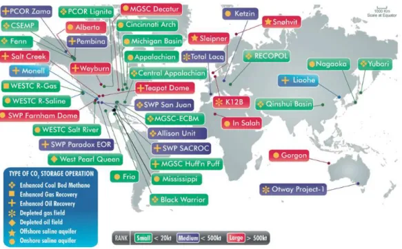
make an effort to mitigate the consequences its increase in the atmosphere may have to future generation. CO2 capture can be achieved in two ways: straight from the
atmosphere or directly from a source and it can either be converted into useful chemicals (Figure I.4) or sequestered (Figure I.5).
Industrial Processes, 6%
Agriculture, 8%
Waste, 3%
Energy, 83%
Other, 2%
N2O, 6%
CH4, 16%
Figure I.4 – Possible chemical transformations of CO2. Adapted from [7].
Figure I.5 – Carbon capture and storage facilities implemented throughout the World.
Adapted from [8].
The main challenge in converting CO2 into useful products, such as industrially
is still quite challenging since it requires a large energy input and suitable catalysts. The response to such a scientific challenge derives from several fields of knowledge, i.e., chemical catalysis, photochemistry, electrochemistry and semiconductor physics and engineering [5].
Nonetheless, the possibility of recovering CO2 directly from the atmosphere poses
a serious challenge due to its concentration, and in situ capture technologies are yet to overcome the difficulties presented by the great amount of energy required to purify, transport and storage it [9,10]. The more traditional solutions, as the ones cited, also pose other drawbacks, such as low selectivity and non-specificity that lead to the production of mixtures [11].
In any case, although the capture of CO2 for posterior activation and conversion to
a biofuel is not a straightforward process, it poses a great opportunity for solving the problems of fossil fuel shortage and global warming. There are numerous advantages of using CO2 as a fuel source, namely its unlimited availability, and the fact its production,
as opposed to the biofuels we are currently depending on, is not dependent of arable land.
In order to seriously consider CO2 as an alternative fuel source we need to find a
way to outstrip these classical approaches and arrive at an efficient and selective way to reduce CO2 that allows for a reduction on our dependency of fossil fuels while aiding to
balance the economic and environmental sustainability.
I.2.
Reduced carbon dioxide as a novel source of energy
In the last decade, a major effort has gone into the research of a novel carbon-neutral energy source, with the main goal being the capture and recycling of atmospheric CO2. The main research focus is its reduction into various energy rich
progresses have been made in this field, metal catalysis usually continues to require extreme conditions in terms of pressure and temperature, and suffers from by-product formation [13–15]. To try to overcome the disadvantages of this latter technique, researchers are now aiming at the development of high-efficient photo-catalysts. However, although selectivity was greatly improved, efficiency continues to fall short [16,17]. Another approach that has been gaining prominence is the application of electrochemistry in the conversion of CO2 into biofuels and other molecules of
commercial interest. Nonetheless, this is still a recent technology that needs to be improved before it can become viable. The main drawback, besides being energetically unfavorable, is the production of multiple end-products as a result of cross reactions [18]. According to a 2008 report of the North American Department of Defense, “The major obstacle preventing efficient conversion of carbon dioxide into energy-bearing products is the lack of catalysts...” [19]. This being the case, biocatalysts are now viewed as an attractive research focus, as they open the possibility to circumvent the drawbacks of the more classical approaches.
I.3.
Enzymes: from bioremediation and chemicals to biofuels
The use of enzymes in bioremediation processes is nothing new. However, their utilization in energy production, or in the valorization of by-products resulting from human activities, is yet to be established at an industrial scale mainly due to their requirements for intricate growth conditions, such as strict anaerobicity and complex purification procedures [20].
In Nature all biological systems need to be able to produce energy from their surroundings in order to sustain metabolic processes that are crucial to life. Nonetheless, as life is very adaptable these systems had to evolve and adjust to diverse conditions. CO2 fixation pathways have evolved for millions of years and in this process diverse
mechanisms and enzymes have been fine-tuned to perform this task. Carboxylases and dehydrogenases are among the enzymes that are able to catalyze the fixation of CO2.
Carboxylases allow the creation of new carbon-carbon bonds by introducing HCO3
- or CO
undergoes the biological carbon cycle is fixated by carboxylases [21]. The predominant mechanism, employed by plants and many prokaryotes to fix CO2 is the reductive
pentose phosphate (Calvin-Benson-Bassham) cycle. The cycle is initiated with the carboxylation of a five-carbon sugar, 1,5-ribulose bisphosphate, by the enzyme ribulose bisphosphate carboxylase/oxygenase (RuBisCO) to form two molecules of 3-phosphoglycerate that latter undergoes a succession of interconversions to form a six-carbon sugar, fructose-1,6-bisphosphate. Globally, this cycle catalyzes the following reaction [22,23].
(Eq. I.1)
3CO2 + 5H2O + 6NADPH + 9ATP →
!!!!!!!!!!!!!!!!!!!!!!!!!!!!!!!!!!!!!!!!!!!!!!!!!!→ 3-phosphoglyceraldehyde + 6NADP + 9 ADP + 8Pi
Nonetheless, other enzymatic CO2 reduction reactions exist. In methanogenensis,
an eight-electron reduction of CO2 to methane is performed [24], and in the
folate-dependent one-carbon metabolism, CO2 is converted to methyltetrahydrofolate,
which is a key component of the reductive acetyl-CoA pathway [25].
Dehydrogenases are amid the best catalysts found in Nature [26–30]. Among these, CO dehydrogenases (COdhs) and formate dehydrogenases (Fdhs) are of special interest for CO2 fixation. COdhs can be divided into two groups. The first groups
O2-sensitive enzymes, found in obligatory anaerobes, with [Fe4S4Ni] active sites. The
other group encompasses all air-stable COdhs, found in anaerobes, that have an [MoSCu] active site. Fdhs are a heterogeneous group of enzymes that will be the focus of this work. These enzymes, found in both prokaryotes and eukaryotes catalyze the oxidation of formate to CO2 and H
+. Aerobic organisms have, mainly, NAD+-dependent
pyruvate and acts as a major electron donor to an assortment of inducible respiratory pathways that rely on NAD+-independent enzymes containing several oxygen sensitive
redox centers and transition metals, such as tungsten, molybdenum and iron [37]. It is also an important precursor for the production of biological fuels in the form of hydrogen, methane and potentially methanol [38].
I.4.
The molybdenum and tungsten containing enzymes
Although molybdenum (42Mo) and tungsten (74W) are trace elements of the
Earth’s crust, they are almost ubiquitous in all living organisms [39–41]. These metals, when inserted in a cofactor on the active center of several enzymes, are responsible for catalyzing key reactions of the biogeochemical cycle of sulfur (sulfite oxidase, polysulfide reductase), nitrogen (nitrate reductase, nitrogenase) and carbon (formate dehydrogenase, carbon monoxide dehydrogenase) [39].
The incorporation of these metals in different cofactors, along with minor differences in the substrate-binding pocket, allows them to be fine-tuned to perform completely different functions in living cells. Exception made for the multinuclear heterometallic [MoFe7S9] cluster, found only in prokaryotic nitrogenases, all other
Figure I.6 – Pyranopterin cofactor present in mononuclear Mo/W-containing enzymes
[39,40]. Top: Structure of the pyranopterin cofactor. Bottom: The cofactor can be found in the simplest monophosphate form (R is a hydrogen atom), or esterificated with different nucleotides (R can be one cytosine monophosphate or guanosine monophosphate).
These mononuclear enzymes can be sorted in four families, according mainly to their active site structure [39,40,42,43]:
The xanthine oxidase (XO) family harbors the molybdenum ion coordinated by one pyranopterin monophosphate (PMP) or pyranopterin cytosine dinucleotide (PCD) molecule. Oxygen, sulfur or selenium completes the coordination sphere of molybdenum in a distorted square pyramidal geometry, as shown in Figure I.7. A
Pyranopterin Mono Phosphate (PMP)
Dithiolene Pyrano Pterin O N H H N NH N NH2 O
-S
S- O
P O O -O R HN N O N N
H2N
O O P O -O -O HO OH R =
N N O
O P O -O -O HO OH O H2N
H
Pyranopterin Guanine
Dinucleotide (PGD)
Pyranopterin Cytosine
common and distinguishable feature in the cofactor of members of this family is the inexistence of a covalent attachment to the polypeptide chain.
The sulfite oxidase (SO) family enzymes, as opposed to those of the XO family, have a sulfur atom from a cysteine residue coordinating directly to the molybdenum and a single pyranopterin cofactor that, together with the cysteine residue, anchors the molybdenum to the protein. An oxo and hydroxo groups, complete the coordination sphere of the molybdenum ion, as depicted in Figure I.7.
The tungsten aldehyde oxidoreductase family comprises enzymes with tungsten at their active sites. As Figure I.7 shows, the metal is coordinated by two pyranopterin guanine dinucleotide (PGD) or two PCD cofactors in a similar fashion to those of the dimethyl sulfoxide reductase family.
o Subfamily I includes enzymes whose active sites are coordinated
by a cysteine or selenocysteine such as periplasmatic nitrate reductases and formate dehydrogenases.
o Subfamily II groups enzymes in which the coordination sphere of
the metal is completed by one or two oxygen atoms from an aspartate residue. Membrane-bound respiratory nitrate reductase and ethylbenzene dehydrogenase are examples of enzymes belonging to this subfamily.
o Subfamily III accounts for enzymes in which a serine side chain
occupies the fifth coordination position of the metal ion. Examples of enzymes belonging to this subfamily are the DMSO reductase from
Rhodobacter capsulatus and the trymethylamine N-oxide reductase from
Xanthine Oxidase Family
Sulfite Oxidase Family
DMSO Reductase Family
Tungsten Aldehyde Oxidoreductase Family
Figure I.7 – Active site structures amongst the different families of Mo and W pyranopterin-dependent enzymes [39,40]; (Xanthine Oxidase Family)Ndh: nicotinate dehydrogenase; Aor: aldehyde oxidoreductase; Xo/Xdh: xanthine oxidase/xanthine dehydrogenase; COdh: carbon monoxide dehydrogenase; (Sulfite Oxidase Family) So: sulfite oxidase; Sdh: sulfite dehydrogenase;
S Mo S Se OH O S Mo S O OH O S Mo S S OH O S X S S S
Y Y1/SH/OH
S Mo S S S O OH O S Mo S S S CysSe SH S Mo S S S CysSe OH S Mo S OH O S Cys S Mo S OH O S Cu SCys S W S S S OH O S Mo S S S S S Cys S Mo S S S O O S W S S S CysSe SH
Ndh Aor Xo/Xdh
PMP or PCD COdh
PMP So, Sdh, Euk-Nr
X: Mo, W
Y: Ser, Asp, Cys, SeCys
bis-PGD
Dg Fdh Dd Fdh Ec Fdh-H Oxidised
Dd NapA Aa EBdh Ec NarGHI
Euk-Nr: eukaryotic nitrate reductase; (DMSO Reductase Family): the metal at the active site (X) can be molybdenum or tungsten. The ligand Y can be a Serine (Ser), Aspartate (Asp), Cysteine (Cys) or a Selenocysteine (SeCys) residue and Y1 can be a second ligand from the Y amino acid. Dg Fdh: Desulfovibrio gigas formate dehydrogenase; Dd Fdh: Desulfovibrio desulfuricans ATCC 27774 formate dehydrogenase; Ec Fdh-H: Escherichia coli formate dehydrogenase H; Dd NapA: Desulfovibrio
desulfuricans ATCC 27774 periplasmatic nitrate reductase A; Aa EBdh: Aromateleum aromaticum
ethylbenzene dehydrogenase; Ec NarGHI: Escherichia coli nitrate reductase GHI; Pf Aor: Pyrococcus
furiosus aldehyde ferrodoxin:oxidoredutase. The pyranopterin cofactor coordinating the metal, in each
family, is indicated on the bottom-left corner: PMP: pyranopterin monophosphate; PCD: pyranopterin cytosine monophosphate; PGD: pyranopterin guanosine monophosphate.
Figure I.8, depicts a schematic representation of the different coordination of the metal in the three subfamilies. Molybdenum or tungsten are hexa-coordinated, bound to four sulfur atoms from two dithiolene moieties from the two pyranopterins molecules and two other ligands, that account for their classification into one of the three subfamilies.
Figure I.8 – Different molybdenum coordination in the three subfamilies within the DMSOr family of Mo/W-enzymes.
Being the most diverse of the four families, both structurally and catalytically, there are members of the DMSOr family that, due to their singular characteristics, cannot be included into any of the three subfamilies. Examples of this diversity can be found in the arsenite oxidase from Acaligenes faecalis, which has no amino acid side chain coordinating the molybdenum atom, and the pyrogallol-phloroglucinol transhydroxylase from Pelobacter acidigallici that catalyzes non-redox reactions.
Mo S S S S S/Se Cys S Mo S S S S OHn O Ser Mo S S S S O O C Asp Mo S S S S OHn O O Asp
Subfamily I Subfamily II Subfamily III
H
I.4.1. Formate dehydrogenase
Formate oxidation and CO2 reduction are interconvertible processes that are
catalyzed by a family of ubiquitous enzymes that can be found throughout all the domains of life [37,41,44,45].
The formate dehydrogenase family branches in two groups. The first group encompasses metal devoid NAD+-dependent Fdhs. These enzymes are found mainly in
aerobic organisms and catalyze, in vivo, the irreversible oxidation of formate to CO2
coupled with the reduction of NAD+ to NADH, according to Equation I.2 [25,46].
(eq. I.2)
!"##!+!!"#! →!"
!+!"#$
Apart from playing an important role in energy conversion reactions in plants, fungi and methylotrophic aerobic bacteria, metal devoid NAD+-dependent Fdhs have
been of central importance since the nineteen seventies, when they were used to solve a NADH regeneration problem that presented itself when redox enzymes were being used in the synthesis of organic chemicals [25]. Nowadays, these Fdhs are widely used for regenerating NADH in enzymatic-aided synthesis of optically active compounds. Degussa, a German company has developed an industrial scale process for the production of ter-L-leucine that relies on Fdh as a catalyst for NADH regeneration, in
one of the largest enzymatic processes in pharmaceutical chemistry [47–49].
The second group of proteins includes all formate dehydrogenases that contain transition metals, such as molybdenum or tungsten associated with a PGD cofactor and iron in the form of iron-sulfur centers and/or hemes. Enzymes belonging to this group are mainly found in anaerobic prokaryotes, where they catalyze the two-electron oxidation of formate to CO2 as follows [50]:
(eq I.3)
HCOO– →!"
!+2!!+!! !
Among all enzymes that may catalyze CO2 reduction, formate dehydrogenase is
one of the most interesting to be used as a biocatalyst in biotechnological processes [25,34,51,52], and it is also one of the most promising catalysts for CO2 scavenging and
conversion into energy-bearing products [25]. Employing formate dehydrogenase as a biocatalyst for formate oxidation would result in safer fuel cell systems [52] whereas its application as a CO2 reduction catalyst would turn formate into an alternative and safer
biofuel [11,37,53–55]. When compared to hydrogen, formate is a non-flammable energy source, making its storage and transportation a more straightforward task. Additionally, the removal of CO2 from the atmosphere by this process can be viewed as
a bioremediation process, as it involves the conversion of an environmentally hazardous compound into a nonhazardous one [56]. The versatility of this enzyme is demonstrated by its employment in several research fields. Besides the research currently being performed, aiming at its use as a biocatalyst in bioremediation processes and biofuel production, it has also been key in the development of biosensors [57] and has found its way into organic chemistry were as a biocatalyst it allows for milder reaction conditions and improved selectivity [58].
The Fdh used in this work was isolated from Desulfovibrio desulfuricans ATCC 27774 (Dd), a sulfate reducing bacteria (SRB), belonging to the DMSOr family of Mo/W-enzymes. So, throughout this dissertation, NAD+-independent Fdh enzymes will
be abbreviated to Fdh.
I.4.1.1. Formate dehydrogenase – Structural Studies
Three Fdh crystallographic structures have been determined. Fdh-H isolated from
conclude this residue was not a molybdenum ligand, with implications to the reaction mechanism proposed [60].
Figure I.9 depicts the three-dimensional structure of the enzyme and the arrangement of the redox centers.
Figure I.9 – Fdh-H from E. coli. Left: Three-dimensional view of Fdh-H. Right: Arrangement of the redox centers shown in the same orientation. The structures shown are based on PDB file 2IV2 and were produced with Chimera v1.9rc.
Figure I.10 represents the active site of E. coli formate dehydrogenase, after being reduced by formate, and the conserved residues SeCys140 (unbound), His141 and Arg333.
Figure I.10 - The Mo active site of E. coli Fdh-H, and conserved residues SeCys140, His141 and Arg333. The structure showed is based on PDB file 2IV2 and was produced with Chimera v1.9rc.
The second structure solved, depicted in Figure I.11, belongs to the membrane-bound Fdh-N, also a Mo-enzyme isolated from E. coli. It is a 510 kDa
Arg333 His141
SeCys140
Mo bis-PGD
(αβγ)3 heterotrimer whose subunits have 113, 32 and 21 kDa, respectively. In this
enzyme the α subunit has two roles; as it harbors the Mo center and the bis-PGD cofactor it is the catalytic subunit. And, as it also harbors a [4Fe-4S] cluster it is also part of the electron transfer pathway. This pathway also encompasses four [4Fe-4S] clusters found in the β subunit and is completed by two heme b groups in the integral membrane γ subunit. Although the catalytic α domain of Fdh-N is substantially larger when compared to that of Fdh-H, its three-dimensional Mo domain is quite similar [61]. It is also composed of a molybdenum atom that adopts a distorted trigonal prismatic geometry and is coordinated by four dithiolene sulfur atoms, from the two PGD cofactors, a selenium atom, from a conserved SeCys residue, and a sulfur ligand.
Figure I.11 – Fdh-N structure from E. Coli. Left: Three-dimensional view of Fdh-N. Right: Arrangement of the redox centers that compose the electron transfer pathway. The structures shown are based on PDB file 1KQF and were produced with Chimera v1.9rc.
In the active site of Fdh-N, as in the active site of Fdh-H, there are three conserved amino acid residues, SeCys196, His197 and Arg446. The active site of Fdh-N and
the conserved amino acid residues are represented in Figure I.12.
α subunit
β subunit
γ subunit heme b
heme b
[4Fe-4S]
[4Fe-4S]
[4Fe-4S]
[4Fe-4S] [4Fe-4S]
Membrane Periplasm
Figure I.12 – The molybdenum-containing active site of Fdh-H, isolated from E. coli, and the conserved residues SeCys196, His197 and Arg446. The structure presented is based on PDB file 1KFQ and
was produced with Chimera v1.9rc.
The third Fdh structure solved belongs to the Fdh of the SRB Desulfovibrio gigas
(Dg). This enzyme is a αβ heterodimer whose subunits have 92 and 29 kDa. Its larger
αsubunit harbors the W active site. In it, the tungsten is coordinated by two PGDs, a SeCys and one sulfur ligand, adopting a distorted trigonal prismatic geometry. Theα
subunit of this enzyme encompasses the catalytic center, where the molybdenum atom is located, and is also the starting point for the electron transfer pathway via its [4Fe-4S] cluster. This pathway is completed by three additional [4Fe-4S] clusters in the β subunit (Figure I.13) [62–64].
SeCys196 His197
Figure I.13 – Fdh structure from D. gigas. Left: Three-dimensional view of W-Fdh from D. gigas. Right: Arrangement of the redox centers that compose the electron transfer pathway in the same orientation. The structures shown are based on PDB file 1H0H and were produced with Chimera v1.9rc.
Figure I.14 depicts the W active site of D. gigas Fdh and the conserved amino acid residues, SeCys158, His159 and Arg407.
Figure I.14 – The W active site of D. gigas Fdh and the conserved residues SeCys158, His159 and Arg407. The structure presented is based on PDB file 1H0H and was produced with Chimera v1.9rc.
Although, no crystallographic structure for the D. desulfuricans Fdh, has been determined, it has already been extensively characterized in previous studies. It is reported to be a αβγ heterotrimer of 88, 29 and 16 kDa, respectively. The α subunit
Arg407 His159
SeCys158
W bis-PGD
[4Fe-4S]
[4Fe-4S] [4Fe-4S] [4Fe-4S]
α−subunit
includes the molybdenum site and one [4Fe-4S] cluster, the β subunit holds one [4Fe-4S] cluster and finally the γ subunit harbors four c-type hemes [44,50,65].
As the structures of the three Fdhs were found to exhibit a high similitude between their α subunits, it is reasonable to assume that the α subunit of the Fdh from
D. desulfuricans should also present the same overall fold [40,61,66]. As it is reasonable to assume that other structural motifs might also be present. Such motifs may include the formate cleft, a positively charged lined funnel-shaped channel used by formate to reach the active site, a putative proton channel (identified in Fdh-H and in the W-Fdh from Desulfovibrio gigas), oriented perpendicular to the formate cleft, coated with protonatable glutamic and aspartic acid side chains, and a hydrophobic channel, that may be responsible for the release of CO2 [62].
Figure I.15, depicts an alignment of the three structures referred earlier and of the conserved amino acids residues at the active site a SeCys, a neighboring His and an Arg that is thought to stabilize the negatively charged substrate in the active site [39,62,66]. The root-mean-square deviation (RMSD) of the amino acid chain for this alignment is 1.1 Å, demonstrating their overall similarity.
Figure I.15 – Alignment of the α subunits of Fdh-H (purple) and Fdh-N (beige) from E. coli
with the α subunit of Fdh from D. gigas (blue) exhibiting an RMSD of 1.1 Å. Left: Overall superimposition of the three α subunits. Right: Detail of the active centers and of the conserved residues.
bis PGD [4Fe-4S]
His
I.4.1.2. Formate dehydrogenase – Mechanistic Studies
NAD+-independent formate dehydrogenases catalyze the oxidation of formate to
carbon dioxide, according to the reaction depicted below.
(eq. I.4)
HCOO– →!"
!+2!!+!! !
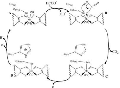
As was showed in Figure 1.7, the metal in the active center of Desulfovibrio desulfuricans Fdh is hexa-coordinated in a distorted trigonal prismatic geometry. Thus, no free coordination position exists for the substrate to bind and interact with the molybdenum ion. Three reaction mechanisms for formate oxidation by Fdh were proposed, by Boyington et al. (1997) [59], Raaijmakers et al. (2006) [60] and Mota et al. (2011) [66]. The differences between the three proposals are the occurrence, or not, of a direct coordination of the SeCys residue to the Mo ion throughout the catalytic cycle and the role of the conserved amino acids in the substrate-binding pocket. The first reaction mechanism, proposed by Boyington et al., was based on crystallographic data obtained for Fdh-H isolated from E. coli. This reaction mechanism proposes that catalysis is initiated with the coordination of the oxygen from formate to the oxidized Mo ion, displacing the –OH ligand while formate is being stabilized by the conserved residues His141 and Arg333 (Figure I.16, A!B). The Se atom captures the α-proton of
formate, two electrons are transferred to the Mo ion and CO2 is released (Figure I.16,
B!C). Active site regeneration starts with an electron transfer from the Mo ion to the
[4Fe-4S] cluster through the PGD moiety (Figure I.16, C!D) and is completed with the
oxidation of the Mo ion from Mo(V) to Mo(VI). This oxidation is achieved with a proton transfer from the SeCys140 residue to the His141 residue followed by a second
Figure I.16 – Reaction mechanism of formate oxidation by Fdh, proposed by Boyington et al.
[59].
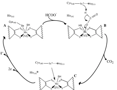
The second reaction mechanism proposed was based in the revised crystallographic structure of the formate-reduced Fdh-H from E. coli. In the original mechanism, proposed by Boyington et al., the SeCys residue was bound to molybdenum after reduction of the enzyme by formate. Raaijmakers et al. re-evaluated the crystallographic data and found that a loop close to the molybdenum active site was mistraced leading to the wrongful placement of catalytic relevant residues such as SeCys140. After data re-evaluation it was found that this residue was no longer bound to
the metal after the reduction of the enzyme with formate. As this interpretation was incompatible with the originally proposed reaction mechanism a new mechanism was proposed (Figure I.17). Before being reduced by formate, the SeCys residue is coordinated to the Mo ion (Figure I.17, A). Formate approximation, frees a coordination position for its binding to the active site, as it triggers the release of SeCys and the stabilization of its selenol group by the Arg residue. Then, the selenol group from the SeCys abstracts the α-proton of formate which is readily transferred to the His residue. CO2 is released while two electrons are transferred to the Mo ion (Figure I.17, B!C).
Active site regeneration is achieved through binding of the selenol group to the Mo ion and its oxidation with electron transfer to the [4Fe-4S] cluster (Figure I.17, C!A).
Mo S S S S O Se Cys140 C O H (VI) His141 Mo S S S S OH Se Cys140 (VI) His141 Mo S S S S Cys140 Se (V) Mo S S S S SeH Cys140 (IV) CO2 HCOO -OH -e -H+ e -A
D C
Figure I.17 - Reaction mechanism for formate oxidation by Fdh, proposed by Raaijmakers et al. [60].
Leopoldini et al. [67] assessed the energy barriers involved in each of these two proposed mechanisms and concluded that the second proposal is kinetic and thermodynamically more favorable, and that the reaction of proton abstraction from formate to the Se atom is more efficient when the SeCys residue is not a ligand at the Mo site.
The latest reaction mechanism to explain the two electron oxidation of formate to CO2 was proposed by Mota et al. (2011) [66]. This mechanism requires the
rearrangement of the sulfur atom coordinated to the metal, through a process known as the sulfur-shift, depicted in the top of Figure I.18.
Mo S S S S His141 SH
Se Arg333
(IV) Cys140 Mo S S S S His141 O HS C O H Se Arg333
(VI) Cys140 Mo S S S S SH Se Cys140 His141 (VI)
A B
CO2 HCOO
-H+
2e
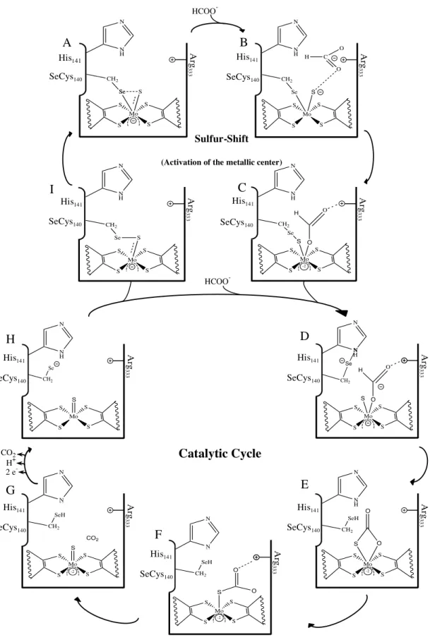
Figure I.18 – Reaction mechanism proposed for Fdh activation and formate oxidation as proposed by Mota, et al. (2011). Adapted from [66]. Top: Activation of the metallic center of Fdh via sulfur-shift. Bottom: Catalytic cycle for formate oxidation by Fdh.
HCOO
-A B
C I HCOO -N H N Ar g 33 3
His141
SeCys140 Mo S S S S Se S Se CH2 N H N Ar g 33 3 His141 SeCys140 Mo S S S S Se S CH2 C O O H Mo S S S S Se S CH2 O O H N H N Ar g 33 3 His141 SeCys140 -2 N H N Ar g 33 3 His141 SeCys140 Mo S S S S Se S CH2 Sulfur-Shift
(Activation of the metallic center)
G Se CH 2 N Ar g 33 3 His141 SeCys140 Mo S S S S HS O O H Ar g 33 3 His141 SeCys140 Mo S S S S SeH S CH2 O O -2 SeH CH2 O N H N Ar g 33 3 His141 SeCys140 Mo S S S S S O O -2 SeH CH2 O N H N Ar g 33 3 His141 SeCys140 Mo S S S S S CO2 -2 Ar g 33 3 His141 SeCys140 Mo S S S S S Se CH 2 CO2 H+ 2 e
In its oxidized, and inactive, form the active center of Fdh has a molybdenum ion in a Mo(VI) oxidation state. In this state, the sulfido (Si) and SeCys ligands form a
quasi-covalent bond with the molybdenum ion (Figure I.18, scheme A). As the formate anion reaches the substrate-binding pocket its negative charge is buffered by the conserved Arg residue and by the two pyranopterins (Figure I.18, scheme B). Further approach from formate to the Mo (VI) ion triggers the insertion of the Si between the
bond of the selenium atom and the molybdenum ion with the simultaneous bond of formate to the molybdenum ion. (Figure I.18, scheme C). Throughout these activation steps the metal remains hexa-coordinated and at the final step the sixth ligand is formate and Fdh becomes active. Formate oxidation involves several steps. The first being the cleavage of the bond between the selenol group from the SeCys and the Si followed by
simultaneous establishment of a hydrogen bond between the selenol group and the conserved His residue (Figure I.18, scheme D). Next, the selenol group from the SeCys abstracts the proton of formate, leading to the cleavage of the hydrogen bond to the conserved His while simultaneously forming a new bond between Si and the carbon
from carbon dioxide (Figure I.18, scheme E). The first step for the release of carbon dioxide involves breaking the Mo–O bond, while maintaining the carbon dioxide molecule connected to the active site through a Si–C bond. (Figure I.18, scheme F). The
release of the carbon dioxide molecule is accomplished through the cleavage of the Si–C
bond and the formation of a double bond between the Si and Mo (Figure I.18, scheme
G). The next step, involves the transfer of the proton attached to the SeCys residue and active site oxidation from Mo(IV) to Mo(VI) (Figure I.18, scheme H). Following this step, active site regeneration may follow two paths. If another formate molecule is present the Si–Mo bond is displaced and formate will bind to the penta-coordinated
molybdenum (Figure I.18, scheme D). However, if no formate molecule is available the SeCys residue binds to the Si (Figure I.18, scheme I) and the SeCys–Mo bond is
I.5.
Subject and objective of this work
The need for CO2 valorization instead of its storage is necessary to help us
overcome the energy crisis derived from both, the near end of fossil fuels and the increasing energy demand in emergent nations. To accomplish this goal, more research will have to focus on CO2 sequestering and in finding carbon-neutral or carbon-negative
strategies to handle with declining fossil fuel supplies and with the environmental impact caused by their usage.
The use of formate dehydrogenase in the conversion of CO2 into a biofuel could
lead to significant changes in the economic paramount, while at the same time significantly reduce GHG emissions. Although the use of enzymes at an industrial scale poses a serious difficulty due to the fragile nature of these systems when compared with synthetic catalysts, there is an increasing demand for catalytically perfect systems, with high turnover constants and low running costs. And one expects this will act as a driving force, attracting investment for this field of study and eventually lead to significant improvements in the development of robust synthetic catalysts.
The conversion of CO2 to formate has been reported and studied in a limited
number of NAD+-independent formate dehydrogenases and it was thought to be
exclusive of W-containing Fdhs. The explanation for this was linked to tungstoenzymes ability to catalyze low potential reduction reactions leading to the assumption that reduction of CO2 to formate is W-dependent [11,61,68,69].
The main purpose of this work was to demonstrate that NAD+-independent
molybdoenzymes could also catalyze the reduction of CO2 to formate. To accomplish
this, a Mo-Fdh will be isolated from the SRB Desulfovibrio desulfuricans ATCC 27774. As a large amount of pure enzyme will be needed it will be necessary to determine which type of culture media allows for a greater development of the bacterial cells. Afterwards, the media will be narrowed to the one that allows a superior Fdh expression and the harvest conditions will be optimized to permit a higher Fdh recovery from the growth extracts. Following growth optimization solution, kinetic assays will be performed to determine the kinetic parameters KM and kcat for the oxidation of formate
catalyze the inverse reaction, carbon dioxide reduction to formate, allowing for its use as a biocatalyst in an energetically efficient CO2 fixation process that might be directed
II.
M
ATERIALS AND METHODS
All reagents used throughout this work were of analytical grade. Table VI-18, in appendix, summarizes the brand and the purity of the reagents used.
II.1.
Bacterial strain, culture media and growth conditions
D. desulfuricans ATCC 27774 (Dd) cells were used in all the experiments. These were grown in several media at 37ºC under strict anaerobic conditions. To establish anaerobiosis, the flasks containing the medium were flushed with argon (Praxair Pure Argon-3X) before being autoclaved, making its gas phase 100% argon.
Figure II.1 – Schematic representation of the acclimatization procedure of D. desulfuricans
cells to the different media tested. Each arrow represents an inoculation that is 10% of the total growth volume. X media represents each of the media tested, ATCC 42, ATCC 1249, ATCC 2755, ATCC 27774 and LYSC. Between each involution the cells were allowed a 24-hour incubation period at 37ºC.
After adapting the cells to each of the test media, a first screening was done to determine which of the test media, ATCC 42, ATCC 1249, ATCC 2755, ATCC 27774, LYSC and VMN allowed for a higher cell mass to be harvested. Employing an inoculum representing 10% of the total growth volume, growth in each media was evaluated by visual inspection, after a 24-hour incubation period at 37ºC. If there was little propagation the cells were allowed another 24-hour incubation period at 37ºC as schematized below.
Figure II.2 – Schematic representation of the screening done to determine which of the test media would allow for a higher cell mass to be harvested. X media represents each of the media tested, ATCC 42, ATCC 1249, ATCC 2755, ATCC 27774, LYSC and VMN.
If after a 48-hour incubation period there was still little propagation the medium was discarded.
10% inoculum
Dd cells X medium
24h
24h
Dd cells X medium
Dd cells X medium
Visua l inspe
ction
Visua l inspe
ction
Dd cells VMN
10% inoculum
90% VMN 10% X medium
10% inoculum
80% VMN 20% X medium
10% inoculum
70% VMN 30% X medium
10% inoculum
50% VMN 50% X medium
10% inoculum
25% VMN 75% X medium
10% inoculum
10% VMN 90% X medium
10% inoculum
Table II-1 – Culture media compositions, per liter.
VMN* ATCC 1249* ATCC 2755** M*
KH2PO4 0.5 g - - -
K2HPO4.3H2O - 0.66 g 0.66 g 0.47 g
MgSO4.7H2O - 4.1 g 2.0 g -
NaNO3 2.4 g - - 2.28 g
Na2SO4 - - 1.0 g -
NH4Cl 1.0 g 1.0 g 1.0 g 1.9 g
MgCl2.6H2O 0.05 g - - 1.55 g
CaCl2.2H2O 0.04 g - 0.1 g 0.2 g
CaSO4.2H2O - 1.3 g - -
Na-Lactate 6.0 g 3.5 g 2.0 g 10 mL
Na-Citrate 0.3 g 5.7 g - -
FeCl2.4H2O 0.003 g - - 0,67 g
Fe(NH4)2(SO4)2 - 2% (v/v)
"
- -
FeSO4.7H2O - - 0.5 g -
Wolfes Elixir 1 mL - - -
NZCYM Broth 2.0 g - - -
Triptone 2.0 g - - -
Vitamin Solution 2 mL - - -
Yeast Extract - 1.0 g 1.0 g 0.95 g
Na-thioglycolate - - 0.1 g -
Ascorbic Acid - - 0.1 g -
Na2S.3H2O - - - 10 mL
#
“Fauque”
Oligoelements - - - 10 mL
* – pH adjusted to 6.5 ± 0.05; ** – pH adjusted to 6.8 ± 0.05; # – 1% (w/v) Na2S.3H2O solution;
"
The bacterial cells were cultivated at 37ºC, in anaerobic flasks with volumes ranging from 10 to 100 mL of medium with inoculums ranging from 2 to 10% of the total flask volume and cell harvest was done at mid exponential or stationary phases. In either case, growth was accompanied by optical density (O. D.) value measurements at 600 nm using a Shimadzu UV 160A spectrophotometer.
Growths under hydrogen were done inside a sealed anaerobic bag at 37ºC, in 100 mL anaerobic flasks under a continuous flow of 100% H2.
Cultures were periodically checked for possible contamination employing optical microscopy. Figure II.3, depicts a pure culture judging from the similar morphology of the cells.
Figure II.3 – D. desulfuricans cells, magnified 1000x.
To obtain enough Fdh to perform the kinetic assays, a 200 L reactor growth was outsourced. This growth was done with an inoculum that was 2% of the total growth volume, and cells were harvested at the end of the exponential phase.
II.2.
Soluble extract preparation
centrifuge. After centrifugation the pellet was resuspended in 10 mM Tris-HCl buffer (pH 7.6) to a cell density of 0.1 g cells (wet weight) per mL.
Periplasmatic soluble extracts were prepared by subjecting this cell suspension to 4 freeze-thaw cycles followed by centrifugation at 7000 g for 40 minutes to remove spheroplasts. The resulting periplasmatic supernatant was later used for activity measurements.
To obtain the cell soluble extract the cells were resuspended in 10 mM phosphate buffer (pH 7.6), to a cell density of 3 g cells (wet weight) per mL. These cells were then subjected to a pressure of 20000 psi in a French Pressure Cell Press (Thermo Electron Corporation) and this sample, total cell soluble extract, was also used for activity measurements. In some samples, 1 mM phenylmethylsulfonyl fluoride (PMSF) and 1 mM dithiothreitol (DTT) were also added to the ressuspension buffer before disrupting the cells in the French Press. Such samples are clearly identified in the results section. A flowchart of these procedures is depicted in Appendixes VI.3 and VI.4.
II.3.
In gel activity assays
In Appendix VI.2, a detailed description of the solutions employed for the preparation of the polyacrylamide gels used throughout this work is given.
II.4.
Fdh purification
Dd Fdh was isolated as described in [65] with some minor modifications as reported in [50]. The cells were cultured in VMN medium and collected by centrifugation at the end of the exponential phase. Then the cells (170 g wet weight, from a outsourced 200 L reactor) were resuspended in 10 mM Tris-HCl buffer and ruptured in a high-pressure homogenizer at 9000 psi. After centrifugation (10000 g; 45 minutes; Sigma 3K30 centrifuge) and ultracentrifugation (180000 g; 60 minutes; Beckman L-70 ultracentrifuge) the supernatant was dialyzed overnight against 10 mM Tris-HCl buffer and loaded onto an anionic exchange column (DEAE Bio Gel, equilibrated with 10 mM Tris-HCl). Elution was achieved with a linear gradient (10 to 300 mM Tris-HCl) in 3 column volumes. Fdh activity was determined in each collected fraction following the procedure described in Section II.3 placing equal volumes of each of the fractions on 7.5% polyacrylamide gels.
At this point, no other purification procedures were done. Further purification steps are presented in a flowchart that is depicted in appendix VI.5.
II.5.
Protein content quantification
Protein quantification was done employing the Lowry method, with bovine serum albumin as standard. This procedure is described in detail in [70].
![Figure I.2 – Atmospheric carbon dioxide concentrations, in ppm, since record began at Mauna Loa Observatory in 1958 [2]](https://thumb-eu.123doks.com/thumbv2/123dok_br/16497421.733638/27.892.161.675.469.777/figure-atmospheric-carbon-dioxide-concentrations-record-mauna-observatory.webp)
![Figure I.6 – Pyranopterin cofactor present in mononuclear Mo/W-containing enzymes [39,40]](https://thumb-eu.123doks.com/thumbv2/123dok_br/16497421.733638/34.892.163.804.168.751/figure-pyranopterin-cofactor-present-mononuclear-mo-containing-enzymes.webp)
![Figure I.7 – Active site structures amongst the different families of Mo and W pyranopterin-dependent enzymes [39,40]; (Xanthine Oxidase Family) Ndh: nicotinate dehydrogenase;](https://thumb-eu.123doks.com/thumbv2/123dok_br/16497421.733638/37.892.99.755.122.1026/structures-different-families-pyranopterin-dependent-xanthine-nicotinate-dehydrogenase.webp)