Extracellular matrix and angiogenic
factors in hematological diseases
Daniela Filipa Nobre Salvador
Mestrado em Ciências Biomédicas
2011
U
niversidade do Algarve
Extracellular matrix and angiogenic
factors in hematological diseases
Dissertação orientada pelo Doutor Sérgio Jerónimo
Rodrigues Dias e pela Doutora Ana Sofia Cachaço
Daniela Filipa Nobre Salvador
Mestrado em Ciências Biomédicas
2011
U
niversidade do Algarve
i
C
ONTENTSii CONTENTS Contents……….... i Agradecimentos……….………… v Resumo………..……. vii Abstract………..……. xi Abbreviations……….. xiii Introduction ……….……… 1
Angiogenesis: key players and biological importance……… 2
Major growth factors involved in the angiogenic process………. 2
Proteolytic degradation of the extracellular matrix: also part of the angiogenesis processes and the remodeling of tissues………… 4
Metabolic changes underlying the angiogenic process: hypoxia as an example...………..……. 5
Clinical relevance for angiogenesis: possible therapeutic targets . 5 BM microenvironment in hematopoiesis: creating niches for hematopoietic stem cells………..………. 6
ECM in the BM microenvironment ……….…….. 7
BM diseases: from BM dysfunctions to malignancy……….…… 12
BM microenvironment in hematological diseases: the deregulation of stem cell niches..………..… 15
Angiogenesis: a link between BM microenvironment and BM diseases…….… 17
References ……… 20
Aims………..………. 37
Methods……….………... 39
Human samples ………..………..……….. 40
iii CONTENTS
cDNA synthesis………..……….. 41
Real-time polymerase chain reaction (RT-PCR)……… 41
Quantitative real time polymerase chain reaction (RQ-PCR)……… 42
Protein quantification……… 42
Enzyme Linked Immunosorbent Assay (ELISA)……… 43
Western-Blotting………..………….. 43 Co-immunoprecipitation……….…………. 45 Gelatin Zymography………. 46 Imunohistochemistry……….… 46 Immunofluorescence………….………. 47 In vitro assays……… 47 Inhibition of MMPs……….……… 48
Inhibition of Notch pathway ……… 48
Statistic analysis……….. 48
Results ………. 49
VEGF is bound to FN in MDS and AML BM patient samples……… 50
MMPs are important for the release of VEGF bound to FN……… 51
Soluble VEGF is more abundant in MDS patients BM samples……… 53
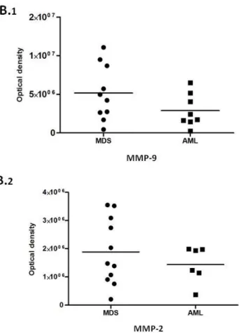
MMP-9 and MMP-2 are more concentrated in BM biopsies from MDS patients………. 54 MMP-9 levels are correlated with soluble VEGF in BM from MDS patients… 56 MDS patients have more FN in BM than AML patients……… 56
BM from MDS patients exhibit a more angiogenic phenotype than from AML patients ……… 58 Notch pathway is activated in both MDS and AML patient BM samples……… 60
iv CONTENTS
Notch pathway activity correlates with FN expression in AML patient BM samples………..
62
Activation of NOTCH pathway increases FN expression in AML patient BM
samples……….. 64
Discussion ………..………..……… 66
References ……….……… 73
v
A
GRADECIMENTOSvi AGRADECIMENTOS
Agradeço:
em primeiro lugar aos meus pais, irmã e Sofia, por todo o amor, compreensão, preocupação e apoio, sem vocês nada disto seria possível. Um especial obrigado por tudo.
ao Doutor Sérgio Dias pela oportunidade que me proporcionou de participar neste grupo de investigação, assim como por todas as ideias que permitiram que este trabalho fosse possível.
à Ana Sofia, por todo o conhecimento que partilhou comigo, pela generosa assistência na elaboração desta tese e, claro, pela amizade. Um profundo obrigado.
a todos os colegas do grupo da Angiogénese pela amizade, paciência, apoio, disponibilidade e por todos os momentos passados no laboratório. Agradeço às meninas mais novinhas, à Joana pela cantoria, à Inês pelas arrumações, às duas pela hora do gelado, à Telma por partilhar a minha bancada, à Sofia, a todas agradeço essencialmente pela amizade, companhia e compreensão. Germana para ti vai um obrigado pelo apoio, compreensão e amizade, à Ana Magalhães por aquele conselho que vou tentar não esquecer, à Leonor pelas chamadas de atenção, por me ensinar o mundo das imunos no visível, e pelas bolachas. À Ana Costa, pelas amostras e pelo esclarecimento das mais diversas dúvidas, ao Francisco, o homem do grupo, simplesmente pela presença e disponibilidade. Fernanda, a menina que sempre se disponibilizou para me trazer lâminas, um obrigado especial pela atenção e apoio. Tânia pela disponibilidade. Jacinta pelas arroxadas, e às doutoras do grupo, Catarina e Ana Bastos pela disponibilidade e apoio. Muito obrigado.
aos restantes elementos do CIPM pela simpatia, disponibilidade e compreensão.
ao laboratório de Anatomia Patológica, ao doutor José Cabeçadas, por toda a disponibilidade e ajuda nos cortes histológicos.
a todos os meus amigos pela amizade, em especial, à Vera e à Maggie, por todo o amor, apoio, compreensão e por todos os momentos juntas, enfim por tudo , ao André e à Vanessa, por todos os momentos, ao Miguel por me conhecer, compreender e ajudar, à Tixa (Ana Patricia), pela companhia, ao David, à Carica (Ana) por esse feitiozinho especial, à Maria, ao José, à Kika (Francisca), ao Hugo, à Agri, ao meu cunhadinho (Miguel). Obrigado a todos os que fazem parte da minha vida e que permitem que os meus dias estejam completos.
vii
R
ESUMOviii RESUMO
A angiogénese consiste na formação de novos vasos sanguíneos através de vasos pré-existentes. Nos adultos, a vasculatura mantêm-se quiescente, excepto em processos altamente controlados, como nos ciclos reprodutores femininos e na cicatrização. No entanto, muitas doenças evoluem graças à angiogénese, como a artrite reumatóide, a neovascularização ocular, o crescimento tumoral e a metastização. Em tumores, a angiogénese é induzida sobretudo pelas condições de hipóxia que ocorrem nestes tecidos, levando à produção de vários factores pró-angiogénicos como o VEGF (o mais importante), PDGF, FGF, angiopoitinas, entre outros. O VEGF pode sofrer “splicing” alternativo, levando à expressão de quatro isoformas que variam no número de aminoácidos e na presença ou ausência de locais de ligação à heparina. VEGF165, VEGF189 e VEGF206 podem ligar-se às membranas
celulares ou à matriz extracelular (MEC) através dos domínios de ligação à heparina; o VEGF121 é uma proteína não ligada à heparina, apresentando-se numa forma solúvel,
livremente difusível no meio. Para além destes factores pró-angiogénicos, as metaloproteases de matriz (MMPs) também são importantes na degradação da membrana basal que envolve os vasos, permitindo a migração das novas células endoteliais. A angiogénese é importante na progressão de uma patologia crónica para uma aguda, mais agressiva, estando bastante estudada em tumores sólidos, sendo, inclusivamente, um dos principais alvos terapêuticos nestas doenças. No entanto, só mais recentemente se começou a atribuir importância a este processo em tumores hematológicos, que ocorrem na medula óssea (MO) e órgãos linfáticos.
As disfunções da MO são doenças clonais resultantes da transformação neoplásica de células estaminais/progenitoras hematopoiéticas (CEPH). Entre elas, a leucemia mielóide aguda (LMA) é caracterizada por um rápido aumento de células sanguíneas imaturas, os blastos, devido a uma inibição nas vias apoptóticas destas células. Tal interfere com a homeostasia da MO e inibe a produção normal de células sanguíneas. As síndromes mielodisplásicas (SMD) são um conjunto heterogéneo de doenças hematológicas que apresentam frequentemente citopénias no sangue periférico, apesar de a MO poder apresentar-se normocelular ou hipercelular. Esta situação pode estar relacionado com um aumento da apoptose que se verifica numa
ix RESUMO
ou em várias linhagens hematopoiéticas. As SMD apresentam um risco elevado de progredir para LMA. Apesar das causas genéticas destas doenças estarem bem descritas, sabe-se que determinados componentes do microambiente medular protegem as células malignas da quimioterapia e propiciam a sua proliferação e sobrevivência. No entanto, muito falta saber acerca do microambiente na progressão, ou mesmo iniciação, das doenças hematológicas, nomeadamente na angiogénese que lhes poderá estar associada. Foi reportado um aumento da angiogénese em SMD e LMA, comparativamente a dadores saudáveis, o que sugere um papel deste processo nestas doenças. O microambiente da MO inclui o estroma (fibroblastos, células endoteliais, osteoblastos, macrófagos), factores solúveis (factores de crescimento, citocinas e quimiocinas), MMPs e a MEC. A MEC inclui várias moléculas como a fibronectina (FN), lamininas, colagéneos, glicosaminoglicanos e proteoglicanos, sendo uma estrutura bastante dinâmica com uma composição espacial e temporal bem definidas dentro da MO, o que influencia as propriedades das CEPH. A FN, as MMPs e vários factores de crescimento estão alterados quantitativamente em diversos tumores sólidos.
Assim, o objectivo deste trabalho foi compreender se o microambiente da MO é diferente entre SMD e LMA e que papel este factor poderá ter na progressão destas doenças. Através da técnica de imunoprecipitação, verificámos um aumento significativo de VEGF ligado à FN em MO de doentes com LMA. Por outro lado, a análise por ELISA do sobrenadante de MO revelou um aumento de VEGF solúvel na MO de doentes com SMD. Nesta doença, observámos um aumento da actividade das MMPs comparativamente aos doentes com LMA, que poderá ser responsável pela libertação do VEGF solúvel nas MO de SMD e, consequentemente, ao aumento da angiogénese verificado nesta patologia. As nossas experiências in vitro comprovaram que quando as MMPs são inibidas, há uma diminuição do VEGF solúvel. Estes resultados sugerem que a biodisponibilidade de factores angiogénicos, nomeadamento do VEGF, pode ser controlada pelo seu grau de associação à FN (dependende das MMPs) e que este fenómeno é importante na angiogénese das doenças hematológicas. Por imunohistoquímica, observámos um aumento da FN em
x RESUMO
MO de doentes com LMA e quisemos saber o que poderia estar a regular a expressão desta proteína. Sabe-se que a via de sinalização Notch, é importante na angiogénese de tumores sólidos, mas o seu papel nas doenças hematológicas é bastante controverso. Um estudo em embriões de ratinho demonstrou que a sobrexpressão do receptor Notch leva a um aumento da FN à volta dos vasos sanguíneos em formação. Por PCR quantitativo realizado nas nossas amostras de MO, concluímos que esta via de sinalização está activa em ambas as doenças. No entanto, observámos uma forte correlação entre a expressão de elementos da via NOTCH e a expressão da FN em MO com LMA, o mesmo não se tendo verificado para as SMD. Aliando estes resultados aos anteriores relativos à regulação do VEGF, concluímos que em LMA, o VEGF encontra-se essencialmente ligado à FN, sendo a actividade das MMPs inferior à encontrada na MO com SMD. Nestas condições, a biodisponibilidade do VEGF é mais regulada, tornando a angiogénese menos exuberante. Além disso, a via de sinalização Notch poderá estar a regular a produção/deposição de FN que envolve os novos vasos sanguíneos, contribuindo para a formação de vasos mais estáveis e funcionais. Por outro lado, em SMD, a elevada actividade das MMPs leva a um aumento do VEGF solúvel na MO que, aliado a uma diminuição da FN à volta dos vasos sanguíneos em formação (dependente da via de sinalização Notch), contribui para um aumento da angiogénese e formação de vasos instáveis e menos funcionais. Assim, as diferenças encontradas no microambiente medular de SMD e LMA, nomeadamente os diferentes níveis de VEGF, MMPs e FN, deverão condicionar a angiogénese nestas doenças e, provavelmente, algumas das suas características patológicas: aumento da apoptose em SMD e sobrevivência dos blastos em LMA. Dentro do nosso conhecimento, nada foi descrito sobre a regulação da biodisponibilidade do VEGF na MO em disfunções hematológicas, assim como sobre a sinalização Notch na angiogénese destas doenças.
Palavras-chave: Medula óssea, microambiente, angiogénese, síndromes mielodisplásicas, leucemia mielóide aguda, fibronectina, VEGF, metaloproteases de matriz
xi
A
BSTRACTxii ABSTRACT
Bone marrow (BM) malignancies are clonal disorders resulting from neoplastic transformation of hematopoietic stem/progenitor cells (HSPCs). Among them, acute myeloid leukemia (AML) is characterized by a rapid increase in immature blood cells numbers, due to apoptosis suppression; myelodysplastic syndromes (MDS) are characterized by peripheral cytopenia, related with increased apoptosis, and can develop to AML. Angiogenesis is an important event that mediates the progression from a chronic to a more acute and aggressive pathology, and its significance in hematological malignancies has just beginning to be explored. BM microenvironment, including soluble factors and extracellular matrix (ECM), in particular fibronectin (FN) that has been found increased in solid tumors, may be responsible for BM disease progression, but its precise role in this context has been poorly investigated. Thus, the aim of this thesis was to know if the BM microenvironment differs between MDS and AML and what role might such factor be playing in disease progression. Our results indicate that MDS BM has more soluble VEGF, a pro-angiogenic factor, and higher matrix metalloproteinases (MMPs) activity, which may be responsible for increased angiogenesis occurring in this disease. Notch pathway, known to be involved in solid tumors angiogenesis, does not regulate FN expression in MDS BM, which leads to new formed vessels instability. In AML, VEGF is kept majorly bounded to FN, and MMPs activity is lower than in MDS BM. In such conditions, VEGF bioavailability is more regulated, being angiogenesis less exuberant. In addition to this, Notch pathway regulates FN deposition around new vessels, contributing to the formation of a more functional and stable vasculature in AML. To our knowledge, nothing has been described about the regulation of VEGF bioavailability in BM diseases, as well as regarding the possible effect of Notch signaling in angiogenesis of hematological dysfunctions.
Keywords: Bone marrow, microenvironment, angiogenesis, myelodysplastic syndromes, acute myeloid leukemia, fibronectin, VEGF, matrix metalloproteinases
xiii
A
BBREVIATIONSxiv ABBREVIATIONS
ALL Acute lymphoblastic leukemia AML Acute myeloid leukemia Ang Angiopoietin
BM Bone marrow
BSA Bovine serum albumin
CLL Chronic lymphocytic leukemia CML Chronic myelogenous leukemia DAB Diaminobenzidine
DAPT N-[N-(3,5-difluorophenacetyl)-L-alanyl]-(S)-phenylglycine t-butyl ester DEPC Diethyl pyrocarbonate
Dlk Delta-like
DLL-4 Delta-like ligand 4
DMEM Dulbecco’s Modified Eagle Medium DMSO Dimethyl sulfoxide
dNTPs Deoxyribonucleoside triphosphates DTT Dithiothreitol
ECM Extracellular matrix
EDTA Ethylenediaminetetraacetic acid EGF Epidermal growth factor
ELISA Enzyme Linked Immunosorbent Assay ET Thrombocythemia essential
FAB French-American-British FBS Fetal bovine serum FGF Fibroblast growth factors
xv ABBREVIATIONS
Flk-1 Fetal liver kinase-1 Flt-1 fms-like tyrosine kinase-1 FN Fibronectin
HC Hematopoietic cells
HGF Hepatocyte growth factor/scatter factor HIF1α Hypoxia-inducible factor 1alpha
HRP Horseradish peroxidase enzyme HSC Hematopoietic stem cell
HSPC Hematopoietic stem/progenitor cell HSPG Heparan sulfate proteoglycans
IPSS International Prognostic Scoring System
JAG Jagged
KDR Kinase insert domain receptor LTBM Long-term bone marrow MAP Mitogen-activated protein MDS Myelodysplastic syndrome
MDS-U Myelodisplasic syndrome unclassified MK Megakaryocyte
MMP-2 Matrix metalloproteinase-2 MMP-9 Matrix metalloproteinase-9 MPS Myeloproliferative syndromes
MT2-MMP Membrane type-2 matrix metalloproteinase
NRP Neuropilin
ON Overnight
xvi ABBREVIATIONS
PDGF Platelet-derived growth factor PFA Paraformaldehyde
PMF Primary or idiopatic myelofibrosis PV Polycythemia vera
RA Refractory anemia
RAEB Refractory anemia with an excess of blasts RARS Refractory anemia with ringed sideroblasts RCMD Refractory anemia with multilinage dysplasia
RCMD-RS Refractory anemia with multilinage dysplasia and ringed sideroblast RNase out Ribonuclease inhibitor
RT-PCR Real-time polymerase chain reaction
RQ-PCR Quantitative real-time polymerase chain reaction RT Room temperature
SDF Stromal cell-derived factor SDS Sodium dodecyl sulfate
SDS-PAGE Sodium dodecyl sulfate polyacrylamide gel electrophoresis TBE Tris/Borate/EDTA
TBS Tris Buffered Saline
TGF Transforming growth factor TGS Tris/glycine/SDS
TIMP Tissue inhibitors of metalloproteinases TNF-α Tumor necrosis factor alpha
VEGF Vascular endothelial growth factor VEGFR VEGF receptors
1
I
NTRODUCTION2 INTRODUCTION
Angiogenesis: key players and biological importance
Blood vessels are fundamentally composed of endothelial cells, which interconnect to form the tubes that direct and maintain blood flow and tissue perfusion. The development of blood vessels in embryogenesis occurs by two processes: vasculogenesis, whereby endothelial cells derive primarily from progenitor cells, and angiogenesis, in which new capillaries sprout from existing vessels. In adult mammals, new vessels are produced only through angiogenesis although a role for endothelial progenitors has been shown in several physiological and pathological situations.
In adults, the vasculature is quiescent except for highly organized processes in the female reproductive cycles (ovulation, menstruation, implantation, pregnancy) (Hanahan and Folkman, 1996). However, many diseases are driven by persistent unregulated angiogenesis, like rheumatoid arthritis (Moon et al., 2010), ocular neovascularization, tumor growth and metastasis (Hanahan and Folkman, 1996; Parangi et al., 1996), among others (reviewed in Carmeliet, 2005).
Major growth factors involved in the angiogenic process
The formation of new blood vessels and their permeability is primarily regulated by vascular endothelial growth factor (VEGF) or VEGF-A (after the discovery of other VEGF family members, like VEGF-B, -C, -D and -E) (Connolly et al., 1989; Ferrara and Henzel, 1989). VEGF is a dimer composed by two subunits that vary 18 to 34kDa subunits (Connolly et al., 1989). Four different transcripts of this protein have been identified resulting from alternate splicing of exon 6 and 7, which alters their heparin-binding affinity, and amino acid number: originating four isoforms, 206, 189, 165 and 121 (Tischer et al., 1991). In addition, inclusion or exclusion of exons 6 and 7 mediate interactions with heparan sulfate proteoglycans (HSPGs) and neuropilin (NRP) co-receptors on the cell surface, enhancing their ability to bind and activate the VEGF receptors (VEGFR). This molecule had specific mitogenic effect on vascular endothelial cells, but not in adrenal cortex cells, lens epithelial cells, corneal endothelial cells, keratinocytes or fibroblasts (Ferrara and Henzel, 1989). Recently, a novel group of iso-
3 INTRODUCTION
forms, the so-called “b-isoforms” or “VEGFxxxb” isoforms, have been described. These transcripts of the VEGF-A gene code for polypeptides with the same length as the classical ones, because exon 8 (present in all the formerly known isoforms) is substituted by an alternatively spliced exon of the same size (exon 8b) (Ladomery et
al., 2007; Catena et al., 2010).
VEGF acts via two receptors protein tyrosine kinases, flt-1 (fms-like tyrosine kinase, VEGFR1) or flk-1 (fetal liver kinase-1; KDR, VEGFR2) (de Vries et al., 1992; Quinn
et al., 1993). The binding of VEGF to VEGFR, in particular to flk-1/KDR, is responsible
for activating several signaling cascades in endothelial cells, within different physiological functions, including survival (Gerber et al., 1998), proliferation (Thakker
et al., 1999), and vascular permeability (Gille et al., 2001). Moreover, VEGF can also
bind to NRP-1, acting as a co-receptor for flk-1/KDR, and enhancing the VEGF signaling (Herzog et al., 2011).
Platelet-derived growth factor (PDGF) is a family of heterodimeric or homodimeric isoforms of A- and B- polypeptide chains, synthesized as precursor molecules undergoing proteolytic maturation. These molecules are produced by different cell types and act via protein kinase receptors (Heldin and Westermark, 1999). PDGF family has a complex role in regulation of blood vessel formation (Cao et
al., 2002).
Fibroblast growth factors (FGFs) are a family of 20 heparin-binding growth factors. FGFs exert their pro-angiogenic activity by interacting with various endothelial cell surface receptors, including tyrosine kinase receptors, HSPGs, and integrins (Kan et
al., 1993). The binding of FGF isoforms to heparin sites protect growth factors from
acid, heat and degradation by circulating proteases, as thrombin 1), trypsin (FGF-2) or plasmin, affecting biological signaling (Powers et al., 2000). Several isoforms of this family are mitogenic for endothelial cells, like FGF-4 (Delli-Bovi et al., 1988), but they have pleiotropic effects, stimulating the growth of several other cells, as smooth muscle cells, fibroblasts, and certain epithelial cells (Folkman and Shing, 1992). Furthermore, FGFRs have variable activity in promoting angiogenesis, with FGFR-1 sub-
4 INTRODUCTION
group being associated with tumor progression and FGFR-2 subgroup with either early tumor development or decreased tumor progression (reviewed in Korc, 2009).
The angiopoietin (Ang) family of growth factors is also important for blood vessel formation (Valenzuela et al., 1999). Ang-1 and Ang-4 are angiogenic factors that signal through the Tie-2 receptor tyrosine kinase, essential for normal vascular development in the mouse (reviewed in Peters et al., 2004; Fukuhara et al., 2009, 2010). On the other hand, Ang-2 and Ang-4 bind to Tie-2 receptor and act as antagonists of Ang1, what is unusual for a family of ligands (Maisonpierre et al., 1997). The biological action of angiopoietin family depends on the unique mixture of angiogenic factors and their receptors operating in tissue microenvironment (Olsen et
al., 2006).
Proteolytic degradation of the extracellular matrix: also part of the angiogenesis processes and the remodeling of tissues
Proteolytic degradation of extracellular matrix (ECM) is implicated in many steps of the angiogenic cascade, not only by degradation of vessel basement membranes, but also in cell migration, ECM invasion and capillary morphogenesis (Maciag et al., 1982; Bellon et al., 2004; reviewed in Bourboulia and Stetler-Stevenson, 2010). ECM proteolysis results from secretion and activation of matrix metalloproteinases (MMPs) in response to exogenous signals, such as cytokines, growth factors and cell-matrix interactions (Bellon et al., 2004). MMP-9 is upregulated during the carcinogenic process in pancreatic islets, having an effect on the switch from vascular quiescence to angiogenesis. It mobilizes VEGF stored in ECM, leaving it available to its receptor (Bergers et al., 2000). Suppression of MMP-2 reduced angiogenesis, inhibiting the transition from the pre-vascular to the vascular stage of tumor development in a chondrosarcoma model (Fang et al., 2000). Moreover, several endogenous molecules are produced by partial proteolysis of ECM such as angiostatin derived from plasminogen, endostatin from collagen XVIII, anastellin from fibronectin (FN) and many others. All these molecules have pro- and anti-angiogenic activities (reviewed in Heissig et al., 2003; Bellon et al., 2004; Rundhaug, 2005; Milkiewicz et al., 2006).
5 INTRODUCTION
Metabolic changes underlying the angiogenic process: hypoxia as an example
Hypoxia is accepted as one of the basic mechanism that initiates and regulates angiogenesis in tumors. It mediates the up-regulation of pro-angiogenic factors, like VEGF, and the downregulation of anti-angiogenic factors (Messmer-Blust et al., 2009), leading to blood vessel growth and, eventually, metastasis. The hypoxia-inducible factor 1alpha (HIF1α) is activated under hypoxia conditions and, in a glioma model, it stimulated the recruitment of CD45+ BM-derived cells to tumor areas. These cells produce MMP-9 that is essential to initiate the angiogenic switch and also regulates tumor invasiveness (Du et al., 2008). Also under hypoxia, the membrane type-2 MMP (MT2-MMP) is upregulated in pancreatic cancer cells, non-small cell lung cancer cells and cervix cancer, cells and this could confer resistance to hypoxia-induced apoptosis and increase invasiveness of cancer cells (Zhu et al., 2011).
Clinical relevance for angiogenesis: possible therapeutic targets
Given the biological relevance attributed to angiogenesis, in tumors but also in other diseases, soon clinical researchers began to attempt at targeting this complex process for therapeutic purposes. The inhibition of VEGF–VEGFR interactions shows a significant improved survival and disease-free survival in patients with cancer, decreasing tumor growth. However, in several cancers, this inhibition is less effective because patients acquire resistance during treatment or originally the tumor is less sensitive to VEGF signaling. Alternatively, drugs against targets related to VEGF-independent regulation of angiogenesis could be good candidates to treat these patients. This may include several molecules, such as angiostatin and endostatin that were reported to be natural anti-angiogenic proteins in the body (Matter, 2001; Shibuya, 2008). Recent studies have shown that FGFs can act synergistically with VEGF to amplify tumor angiogenesis, highlighting that targeting of both the FGF and VEGF pathways may be more efficient in suppressing tumor growth and angiogenesis than targeting either factor alone (reviewed in Korc, 2009). Even though the importance of angiogenesis was first shown in setting of solid cancer progression many years ago (Folkman, 1971), it has also been associated with hematological diseases (Perez- Atayde et al., 1997; Aguayo et al., 1999). VEGF and basic FGFs expression on hemato-
6 INTRODUCTION
poietic organs (bone marrow (BM) and lymphatic organs) define some clinical characteristics in leukemias and non-Hodgkin’s lymphoma, and their levels in serum/plasma patients are predictors of poor prognosis (Fiedler et al., 1997; Perez-Atayde et al., 1997; Aguayo et al., 2000; De Bont et al., 2001; Fragoso et al., 2007; reviewed in Albitar, 2001; Moehler et al., 2003).
In hematological tumors, MMPs expression has been shown to be upregulated in some leukemia, lymphomas and multiple myeloma (Barillé et al., 1997; Kossakowska
et al., 1999; Kuittinen et al., 2001; 2003; Pennanen et al., 2008). Similar to solid tumor,
in hematological tumors the BM microenvironment (stroma cells, ECM and soluble factors) must play a role in the angiogenic switch, but its actual importance on this process is not completely understood. Litwin and colleagues, comparing the low angiogenic potential of acute myeloid leukemia (AML) blasts in vitro with the high vessel density in BM patients in vivo, concluded that angiogenesis in AML likely represents a response to microenvironmental factors in vivo, rather than being an intrinsic property of leukemic cells (Litwin et al., 2002). It is, thus, important to understand how angiogenic factor bioavailability, in particular of VEGF, can be modulated by the BM microenvironment, which can help improving anti-angiogenic therapies in hematological diseases.
BM microenvironment in hematopoiesis: creating niches for hematopoietic stem cells
The BM is the major hematopoietic organ in adult and it is found in the interior of long (e.g. humerus, femur, tibia, fibula) and flat bones (e.g. ribs, vertebrae, cranium, hip). It is a trabecular structure irrigated by large vessels and sinusoids, and filled with stromal and hematopoietic cells.
BM microenvironment comprises stromal cells (as osteoblasts, adypocytes, endothelial cells, fibroblasts, macrophages), soluble factors (growth factors, cytokines and chemokines) produced by stromal and hematopoietic cells (HCs), and the ECM. Within BM microenvironment, two well defined regions called niches were considered
7 INTRODUCTION
to provide hematopoietic stem/progenitor cells (HSPCs) with critical instructions to self-renew, proliferate, differentiate, homing, migrate and survival (reviewed in Li and Xie, 2005; Kacena et al., 2006; Moore and Lemischka, 2006; Wilson and Trumpp, 2006; Yin and Li, 2006; Arai and Suda, 2005; Scadden, 2007). HSPCs are believed to be located near bone surfaces (the osteoblastic niche) or associated with the sinusoidal endothelium (the vascular niche); the molecular signals generated by these two niches have been extensively studied (in particular for the osteoblastic niche). Osteoblasts produce important signaling molecules like osteopontin and Ang that interact with their receptors on HSPCs, keeping these cells in a quiescent state (Grassinger et al., 2009; reviewed in Arai and Suda, 2007; Suda et al., 2005). On the other hand, the vascular niche is considered to promote proliferation and further differentiation of HSPCs (Kopp et al., 2005); it produces FGF-4 and chemokines such as stromal derived factor (SDF-1) (reviewed in Yin and Li, 2006) that recruits the HSPCs from the osteoblastic to vascular niche. Recently, with new imaging approaches, it is becoming evident that endosteal and vascular compartments may not be mutually exclusive in terms of their role on HSPC fate (Kiel and Morrison, 2008).
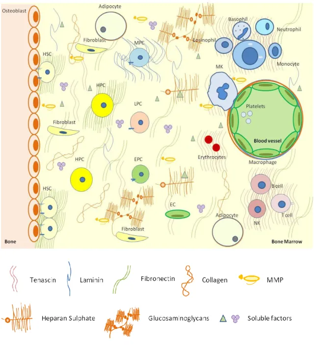
ECM in the BM microenvironment
The ECM component of the BM can be seen as a very dynamic structure, which spatial and temporal composition within BM creates niches that highly influence the HSPCs proprieties (Figure 1). These niches also function as a soluble factors reservoir, controlling their availability to adjacent cells. To acquire proper information from the surrounding ECM, hematopoietic cells express adhesion molecules in a regulated fashion, being integrins and proteoglycans the major receptors for ECM (reviewed in Campbell and Humphries, 2011; Kim et al., 2011). Adhesion molecules participate in a range of signal transduction processes involving not only cell adhesion, but also migration, proliferation and apoptosis. In more detail, the BM ECM is an intricate network of proteins (e.g. collagens), glycoproteins (FN, laminins) glycosaminoglycans (e.g. hyaluronan) and proteoglycans (e.g. syndecans) whose turnover is tightly controlled by the adjacent cells (reviewed in Kim et al., 2011). ECM molecules are pro-
8 INTRODUCTION
duced in response to diverse environmental stimuli which are intrinsic to organ function, or may be present in stress situations involving tissue turnover such as during wound healing processes, or in malignancy (reviewed in Hynes, 2009). Nevertheless, the contribution of ECM in regulating cell-tissue-organ function, namely its role in BM homeostasis, has received little attention, and the majority of studies have focused on their receptors integrins and the signaling pathways activated through them. Also, most of studies have been made in vitro. The effect of ECM in hematopoiesis varies accordingly to cell lineage and maturation stage of the progenitor cell (reviewed in Lam and Adams, 2010). In long-term (LT) BM cultures, progenitors that adhere to FN proliferate significantly less than non-adherent progenitors (Hurley and Verfaillie, 1995). Collagen significantly increases the adhesion of human long term bone marrow (LTBM) cells and induces a decreased in myeloid progenitor cell production, whereas FN increases myelopoiesis (Hassan et al., 1997). Laminin-10/11 is highly adhesive to lineage-committed myelomonocytic and erythroid progenitor cells and several lymphoid and myeloid cell lines, whereas laminin-8 is less adhesive. Ex vivo culture of murine HSCs in the presence of FN and laminin resulted in expansion of primitive stem cells and improvement in the marrow engraftability (Sagar et al., 2006). Other ECM molecules seem to modulate HSPCs microenvironment. Heparan sulphate is produced by stromal cells and it presents cytokines to HSPCs, as well as promoting their ligation to the ECM molecules like thrombospondin. This leads to the formation of discrete niches, thereby orchestrating the controlled growth and differentiation of stem cells (Coombe, 1996; Gupta et al., 1998). Tenascin is co-localized with other ECM molecules such as FN and collagen type III in the microenvironment surrounding the maturing hematopoietic cells and has a role in the retention of HSPCs in the stroma (Klein et al., 1993). Osteopontin negatively regulates HSC numbers, as evidenced by increased of these cells in the osteopontin-null microenvironment (Calvi et al., 2003; Stier et al., 2005). ECM is also essential in megakaryocyte (MK) differentiation. Depending on which integrin is expressed at their surfaces, these cells exhibit different affinities for ECM ligands, which in turn influence their fates (Zweegman et al., 2000; Inoue et al., 2003; Balduini et al., 2008; Mazharian et al., 2011). Additionally, MKs are important producers of MMPs, being MMP-9 secretion essential for MK migration towards blood
9 INTRODUCTION
Figure 1: Schematic representation of major steps in hematopoiesis within BM microenvironment. The image represents BM niches: the osteoblastic niche, where quiescent HSPC localize, and the vascular niche, where HSPC differentiation occurs. Myeloid progenitor cells (MPC) originate basophils, neutrophils, eosinophils, monocytes, MKs (give rise to platelets), and erithrocytes, and lymphocyte progenitor cells (LPC) originate lymphocytes B and T, and natural killer cell (NK) cell. BM microenvironment is composed by ECM proteins (collagen), glycoproteins (FN and laminin) and glycosaminoglycans, soluble factors (e.g. growth factors, MMPs), and stroma (endothelial cells, osteoblasts, fibroblasts, macrophages, adipocytes).
10 INTRODUCTION
vessels and posterior platelets release (Lane et al., 2000). MMPs production by MKs may also modulate BM niches in terms of ECM remodelling, and thus, the fate of hematopoietic cells. Cell adhesion events are also very important to assure the success of transplantation (Chute, 2006; Lam and Adams, 2010). Absence of β1-containing integrins (in particular, α4β1 integrin) resulted in sequestration of HSCs in the circulation and their reduced adhesion to endothelial cells (Hirsch et al., 1996; Arroyo
et al., 1999; Dave et al., 2000; Potocnik et al., 2000; reviewed in Imai et al., 2010),
impairing their entrance in BM.
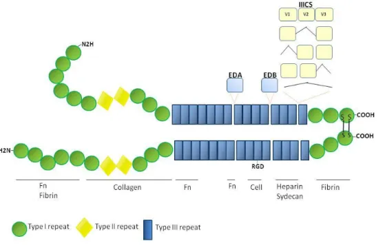
FN, a major constituent of ECMs, plays an important role during embryogenesis, wound healing and cancer invasion by promoting cell adhesion, motility, cell cycle progression and cell survival (Hynes and Yamada, 1982; Humphries, 1989; Frisch and Ruoslahti, 1997). Reduction or loss of FN expression occur in many transformed cells in culture (Ruoslahti, 1989), but its overexpression is also associated with various human tumor cells like colorectal cancer, breast carcinoma, head and neck squamous carcinoma, and metastatic melanoma (Zhang G, 1997; Bittner et al., 2000; Jiang et al., 2002; Al Moustafa, 2002). FN and their integrin receptors are also key regulators of endothelial growth (Hynes et al., 1999; Dvorak, 2005). While laminins and collagens appeared early in evolution, FN has been found only in vertebrates and its appearance in evolution correlates with the appearance of organisms with endothelial cell-lined vasculature (Hynes and Zhao, 2000; Whittaker et al., 2006; Astrof and Hynes, 2009). It is a high-molecular weight (440KDa) molecule, consisting of two nearly identical monomers linked by a pair of disulfide bonds. The FN protein is produced from a single gene, but alternative splicing of its pre-mRNA leads to the creation of several isoforms (Figure 2). There are two types of FN: the soluble plasma FN, a major protein component of blood plasma (300μg/ml) and produced in the liver by hepatocytes, and the insoluble cellular FN, a major component of ECM (reviewed in Pankov and Yamada, 2002). Within BM, FN is localized in the osteoblastic niche, the region for which HSPCs have high affinity, but has also a wide distribution in central BM region (Nilsson et al., 1998). Its broad allocation suggests that FN may be important in creating specific niches for HCs, and some roles of this molecule in hematopoiesis have been revealed, mostly on in vitro studies (see above).
11 INTRODUCTION
Figure 2. Schematic representation of human dimeric FN and its several binding domains. Each 220kDa FN monomere comprises multiple type I (green circles), type II (yellow lozenge) and type III (blue rectangle) repeats. Several FN isoforms can be obtained by alternative splicing: the entire EDA and EDB domains are independently included or excluded by exon skipping, whereas the IIICS domain undergoes complex splicing of mRNA transcribed from a single exon (exon subdivision is indicated). The binding sites for interaction between FN and collagen, fibrin, heparin, cells, and to other FN molecules are also represented. Cell-binding sites include the RGD domain (for α5β3 and αVβ3 integrin interaction) and the IIICS domain (for α4β1 integrin interaction).
Nevertheless, not too much is known about its importance in hematological diseases, in particular in the angiogenic processes associated with these malignancies. In this context, most of the studies only concern ECM remodeling by MMPs, but which factors regulate FN expression during this process are still to identify. Also in other biological systems, the regulation of FN has not been totally explored. Hepatocyte growth factor/scatter factor (HGF) induces FN expression and extracellular assembly on the surface of melanoma cells through activation of mitogen-activated protein (MAP) kinase pathway (Gaggioli et al., 2005). The pleiotropic cytokine transforming growth factor-β (TGF-β) is the well known regulator of FN, being studied in epithelial-to-mesenchyme transition contexts, like in embryogenesis and in fibrosis (as an example, see Sureshbabu et al., 2011). A recent study on mouse embryonic angiogene-
12 INTRODUCTION
sis revealed that overexpression of the Notch ligand Dll4 increases the deposition of FN around the vessels, although the direct role of Notch signaling on FN production has not been addressed (Trindade et al., 2008). In BM, nothing is known about the regulation of FN (or other ECM molecules) expression, or in homeostasis, or in disease.
BM diseases: from BM dysfunctions to malignancy
BM malignancies are clonal disorders resulting from neoplastic transformation of HSPCs. They include leukemias, lymphomas and multiple myeloma. Other related BM diseases, but not considered cancer forms, comprise myeloproliferative syndromes (MPS) and myelodisplastic syndromes (MDS).
Leukemias are classified accordingly to the hematopoietic cells which undergo the clonal transformation: lymphoblastic or lymphocytic leukemias – involving lymphocyte precursors - and myeloid or myelogenous leukemias – involving myeloid cell but also erythrocyte or platelet precursors. They are also subdivided in acute or chronic leukemias. Acute leukemias are characterized by a rapid increase in the numbers of immature blood cells, making the BM unable to produce healthy blood cells. It requires immediate treatment due to the rapid progression of the disease that leads to the exit of malignant cells into the bloodstream and spread to other organs of the body. Chronic leukemias are characterized by the excessive production of relatively mature, but still abnormal, leukocytes. Typically, they take years to progress, so they are sometimes monitored for some time before treatment to ensure maximum effectiveness of therapy. Combining these two classifications, four main types of leukemias emerge (although other forms of rare leukemias can occur): acute lymphoblastic leukemia (ALL), AML, chronic lymphocytic leukemia (CLL), and chronic myelogenous leukemia (CML) (Swerdlow et al., 2008).
MPS are a group of BM clonal disorders in which excess cells are produced. They are categorized by the presence or absence of Philadelphia chromosome (t9;22) and include, among others primary or idiopatic myelofibrosis (PMF), polycythemia vera (PV),
13 INTRODUCTION
essential thrombocythemia (ET) (Philadelphia chromosome-negative), and CML (Philadelphia chromosome-positive). CML can occur independently or progress from a PMF and is characterized by the presence of HSPC expressing the bcr-abl oncogene, with abnormal release of these clonal cells into the circulation. A complete genetic and clinicopathological classification of this group of disorders can be consulted, for instance, in Michiels et al., 2007; Tefferi and Gililland, 2007, 2006; Michiels and Thiele, 2002.
MDS are a heterogenous group of clonal hematopoietic diseases characterized by peripheral cytopenia (despite a normocellular or hypercellular BM) and with a variable probability to progress to AML (Miyazato et al., 2001; Braun et al., 2006; Nolte and Hofmann, 2008). The incidence of these conditions has risen sharply over the past several years, making them the most common malignant BM disorders. It has become apparent that the ineffective hematopoiesis is largely caused by excessive apoptosis of myeloid precursors (Braun et al., 2007; reviewed in Westwood and Mufti, 2003). Recent observations suggest that downregulation of α4β1 and α5β1 integrins on HSC, correlated with decreased in vitro adhesiveness to FN fragments, could be a newly identified proapoptotic mechanism in MDS (Delforge et al., 2005). Inversely, the evolution of MDS from early relatively chronic phenotype to an aggressive AML is accompanied by a suppression of apoptosis in the malignant cells, mediated by changes in intracellular levels of Bcl-2-family proteins (reviewed in Westwood and Mufti, 2003). MDS-associated AML (secondary AML) is rarely cured by conventional chemotherapy (reviewed in Hamblin, 1992), which contrasts with the somewhat better outcome of de novo AML. The protein Delta-like (Dll), distantly related to Delta-Notch family of signaling proteins, has highly selective expression in the individuals with MDS and lower in secondary AML, being absent from de novo AML. This makes Dll a good candidate molecule to differentiate MDS from AML (Miyazato et al., 2001).
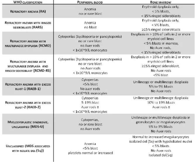
As biopsies from several MDS patients have been used on this project, a brief classification of this disease is given due to the heterogeneity of the samples. The French-American-British (FAB) group established the first classification of MDS in 1982, defining five subtypes based on morphology, the number of blasts (in BM and periphe-
14 INTRODUCTION
ral blood) and monocyte counts. This classification reflects the progression and the clinical course of MDS but, although providing important diagnostic information, revealed some limitations. To overcome them, in 2001, the World Health Organization (WHO) classification suggested new subtypes of MDS (table 1) (Bennett et al., 1982).
Table 1. MDS classification according to WHO
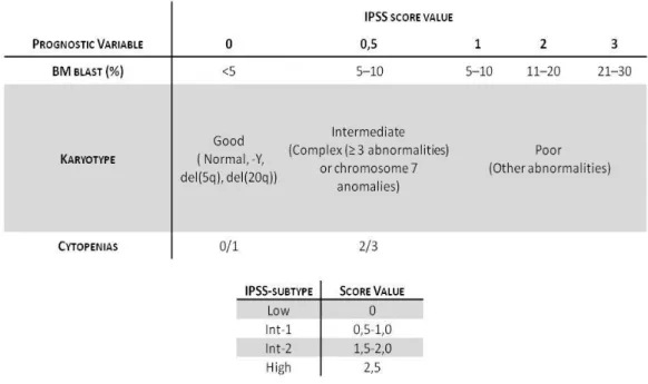
However, this classification did not include important information on genetic abnormalities, a major prognostic factor in MDS patients. So, another classification based on significant prognostic factors, proposed in 1996 by the International Prognostic Scoring System (IPSS), defines four groups of risk (table 2).
15 INTRODUCTION
Table 2. MDS Risk proposed by IPSS
BM microenvironment in hematological diseases: the deregulation of stem cell niches
Malignant hematopoietic cells are known to express particular cell adhesion repertoires that provide them with proliferative and survival advantages within the BM microenvironment. Specific niche composition provides ideal conditions for some leukemic cells to escape from chemotherapy-induced death and acquire drug-resistance. There are several reviews on this subject (e.g. Verfaillie et al., 1997; Rizo et
al., 2006; Konopleva et al., 2009; Lane et al., 2009). Most of the studies on
hematological diseases refer integrins-ECM interactions as key mechanisms involved in tumor progression, whereas well identified genetic hits occurring on HSCs would be the major beginners of the oncogenic process (reviewed in Tefferi and Gilliland, 2007). Presently, the acceptance that the BM microenvironment is important in supporting leukemia stem cells survival has conducted to the rational development of therapies that target microenvironment molecules (reviewed in Konopleva et al., 2009;
16 INTRODUCTION
Lane et al., 2009). For instance, inhibition of α4β1 integrin - FN interaction in AML patients increases their sensibility to chemotherapy (Matsunaga et al., 2003).
Emerging data suggests the balance between ECM production and degradation (turnover) may be crucial for normal (versus malignant) organ function, while it may also represent a way to detect tissue damage (alterations), in particular situations. In BM, mononucleated cells from healthy donors continuously produce MMP-9 and TIMP-1 (a MMP inhibitor), whereas AML and CML blast cells additionally secrete MMP-2, representing a potential marker for dissemination in myeloproliferative malignancies (Ries et al., 1999). Nevertheless, the role of the BM ECM in the pathophysiology of hematological disorders has remained controversial. Although microenvironment may not initiate clonal proliferation, it should somehow favor the progression of the disease. Thus, microenvironment analysis will be important to develop effective regimens that allow for elimination of a specific clone in human patients. For that reason, we will provide some examples on the role of microenvironment, in particular ECM, in BM malignancies.
The MPS PMF is a remarkable model in which deregulation of the stem cell niche is of great importance in disease development (Lataillade et al., 2008). In a typical case of PMF, hematopoietic cells in BM are replaced by collagen fibrosis (although other ECM proteins are also increased), impairing the patient's ability to generate new blood cells resulting in a progressive pancytopenia (Reilly, 1997). An ineffective megakaryocytopoiesis leads to an excessive concentration of abnormal MKs which release specific growth factors (like TGF-β) essential for fibroblast activation and, consequently, collagens, FN and other ECM molecules production (Schmitt et al., 2000; Kuter et al., 2007). Additionally, not only increased production of ECM occurs in PMF but there is also an imbalance between MMPs and TIMPs that may contribute to BM fibrosis (reviewed in Wang, 2005). In CML, imature CML cells have lower adhesion to stromal cells and FN as well as lower engraftment potential than normal HSC (Peled
et al., 2002), a feature that may account for their exit to peripheral blood.
In MDS, alterations in adhesive proprieties of myeloid progenitors (see above) are cell-autonomous and confer cell susceptibility to apoptosis (Raza et al, 1995).
17 INTRODUCTION
However, changes in BM stroma, namely on ECM, have already been detected in some MDS patients, suggesting again a role for microenvironment in MDS progression (Tennant et al., 2000; Tauro et al., 2001). Remarkably, increased cell death in MDS can be followed by increased proliferation, likely of selected and well adapted cell clones, and in this case progression to AML occurs. In normal hematopoiesis, α4β1 and α5β1 integrin-mediated interactions between progenitor cells and FN are critical for progenitor cell survival. Also in AML, adhesion of blast cells to stroma via β1 (principally α4β1) and β2 integrins, seems to inhibit apoptosis in a proportion of cases (Denkers et al., 1992; Liesveld et al., 1993).
Angiogenesis: a link between BM microenvironment and BM diseases
As mentioned above, BM microenvironment has a role in hematological disorders progression, but may also create the appropriate conditions for disease initiation. Of particular interest is the frequent progression from an MDS to an AML, during which the specific and most relevant microenvironment factors involved are largely still unknown. This secondary AML is usually more aggressive that de novo AML, being of outmost importance to uncover the mechanisms behind MDS, de novo AML and secondary AML. These can help in finding new therapeutic approaches appropriated for each type of pathology. Angiogenesis is an important event that mediates the progression from a chronic to a more acute and aggressive pathology, and its significance in hematological malignancies has just beginning to be explored (reviewed in Shadduck et al., 2007). Angiogenic factors may be produced by fibroblasts in the BM stroma and by immune cells (reviewed in Mangi and Newland, 2000), but their availability in BM may be regulated not only at gene expression level, but also by microenvironment elements. Both MDS and AML are associated with a substantial increase in BM vascularity as well as increased levels of various angiogenic factors, in cluding VEGF, basic FGFs, angiogenin, Ang-1, PDGF, HGF, epidermal growth factor (EGF), tumor necrosis factor-α (TNF-α), and TGF-α and TGF-β (Aguayo et al., 2000; Master et al., 2001; reviewed in Albitar, 2001). Malignant cell proliferation, angiogenesis and VEGF expression are linked in AML, as well as MMP-2 and/or MMP-9
18 INTRODUCTION
expression (De Bont et al., 2001). In addition, MMPs correlate with aggressive ALL (Kuittinen et al., 2001). Increasing VEGF levels significantly correlate with shorter survival of patients with MDS and AML (Verstovsek et al., 2002). Nevertheless, higher levels of cellular VEGF and lower levels of its receptor KDR are seen in MDS more than in AML (Verstovsek et al., 2002; reviewed in Albitar, 2001). This fact is in agreement with a significant increase of BM microvascular density in MDS and de novo AML compared with healthy donors. Surprisingly, in MDS, microvascular density significantly decreases upon transformation to AML, which microvascular density was also significantly lower than in de novo AML (Keith et al., 2007).
ECM has a key role in storing/retaining soluble factors, being proteolic enzymes important regulators of soluble factors bioavailability. For instance, VEGF levels correlate with MMP-2 and MMP-9 activity in human breast cancer (Munaut et al., 2003). Also, in glioblastoma, HIF1α, the direct effector of hypoxia, induces the recruitment to tumor area of BM-derived cells that produce MMP-9, which in turn is essential and sufficient to initiate angiogenesis by increasing VEGF bioavailability (Du
et al., 2008).
It is not known if the type or amount of ECM molecules are important in this process, but the existence of appropriate binding sites for certain growth factor on ECM molecules strongly suggest these may be necessary. In this context, FN contains three heparin-binding and syndecan (a heparan sulfate) domains. Interestingly, the two longer forms of VEGF, VEGF189 and VEGF206, are not found freely in the media,
because they are bound to the cell surface or ECM, via heparin-binding sites (Houck et
al., 1992). However, VEGF121 is a nonheparin-bindingprotein, freely diffusible (Houck
et al., 1991). VEGF165 has intermediatedproprieties, being found in a soluble form but
also bounded to ECM. Activity of the serine protease plasmin and MMP-9 can cleave the bounded forms of VEGF, in a colorectal cancer cell line, releasing a soluble factor capable of stimulating endothelial cell growth (Houck et al., 1992; Hawinkels et al., 2008). Also, heparin, heparan sulfate, and heparinase all induce the release of VEGF165
and VEGFl189, in a human embryonic kidney cell line (Hawinkels et al., 2008),
19 INTRODUCTION
these data together suggest that ECM, in particular FN, can regulate VEGF bioavailability in angiogenic processes. It remains to elucidate which signaling pathways regulate ECM and if it is an important issue in angiogenesis and tumor progression in hematological tumors.
A good candidate to regulate FN in vessel formation is the Notch signaling pathway (Trindade et al., 2008). Notch signaling regulates the self-renewal of HSPCs and is also involved in various hematological malignancies (reviewed in Leong and Karsan, 2006), for example, acute T-lymphoblastic leukemia (Weng et al., 2004; Tohda
et al., 2005; Lee et al., 2007) and MDS (Länger et al., 2004; Li et al., 2005; Fu et al.,
2007; Qi et al., 2008). The role of Notch signaling in oncogenesis is controversial, and because most cancers express more than one type of Notch ligand and/or receptor, the overall expression profile of these ligands/receptors may ultimately determine whether this pathway will be oncogenic or in oncosuppressive. Also cell type and the presence of specific soluble factors in tumor microenvironment influence the effect of Notch signaling in oncogenesis (reviewed Leong and Karsan, 2006). This suggests that Notch signaling may be involved in regulating BM microenvironment in hematological diseases (possibly also through FN production or release). HPCs isolated from MDS patients display a disrupted Notch signaling. Furthermore, there was a marked reduction in the plasticity of mesenchymal stem cells of MDS patients compared with those of normal BM donors, in neurogenic and adipogenic differentiation ability and hematopoiesis supporting capacity in vitro (Varga et al., 2007).
As a final remark, BM microenvironment engages a complex mixture of cells, ECM molecules, growth factors and cytokines, which can exert reciprocal influence on each other’s availability. A sum of all these factors will activate (and additionally being activated by) specific signaling pathways that will certainly contribute to hematological dysfunctions. In this Thesis, we have exploited some of the interactions between ECM molecules and angiogenic growth factor bioavailability in the setting of hematological malignancies.
20
R
EFERENCES21 REFERENCES
Aguayo, A,. Estey, E., Kantarjian, H., Mansouri, T., Gidel, C., Keating, M., Giles, F., Estrov, Z., Barlogie, B., and Albitar, M. (1999). Cellular Vascular Endothelial Growth Factor Is a Predictor of Outcome in Patients With Acute Myeloid Leukemia. Blood 94, 3717-3721.
Aguayo, A., Kantarjian, H., Manshouri, T., Gidel, C., Estey, E., Thomas, D., Koller, C., Estrov, Z., O'Brien, S., Keating, M., Freireich, E. and Albitar, M. (2000). Angiogenesis in acute and chronic leukemias and myelodysplastic syndromes. Blood 96, 2240-2245.
Albitar, M. (2001). Angiogenesis in acute myeloid leukemia and myelodysplastic syndrome. Acta Haematol. 106, 170-176.
Al Moustafa, A., Alaoui-Jamali MA., Batist, G., Hernandez-Perez, M., Serruya, C., Alpert, L., Black, MJ., Sladek, R., and Foulkes, WD. (2002). Identification of genes associated with head and neck carcinogenesis by cDNA microarray comparison between matched primary normal epithelial and squamous carcinoma cells. Oncogene
21, 2634-2640.
Arai F., Hirao. A., and Suda T. (2005). Regulation of hematopoiesis and its interaction with stem cell niches. Int J Hematol 82, 371-376.
Arai, F. and Suda, T. (2007). Maintenance of Quiescent Hematopoietic Stem Cells in the Osteoblastic Niche. Annals of the New York Academy of Sciences 1106, 41-53.
Arroyo, AG., Yang, JT., Rayburn, H., and Hynes, RO., (1999). ±4 Integrins Regulate the Proliferation/Differentiation Balance of Multilineage Hematopoietic Progenitors In Vivo. Immunity 11, 555-566.
Astrof, S., and Hynes, R., (2009). Fibronectins in vascular morphogenesis. Angiogenesis 12, 165-175.
Balduini, A., Pallotta, I., Malara, A., Lova, P., Pecci, A., Viarengo, G., Balduini, CL., and Torti, M. (2008). Adhesive receptors, extracellular proteins and myosin IIA orchestrate proplatelet formation by human megakaryocytes. Journal of Thrombosis and Haemostasis 6, 1900-1907.
22 REFERENCES
Barillé, S., Akhoundi, Cl., Collette, M., Mellerin, MP., Rapp, MJ.,Harousseau, JL., Bataille, R., and Amiot, M. (1997). Metalloproteinases in Multiple Myeloma: Production of Matrix Metalloproteinase-9 (MMP-9), Activation of proMMP-2, and Induction of MMP-1 by Myeloma Cells. Blood 90, 1649-1655.
Bellon, G., Martiny, L., and Robinet, A. (2004). Matrix metalloproteinases and matrikines in angiogenesis. Critical reviews in oncology/hematology 49, 203-220.
Bennett, J., Catovsky, D., Daniel, M., Flandrin, G., Galton, D., Gralnick, H., and Sultan, C. (1982). Proposals for the classification of the myelodysplastic syndromes. Br J Haematol. 51, 189-199.
Bergers, G., Brekken, R., McMahon, G., Vu, TH., Itoh, T., Tamaki, K., Tanzawa, K., Thorpe, P., Itohara, S., Werb, Z., and Hanahan, D. (2000). Matrix metalloproteinase-9 triggers the angiogenic switch during carcinogenesis. Nat Cell Biol 2, 737-744.
Bittner, M., Meltzer, P., Chen, Y., Jiang, Y., Seftor, E., Hendrix, M., Radmacher, M., Simon, R., Yakhini, Z., Ben-Dor, A., Sampas, N., Dougherty, E., Wang, E., Marincola, F., Gooden, C., Lueders, J., Glatfelter, A., Pollock, P., Carpten, J., Gillanders, E., Leja, D., Dietrich, K., Beaudry, C., Berens, M., Alberts, D., Sondak, V., Hayward, N., and Trent, J. (2000). Molecular classification of cutaneous malignant melanoma by gene expression profiling. Nature 406, 536-540.
Bourboulia, D., and Stetler-Stevenson, WG. (2010). Matrix metalloproteinases (MMPs) and tissue inhibitors of metalloproteinases (TIMPs): Positive and negative regulators in tumor cell adhesion. Seminars in Cancer Biology 20, 161-168.
Braun, T., Carvalho, G., Coquelle, A., Vozenin, MC., Lepelley, P., Hirsch, F, Kiladjian, JJ., Ribrag, V., Fenaux, P., and Kroemer, G. (2006). NF-kB constitutes a potential therapeutic target in high-risk myelodysplastic syndrome. Blood 107, 1156-1165.
Braun, T., Carvalho, G., Grosjean, J., Ades, L., Fabre, C., Boehrer, S., Debili, N., Fenaux, P., and Kroemer, G. (2007). Differentiating megakaryocytes in myelodysplastic syndromes succumb to mitochondrial derangement without caspase activation. Apoptosis 12, 1101-1108.
Calvi, LM., Adams, GB., Weibrecht, KW., Weber, JM., Olson, DP., Knight, MC., Martin, RP., Schipani, E., Divieti, P., Bringhurst, FR., Milner, LA., Kronenberg, HM., and
23 REFERENCES
Scadden, DT. (2003). Osteoblastic cells regulate the haematopoietic stem cell niche. Nature 425, 841-846.
Campbell, ID., and Humphries, MJ. (2011). Integrin Structure, Activation, and Interactions. Cold Spring Harbor Perspectives in Biology 3.
Cao, R., Brakenhielm, E., Li, X., Pietras, K., Widenfalk, J., O''Stman, A., Eriksson, U., and Cao, Y. (2002). Angiogenesis stimulated by PDGF-CC, a novel member in the PDGF family, involves activation of PDGFR-alphaalpha and -alphabeta receptors. The FASEB Journal 16, 1575-1583.
Carmeliet, P. (2005). Angiogenesis in life, disease and medicine. Nature 438, 932-936.
Catena, R Larzabal, L., Larrayoz, M., Molina, E., Hermida, J., Agorreta, J., Montes, R., Pio, R., Montuenga, LM., and Calvo, A., (2010). VEGF121b and VEGF165b are weakly angiogenic isoforms of VEGF-A. Molecular Cancer 9.
Chute, JP. (2006). Stem cell homing. Current Opinion in Hematology 13, 399-406
Connolly, DT., Heuvelman, DM., Nelson, R., Olander, JV., Eppley, BL., Delfino, JJ., Siegel, NR., Leimgruber, RM., and Feder, J. (1989). Tumor vascular permeability factor stimulates endothelial cell growth and angiogenesis. The Journal of Clinical Investigation 84, 1470-1478.
Coombe, DR. (1996). The Role of Stromal Cell Heparan Sulphate in Regulating Haemopoiesis. Leukemia & Lymphoma 21, 399-406.
Dave B, Watanabe, T., Singh, RK., Ageitos, A., Heimann, DG., Talmadge, JE., (2000). Growth factor mobilization and modulation of progenitor cell adhesion to stromal cells: role of VLA-4. J Hematother Stem Cell Res. 9, 507-515.
De Bont, ESJM., Rosati, S., Jacobs, S., Kamps, W.A., and Vellenga, E. (2001). Increased bone marrow vascularization in patients with acute myeloid leukaemia: a possible role for vascular endothelial growth factor. British Journal of Haematology
113, 296-304.
de Vries, C., Escobedo, JA., Ueno, H., Houck, K., Ferrara, N., and Williams, L.T. (1992). The fms-like tyrosine kinase, a receptor for vascular endothelial growth factor. Science 255, 989-991.
24 REFERENCES
Delforge, M., Raets, V., Van Duppen, V., Vandenberghe, P., and Boogaerts, M. (2005). CD34+ marrow progenitors from MDS patients with high levels of intramedullary apoptosis have reduced expression of [alpha]4[beta]1 and [alpha]5[beta]1 integrins. Leukemia 19, 57-63.
Delli-Bovi, P., Curatola, AM., Newman, KM., Sato, Y., Moscatelli, D., Hewick, RM., Rifkin, DB., and Basilico, C. (1988). Processing, secretion, and biological properties of a novel growth factor of the fibroblast growth factor family with oncogenic potential. Mol. Cell. Biol. 8, 2933-2941.
Denkers, IAM., de Jong-de Boer, TJM., Beelen, RHJ., Ossenkoppele, GJ., and Langenhuijsen, MMAC. (1992). VLA molecule expression may be involved in the release of acute myeloid leukaemic cells from the bone marrow. Leukemia research
16, 469-474.
Du, R., Lu, KV., Petritsch, C., Liu, P., Ganss, R., Passegué, E., Song, H., VandenBerg, S., Johnson, RS., Werb, Z., and Bergers, G. (2008). HIF1± Induces the Recruitment of Bone Marrow-Derived Vascular Modulatory Cells to Regulate Tumor Angiogenesis and Invasion. Cancer cell 13, 206-220.
Dvorak, HF. (2005). Angiogenesis: update 2005. Journal of Thrombosis and Haemostasis 3, 1835-1842.
Fang, J., Shing, Y., Wiederschain, D., Yan, L., Butterfield, C., Jackson, G., Harper, J., Tamvakopoulos, G., and Moses, MA. (2000). Matrix metalloproteinase-2 is required for the switch to the angiogenic phenotype in a tumor model. Proceedings of the National Academy of Sciences 97, 3884-3889.
Ferrara, N., and Henzel, WJ. (1989). Pituitary follicular cells secrete a novel heparin-binding growth factor specific for vascular endothelial cells. Biochemical and Biophysical Research Communications 161, 851-858.
Fiedler, W., Graeven, U., Ergün, S., Verago, S., Kilic, N., Stockschläder, M., and Hossfeld, DK. (1997). Vascular Endothelial Growth Factor, a Possible Paracrine Growth Factor in Human Acute Myeloid Leukemia. Blood 89, 1870-1875.
Folkman, J., Merler, E., Abernathy, C., Williams, G., (1971). Isolation of a tumor factor responsible for angiogenesis. J Exp Med. 133, 275-288.
25 REFERENCES
Folkman, J., and Shing, Y. (1992). Angiogenesis. Journal of Biological Chemistry
267, 10931-10934.
Fragoso, R., Elias, AP., and Dias, S. (2007). Autocrine VEGF loops, signaling pathways, and acute leukemia regulation. Leukemia & Lymphoma 48, 481-488.
Frisch, SM., and Ruoslahti, E. (1997). Integrins and anoikis. Current Opinion in Cell Biology 9, 701-706.
Fu, L., Nara, N., and Tohda, S. (2007). Involvement of Notch signaling in myelodysplastic syndrome. Leukemia research 31, 1160-1161.
Fukuhara, S., Sako. K., Noda, K., Nagao, K., Miura, K., and Mochizuki, N., (2009). Tie2 is tied at the cell-cell contacts and to extracellular matrix by angiopoietin-1. Exp Mol Med. 41, 133-139.
Fukuhara, S., Sako. K., Noda, K., Zhang, J., Minami, M., and Mochizuk,i N., (2010). Angiopoietin-1/Tie2 receptor signaling in vascular quiescence and angiogenesis. Histol Histopathol. 25, 387-396.
Gerber, HP., McMurtrey, A., Kowalski, J., Yan, M., Keyt, BA., Dixit, V., and Ferrara, N. (1998). Vascular Endothelial Growth Factor Regulates Endothelial Cell Survival through the Phosphatidylinositol 3'-Kinase/Akt Signal Transduction Pathway. Journal of Biological Chemistry 273, 30336-30343.
Gille, H., Kowalski, J., Li, B., LeCouter, J., Moffat, B., Zioncheck, TF., Pelletier, N., and Ferrara, N. (2001). Analysis of Biological Effects and Signaling Properties of Flt-1 (VEGFR-1) and KDR (VEGFR-2). Journal of Biological Chemistry 276, 3222-3230.
Grassinger, J., Haylock, DN., Storan, MJ., Haines, GO., Williams, B., Whitty, GA., Vinson, AR., Be, CL., Li, S., Sorensen, ES., Tam, PPL., Denhardt, DT., Sheppard, D., Choong, PF., and Nilsson, SK. (2009). Thrombin-cleaved osteopontin regulates hemopoietic stem and progenitor cell functions through interactions with alpha9beta1 and alpha4beta1 integrins. Blood 114, 49-59.
Gupta, P., Oegema, TR., Brazil, JJ., Dudek, AZ., Slungaard, A., and Verfaillie, CM. (1998). Structurally Specific Heparan Sulfates Support Primitive Human Hematopoiesis by Formation of a Multimolecular Stem Cell Niche. Blood 92, 4641-4651.
Hanahan, D., and Folkman, J. (1996). Patterns and Emerging Mechanisms of the Angiogenic Switch during Tumorigenesis. Cell 86, 353-364.