Joana Margarida de Carvalho Ribeiro
Dissertação para obtenção do Grau de Mestre em Biotecnologia dos Recursos Marinhos
Copyright © Joana Margarida de Carvalho Ribeiro
Escola Superior de Turismo e Tecnologia do Mar – Peniche Instituto Politécnico de Leiria
2011
A Escola Superior de Turismo e Tecnologia do Mar e o Instituto Politécnico de Leiria têm o direito, perpétuo e sem limites geográficos, de arquivar e publicar esta dissertação através de exemplares impressos reproduzidos em papel ou de forma digital, ou por qualquer outro meio conhecido ou que venha a ser inventado, e de a divulgar através de repositórios cientí-ficos e de admitir a sua cópia e distribuição com objetivos educacionais ou de investigação, não comerciais, desde que seja dado crédito ao autor e editor.
Agradecimentos
Ao meu orientador, Doutor Américo Rodrigues, pelo conhecimento e apoio transmitidos, ao longo desta etapa e ainda, pela inteira disponibilidade prestada desde o começo deste traba-lho.
Aos técnicos Pedro Ramalho, Vera Severiano e Cristina Salas pela ajuda dispensada quando necessitei.
Agradeço ao meu colega Marco, por me ter aconselhado e ajudado na realização do trabalho, e também a outros colegas de mestrado com quem convivi durante estes dois anos. Não esquecendo três pessoas muito importantes, Tiago, Fátima e Vanessa por estarem sempre presentes e por todos os momentos bem passados.
Por fim, um agradecimento especial aos meus pais, prima e namorado pelo apoio incondici-onal e os bons conselhos dados.
Resumo
O stress biótico ou abiótico e a falta de nutrientes, têm um efeito negativo muito grande no normal desenvolvimento das plantas. Desta forma, a planta precisa de alterar o seu metabolismo, para produzir energia, através de vias metabólicas alternativas para rees-tabelecer a homeostasia e sobreviver. O complexo SnRK1 (SNF1-related protein kinase) tem um papel muito importante na coordenação desta resposta. Sob condições de stress ener-gético, o complexo SnRK1 é ativado e leva a alteração na expressão de mais de mil genes que permitem, então, à planta alcançar a homeostasia e ter uma resposta adaptativa mais específica.
Apesar da importância que a SnRK1 tem no crescimento e desenvolvimento da planta, o nosso conhecimento atual sobre esta via de sinalização é limitado. O complexo SnRK1 é ortólogo de SNF1, de leveduras, e de AMPK presente em mamíferos, e, portanto, a caracterização do complexo SnRK1 em plantas, baseou-se no conhecimento que já se tinha destes outros complexos. Contudo, ainda que muito valiosa e bem-sucedida, esta abordagem é limitada no que diz respeito à descoberta de novas interações únicas na planta. A fim de identificar alvos a jusante de SnRK1, foi realizado um rastreio de dois híbridos em levedura (Y2H), utilizando SnRK1β1.
Este rastreio foi realizado usando uma biblioteca de cDNA de Arabidopsis (Clon-tech) e permitiu a identificação de 64 proteínas que supostamente interagem com esta subunidade β1. Mais de metade das proteínas identificadas estão relacionadas com o meta-bolismo, estando de acordo com a função de SnRK1 na resposta metabólica à falta de ener-gia. Curiosamente, algumas destas proteínas estão envolvidas em processos de splicing, me-tabolismo do azoto, bem como fatores de transcrição da família das KNATs.
Abstract
Biotic or abiotic stresses, and nutrient starvation have a profound negative effect in the normal plant development. Thus, the plant needs to change its metabolism to produce energy through metabolic alternative pathways in order to re-establish energy homeostasis and to survive. SnRK1 (SNF1-related protein kinase) complex plays an important role in the coordination of this response. Under energy stress conditions, SnRK1 complex is activated and trigger changes in the expression of over one thousand genes that allow the re-establish-ment of homeostasis and the mounting of a more specific adaptive response.
Despite the importance of SnRK1s for plant growth and development, our current knowledge on this signalling pathway is limited. SnRK1s are the orthologs of the yeast SNF1 and the mammalian AMPK, and so far most of the characterization of the SnRK1 system has relied on knowledge from these systems. However, although very valuable and success-ful, this approach is limited in its ability to uncover novel interactions unique to plants. In order to identify downstream targets of the SnRK1, we performed an yeast two hybrid screen using SnRK1β1 as bait.
The Yeast-two hybrid screen assay was done using an Arabidopsis cDNA library (Clontech) and allowed the identification of 64 proteins that putatively interact with the β1-subunit. More than half of the proteins identified are functionally related to metabolism, in line with the important role of SnRK1 in the metabolic response to energy deficit. Interest-ingly, some of the novel interactors identified include proteins involved in the splicing pro-cess, nitrogen metabolism and transcription factors of the KNAT family.
Agradecimentos ... v
Resumo ... vii
Abstract ... ix
Index ... xi
Index of Figures ... xiii
Index of Tables ... xv Abbreviations ... xvii 1. Introduction ... 1 1.1. Stress signalling ... 1 1.2 AMPK/SNF1/SnRK1 Complex ... 2 1.2.1 Mammalian AMPK ... 6 1.2.2 Yeast SNF1 ... 7 1.2.3 Plant SnRK1 ... 9 1.2.3.1 SnRK1 activity regulation ... 12 1.2.3.2 SnRK1 downstream targets ... 12 Aim: ... 15
2 Materials and Methods ... 19
2.1 Construction of Plasmids ... 19
2.2 Yeast two-hybrid screen ... 20
2.2.1 Transformation of Y2HGold with pGBKT7-SnRK1β1 ... 20
2.2.2 Mating and selection of clones ... 21
2.2.3 Determination of transformation and mating efficiency ... 22
xii
2.4 Auxiliaries software’s ... 23
2.4.1 The Arabidopsis Information Resource (TAIR) ... 23
2.4.2 The MIPS Functional Catalogue Database (FunCatDB) ... 24
2.4.3 The Arabidopsis Protein Phosphorylation Site Database (PhosPhAt 4.0) .... 24
3 Results ... 27
3.1 Identification of SnRK1β1 protein interactors ... 27
3.1.1 Functional categorization ... 29
3.2 Yeast-two Hybrid System ... 30
4 Discussion and conclusions ... 39
4.1 Identification of SnRK1β1 protein interactors ... 39
References ... 45
Appendix 1 ... 63
Appendix 2 ... 65
Figure 1: SNF1/AMPK/SnRK1 subunit domain architecture and subunit domains. ... 5 Figure 2: The Arabidopsis SnRK superfamily comprises 3 groups. ... 10 Figure 3: An overview of SnRK1 Complex activity. ... 14 Figure 4: Protein-protein interaction between SnRK1β1 and U1A, U2B” and AT1G06960
(protein with RNA binding motif). ... 31
Figure 5: Protein-protein interaction between SnRK1β1 and KNATs. ... 32 Figure 6: Protein-protein interaction between SnRK1β1 and GLUs. ... 33 Figure 7: Protein-protein interaction between SnRK1.1 as a bait with all proteins studied.
... 34
Figure 8: Protein-protein interaction between SnRK1.1 with all proteins studied under a
Table I: Sequence of the primers used in this work for PCR amplifications. ... 20 Table II: Measurement of the viability of the Prey Library, the Bait and diploids expressing
interacting proteins. ... 22
Table III: Putative SnRK1 β1-subunit interactors identified in this Y2H screen. ... 28 Table IV: The main overrepresented FunCat categories with a P-value < 0.05... 30
Ade (A)/ His (H)/ Leu (L)/Trp (W) – Adenine/ Histidine/ Leucine/ Tryptophan ADP – Adenosine diphosphate
AMP – Adenosine monophosphate Amp – Ampicillin
ATP – Adenosine triphosphate cDNA – Complementary DNA cfu – Colony-Forming Unit
DNA-BD - Plasmid encoding the Gal4 DNA-binding domain EDTA – Ethylenediaminetetraacetic Acid
LiAc – Lithium acetate
mM/mL – millimolar/milliliter PCR – Polymerase Chain Reaction PEG – Polyethylene glycol
SD - Minimal, synthetically defined medium for yeast; is comprised of a nitrogen base, a carbon
source (glucose unless stated otherwise), and a DO supplement
SD -2/SD -4 - Minimal, synthetically defined medium for yeast without Leucine (L) and
Tryp-tophan (W)/ without leucine (L), trypTryp-tophan (W), Adenine (A) and histidine (H)
TE buffer – Tris+EDTA
Tris – Tris(Hydroxymethyl)aminomethane WT – Wild type
Y2H – Yeast Two-Hybrid System
YPD - A blend of yeast extract, peptone, and dextrose in optimal proportions for growth of most
strains of S. cerevisiae
YPDA - YPD medium supplemented with adenine hemisulfate (1X concentration = 120 μg/ml) μM/μL – micromolar/microliter
1. Introduction
1.1. Stress signalling
The ability of an organism to respond to adverse environmental conditions is critical for its survival. Energy supplies varies over the time and it was necessary to develop sophis-ticated mechanisms that perceive fluctuation in nutrient availability and are able to maintain the energy balance at cellular and organism levels. These mechanisms, which respond to energy depletion, are conserved in all eukaryotes and due to its importance they have been extensively studied (Hardie et al., 1998; Xiong et al., 2002; Halford et al., 2003; Hardie et al., 2012; Cabello et al., 2014; Crozet et al., 2014). Specifically in plants, energy deprivation is caused not only by nutrient depletion but also by biotic and abiotic stresses. Indeed, severe environmental factors such as low temperature, drought, high salinity, darkness and patho-gens cause biochemical, molecular and physiological changes in plants that might compro-mise photosynthesis and/or respiration leading, in this way, to energy stress. Drought and salinity are the two major adverse environmental factors because they prevent plants from realizing their full genetic potential and reduce crop productivity being responsible, in severe conditions, for up to 65% reduction in yield, much higher than the losses caused by diseases and insects that, while devastating to individual farmers, globally are generally less than 10% (Serrano et al., 1999; Zhu, 2002; Tuteja, 2007).
Environmental stress and the consequent energy deficit cause cessation of growth, activation of catabolic pathways and a decline of biosynthetic activity as well as the activa-tion of autophagy in order to re-establishment the cellular homeostasis (Baena-Gonzalez and Sheen, 2008; Akpinar et al., 2012).
Introduction
2
1.2. AMPK/SNF1/SnRK1 Complex
Despite the crucial function of other kinases, the AMPK/SNF1/SnRK1 family of pro-tein kinases appears to be the central regulator of the stress response playing an important role as metabolic sensors. These kinases belong to a highly conserved protein kinase family with a N-terminal kinase domain being found throughout Eukaryotes, as represented below in figure 2, among which are the insects, roundworms, mammals, fungi and plants (Hardie et al., 1998; Hardie, 2007; Polge and Thomas, 2007; Hedbacker and Carlson, 2008; Baena-Gonzalez, 2010; Ghillebert et al., 2011). The first experimental evidences of the presence of AMP-activated protein kinase in rat was observed in 1973 by two independent studies (Beg et al., 1973; Carlson and Kim, 1973).This protein was allosterically activated by AMP levels and so it was called AMP-activated protein kinase (AMPK) (Hardie, 2007; Ghillebert et al., 2011). Sucrose non-fermenting 1 (SNF1) is an ortholog of AMPK and was identified in Sac-charomyces cerevisiae in 1981 when the snf1 mutation was discovered among other mutants that were unable to utilize sucrose (Carlson et al., 1981). Later, sequence similarity has re-vealed the existence in plants of three sub-families related to AMPK and SNF1 i.e. SNF1-related protein kinases (Halford and Hardie, 1998; Crozet et al., 2014).
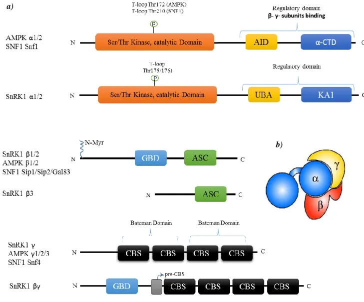
The AMPK/SNF1/SnRK1 has been found in all eukaryotes as heterotrimeric com-plexes comprising one catalytic subunit (α-subunit) and two regulatory subunits (β- and γ-subunits) (figure 1b) (Bouly et al., 1999; Polge and Thomas, 2007; Baena-Gonzalez and Sheen, 2008; Baena-Gonzalez, 2010; Hardie et al., 2012; Crozet et al., 2014). The response of this complex to a specific stimuli depends on the tissue, the development stage and on the precise subunit composition of the complex (Ramon et al., 2013; Nietzsche et al., 2014).
AMPK/SNF1/SnRK1 catalytic α-subunit has a conventional Serine (Ser)/Threonine (Thr) kinase domain that is activated by phosphorylation in a specific conserved Thr residue, present in the activation loop, by upstream kinases (Hardie et al., 2012). The specific residue that is phosphorylated depends on the species, for example in the case of Arabidopsis it is Thr175 (α1) or Thr176 (α2) (Sugden et al., 1999a; Shen et al., 2009; Crozet et al., 2010), in
AMPK is the Thr172 (in human α1) (Hawley et al., 1996) and Thr210 in case of yeast SNF1
(McCartney and Schmidt, 2001). In animals and yeast, the C-terminal region of α-subunit contains an auto-inhibitory domain/sequence (AID or AIS) which inhibits kinase activity (figure 1a) (Hardie et al., 2012; Crozet et al., 2014). Following this region, there is an
α-subunit carboxy-terminal domain (α-CTD) where the β-α-subunit will bind (figure 1a) (Hardie et al., 2012; Crozet et al., 2014). In plants the AID region has not the same role as in animals and yeasts and it is called Ubiquitin-Associated Domain (UBA) (figure 1a) because it seems to mediate the interaction with ubiquitinated proteins (Crozet et al., 2014). With a function similar to animals and yeasts α-CTD, SnRK1α (or AKINα) contains a Kinase Associated 1 (KA1) domain responsible for the interaction with other subunits and the upstream phospha-tases (figure 1a) (Rodrigues et al., 2013; Crozet et al., 2014)
The typical β-subunits (AMPKβ1/β2, SNFsip1/sip2/Gal83 and SnRK1β1/β2) play an essential role in specificity and the recognition by the kinase complex of its targets and they are also responsible for the subcellular localization of the complex which is dependent on the presence of N-terminal myristoylation (N-MYR) of the β1- and β2-subunits (figure 1a) (Warden et al., 2001; Lin et al., 2003; Polekhina et al., 2005; Li et al., 2009; Avila et al., 2012; Liang et al., 2015). N-MYR of the SnRK1β1 appears to negatively regulate nuclear SnRK1 activity by sequestering the complex at the plasma membrane (Pierre et al., 2007). N-MYR is required for the regulation of SnRK1 and N-MYR defect of the SnRK1βs pre-vents shoot apical meristem (SAM) differentiation and leads to development arrest. (Pierre et al., 2007; Traverso et al., 2008). Accumulation of StubGal83 mRNA (potato gene that encodes β-subunit) as well as of SnRK1β1 (Arabidopsis β-subunit) is induced by darkness and this effect is reversed by a light treatment. Moreover, StubGal83 is involved in root and tuber development (Lovas et al., 2003). SnRK1β1 seems to modulate leaf and cotyledon shape in response to metabolic sugars and specifically participates in regulation of Nitrate reductase (NR) and the interaction between SnRK1.1/1.2 (α-subunit) and NR2 (one of the NR genes) (Schmidt and McCartney, 2000; Polge et al., 2008; Li et al., 2009). The β-subu-nits also possess a carbohydrate-binding module (CBM) or Glycogen-binding domain (GBD) (figure 1a) inserted in KIS (Kinase Interaction Sequence) region. In AMPK, GBD serves to bind glycogen, however, in SNF1, GBD motif seems to have not the same function because blocking glycogen synthesis has no impact in SNF1 activity and deletion of the GBD motif in Gal83 (one of the SNF1 β-subunits) increased the activity of SNF1 (Avila-Castaneda et al., 2014; Emanuelle et al., 2015). In plants, the function of GBD motif of SnRK1 β-subunits still remains unclear (Emanuelle et al., 2015). Another important function of the β-subunits is to act as a scaffold keeping the α and γ together through the Association
Introduction
4
with the SNF1 Complex (ASC) domain (Avila-Castaneda et al., 2014) also called Subunit Binding Sequence (SBS) (figure 1a and b) (Polekhina et al., 2005).
The γ-type subunit is composed by four tandem cystathionine β–synthase (CBS) mo-tifs that function as dimers forming two domains called Bateman domain 1 and 2 (figure 1a) that can bind the regulatory adenine nucleotides (AMP/ADP/ATP) providing the primary means of AMPK catalytic regulation. AMP is the only adenylate that directly activates AMPK (Oakhill et al., 2011; Hardie et al., 2012; Emanuelle et al., 2015). However, this allosterically effect of the adenine nucleotides is not observed in the case of yeast SNF1 (Mayer et al., 2011) and plant SnRK1 (Shen et al., 2009). Plant SnRK1γ complements yeast snf4Δ (Bradford et al., 2003; Rosnoblet et al., 2007). In addition, the γ-subunit of Medicago truncatula (MtSNF4b) seems to play an important role in seed longevity and in fruit ripening (Rosnoblet et al., 2007; Bolingue et al., 2010).
Beyond these subunits, which are generally conserved throughout eukaryotes, there are two other subunits (β3 and βγ) that are atypical and are only present in plants (figure 1a) (Polge and Thomas, 2007; Avila-Castaneda et al., 2014; Crozet et al., 2014). SnRK1βγ is able to suppress yeast snf4Δ mutation and it is constitutively expressed in all plants and seems to be more important than SnRK1γ in the SnRK1 complex composition (Lumbreras et al., 2001; Ramon et al., 2013). This subunit is similar to SNF4/AMPKγ with the four tandem CBS motif but contain an extended N-terminal region similar to the KIS domain (containing the GBD region) of AMPK/SNF1 β-type (figure 1a) (Lumbreras et al., 2001; Lopez-Paz et al., 2009). Furthermore, according to the work done by Ramon et al., (2013) Arabidopsis SnRK1γ is not directly involved in SnRK1 signalling since knock-out (KO) of this subunit caused no differences in transcript levels of one of the SnRK1 target genes. Whereas SnRK1βγ RNAi (RNA interference) caused significant differences in target genes expression suggesting that this subunit is needed for formation of the heterotrimeric complex of SnRK1 instead of γ-subunit (Lumbreras et al., 2001; Ramon et al., 2013).
The SnRK1β3 subunit presents a truncated KIS domain and no N-terminal region comprising only the C-terminal domain (ASC) which is closely related to the ASC domains present in SnRK1β1 and SnRK1β2 (figure 1a). This subunit interacts with both α- and γ-subunits and it is also capable to complement β-deficient yeast (sip1Δsip2Δgal83Δ) (Gissot et al., 2004; Polge et al., 2008; Ghillebert et al., 2011; Emanuelle et al., 2015). Additionally
SnRK1β3 transcripts were found in all the organs and at different development stages. A strong interaction is observed between SnRK1β3 and SnRK1βγ but not with SnRK1γ (Gissot et al., 2004).
Figure 1: SNF1/AMPK/SnRK1 subunit domain architecture and subunit domains.
a) The major regulatory phosphorylation site is the T–loop threonine residue in the subunit catalytic domain. The residue that is phosphorylated differs among animals, fungi and plants. UBA - ubiqui-tin-associated domain (just in plants); AID - Auto-inhibitory domain (present in animals and yeasts); KA1 - kinase-associated 1 domain (plants); CTD - α‑subunit carboxy‑terminal domain (an-imals and yeast); N–Myr - N–myristoylation site; CBM - carbohydrate-binding module; CBS - cystathionine β–synthase motif; The plant βγ-subunit has a pre-CBS and GBD domain similar to β-subunits.
b) Complex structure showing how the subunits interact with each other to form the active complex. This structure is common to the three kingdoms. (Adapted from Crozet et al., 2014; Emanuelle et
b) a)
Introduction
6
1.2.1. Mammalian AMPK
So AMPK has been characterized as a mammalian protein kinase that is allosterically activated by AMP leading to the inactivation of the biosynthetic pathways while catabolic pathways such as fatty acid oxidation, glycolysis and autophagy are activated, providing in this manner the energy that the cells need (Bouly et al., 1999; Hardie, 2007; Ghillebert et al., 2011; Hardie, 2011; Oakhill et al., 2011; Hardie et al., 2012; Crozet et al., 2014). Recent studies have shown that AMPK may be sensitive to the intracellular fatty acids availability in an independent manner of the cellular AMP levels (Clark et al., 2004) and this provide an increase activity of AMPK due to increase of AMPK affinity with LKB1, an upstream kinase (Watt et al., 2006). Thus fatty acids are able to allosterically activate AMPK (Clark et al., 2004; Watt et al., 2006). Beyond the allosteric activation, high levels of ADP:ATP ratio and cytosolic Ca2+ are also capable to activate the kinase domain of the AMPK Complex
(Ghillebert et al., 2011; Hardie et al., 2012). So AMPK is also indirectly activated by meta-bolic stresses that inhibit ATP production (hypoxia, glucose deprivation) or stimulate ATP consumption (motor proteins, ion pumps, biosynthetic pathways). Cytokines, ciliary neu-rotrophic factor (CNTF) and certain drugs are also responsible for AMPK activation (Hardie, 2007). Activation of AMPK promotes phosphorylation of metabolic enzymes, transcription factors and co-activators followed by regulation of gene expression (Hardie, 2007).
The activation of the complex is performed in three steps: first, AMP binds to γ-subunit stimulating α-γ-subunit. Next, this binding promotes the phosphorylation of the Thr residue (Thr172) in the catalytic subunit (Hardie, 2007). Third, the activation of the complex requires the N‑terminal myristoylation of the AMPKβ that will regulates subcellular locali-zation and kinase activity acting as a scaffold protein (Warden et al., 2001; Oakhill et al., 2010). The major upstream kinase is LKB1 associated to two accessory subunits, STRAD and MO25, to form the LKB1–STRAD–MO25 complex (identified genetically as a tumor suppressor), and it is responsible for the function of AMPK of inhibiting cell growth and proliferation and promote cell polarity. Ca2+/calmodulin-activated protein kinase kinases (also known as CaMKK2) and TAK1 (transforming growth factor‑β [TGFβ]-activated ki-nase 1) are other two upstream kiki-nases that are able to phosphorylate AMPK, depending on cellular and activation context (Hardie, 2007; Crozet et al., 2010; Hardie et al., 2012; Liang and Mills, 2013; Crozet et al., 2014). CaMKKs, appears to act in cells with an increased
levels of cytosolic Ca2+ and to be able to activate AMPK even in the absence of high AMP levels (Hardie et al., 2012).
More importantly, it is well known the effects of AMPK on metabolism but it may also regulate energy levels by other pathways. Two of those, are regulation of cell cycle and modulation of the nervous system (Hardie et al., 2012). The DNA replication (S phase) and mitosis (M phase) are processes that require energy to go through. The activation of AMPK in cycling cell causes arrest of cell cycle in G1 phase. This event is associated with phos-phorylation of p53 (a tumor suppressor) by AMPK, in turn p53 will activate the expression of cyclin-dependent kinase inhibitor 1A (CDKN1A; also known as p21WAF1) (Jones et al., 2005; Liang et al., 2007; Hardie et al., 2012). This signal leads to cell survival while inhibit cell proliferation (Liang and Mills, 2013; Liang et al., 2015). AMPK is also required for maintenance of nervous system (Tschape et al., 2002; Ghillebert et al., 2011) and in Dro-sophila a mutation in AMPKγ results in a neurodegenerative phenotype (Tschape et al., 2002). Additionally, KO of AMPK in Drosophila melanogaster provokes damages of the nervous system and defects on epithelial integrity and cell division (Tschape et al., 2002; Lee et al., 2007; Spasic et al., 2008).
1.2.2. Yeast SNF1
In yeast, the AMPK ortholog, Sucrose non-fermenting 1 (SNF1) is activated mainly by glucose absence leading to the subsequent activation of a plethora of genes allowing yeast to utilize alternative carbon sources (e.g. sucrose, glycerol or ethanol) (Carlson et al., 1981; McCartney and Schmidt, 2001). Besides sugar signalling, there are many types of conditions that induce SNF1 activation such as sodium ion stress, oxidative stress, alkaline pH and inhibitors of respiratory chain (Hedbacker and Carlson, 2008; Ghillebert et al., 2011). Con-trary to what happens with the AMPK, SNF1 is not allosterically activated by AMP levels (Ghillebert et al., 2011) while ADP appears to regulate SNF1 T-loop phosphorylation pro-tecting the kinase from dephosphorylation (Mayer et al., 2011). In particular, the β subunits of SNF1 complex exhibits also very important role as each one alone (sip1 or sip2 or gal83) is sufficient for yeast growth in media with different carbon sources like glucose, galactose, synthetic complete media with raffinose or glycerol-ethanol (Schmidt and McCartney, 2000). Additionally, the N-terminal domain of the β1- and β2-subunits confers unique
cel-Introduction
8
are required for substrate definition (Schmidt and McCartney, 2000; Vincent et al., 2001; Hedbacker and Carlson, 2008). The γ-subunit (Snf4) have the same structure as AMPKγ (figure 1a) with four CBS domains but Snf4 has a substitution in a residue which is determi-nant for allosterically connection of AMP to the γ-subunits. However Snf4 is still required for the catalytic activity of the heterotrimeric kinase (Celenza and Carlson, 1984, 1989; McCartney and Schmidt, 2001). If necessary, SNF1 inactivates itself by binding of AID (snf1 regulatory domain) to the snf1 kinase domain or to the Snf4 (Lin et al., 2003; Hedbacker and Carlson, 2008).
SNF1 catalytic activity is regulated by three upstream kinases: Elm1, Tos3 and Sak1 (Hong et al., 2003; Nath et al., 2003; Sutherland et al., 2003; Hedbacker et al., 2004). They exhibit overlapping functions as only the triple mutant of these kinases (elm1Δsak1Δtos3Δ) shows the same phenotype of snf1Δ (Hong et al., 2003). The catalytic domain of the three yeast upstream kinases are similar to those of CaMKKs and LKB1 (Hong et al., 2003). De-spite all three being functionally redundant, they have different contribution to cellular reg-ulation under different growth conditions (Kim et al., 2005) as well as in its cellular locali-zation (Hong et al., 2003; Kim et al., 2005). SNF1 is inactivated by dephosphorylation by the protein phosphatase type 1 GLC7 in association with the regulatory subunit REG1 (Tu and Carlson, 1995; Ludin et al., 1998). Interestingly, it seems that glucose does not change Glc7-Reg1 activity, but rather indirectly controls SNF1 dephosphorylation by changing the ability of the activation to be dephosphorylated by the phosphatase (Ruiz et al., 2011).
Once activated, SNF1 triggers a transcriptional reprogramming achieved by the ac-tivation of various TFs and the inacac-tivation of others. This will allow the expression of a large set of genes (over 400 genes) including many of the glucose-repressed genes through control of the transcriptional repressor Mig1 (Treitel et al., 1998). Under low glucose con-ditions, SNF1 phosphorylates Mig1 leading to its nuclear export (Lin et al., 2003; Hedbacker and Carlson, 2008; Ghillebert et al., 2011; Cho et al., 2012). Other TFs are activated by the SNF1 complex like Cat8 and Sip4 which will activate gluconeogenic genes expression (Hedges et al., 1995; Lesage et al., 1996).
Additionally, SNF1 switch from fermentative to oxidative metabolism and glucone-ogenesis (Bouly et al., 1999; Polge and Thomas, 2007; Baena-Gonzalez, 2010; Hardie et al., 2011; Crozet et al., 2014; Emanuelle et al., 2015) and is also involved in controlling the
biosynthesis of reserve carbohydrates and activates autophagy to recycle macromolecules and organelles (Ghillebert et al., 2011). In addition, SNF1 has an important role in develop-ment processes such as meiosis, ageing, growth (diploid pseudohyphal growth and haploid invasive growth) and proliferation as well as mating and sporulation (Hedbacker and Carlson, 2008; Ghillebert et al., 2011; Crozet et al., 2014). Overexpression of Snf1 and the absence of N-myristoylation of Sip2 promote aging and this effect is unique among Snf1 β-subunits (Lin et al., 2003; Crozet et al., 2014). On the contrary, the absence of Snf4 extends life span (Schmidt and McCartney, 2000; Lin et al., 2003).
1.2.3. Plant SnRK1
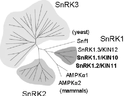
The large Arabidopsis SnRK superfamily is composed of three distinct subfamilies: SnRK1, SnRK2 and SnRK3 (figure 2) (Hrabak et al., 2003; Thelander et al., 2004). The SnRK2 and SnRK3 are known for their involvement in stress and abscisic acid signalling (Hrabak et al., 2003). The SnRK1 subfamily comprises SnRK1.1/SnRK1.2/SnRK1.3 (also known as KIN10/KIN11/KIN12) that are the closest relatives to SNF1 and AMPK com-plexes (figure 2) (Baena-Gonzalez, 2010; Crozet et al., 2014) and it has been studied in several plant species (besides Arabidopsis) such as tomato, rice, potato, tobacco, pea and barley (Li et al., 2010). SnRK1.3 has a very low expression during Arabidopsis development as compared to the other two catalytic subunits (Fragoso et al., 2009). SnRK1.1 and SnRK1.2 are 81% identical. However SnRK1 activity in plant cells is mostly held by SnRK1.1 (Jossier et al., 2009), while the SnRK1.2 may plays a restricted role in regulation of metabolism and stress signalling (Williams et al., 2014).
Introduction
10
SnRK1 was identified in Arabidopsis and in other plants and appears to have plei-otropic roles in response to hormonal, nutritional and environment stresses (figure 3) (Hardie et al., 1998; Wang et al., 2003; Baena-Gonzalez, 2010; Ghillebert et al., 2011). SnRK1 com-plex is activated and required to respond to sugar depletion or to long darkness period but not by an increase of the AMP:ATP ratio, as happens with AMPK complex (Baena-Gonzalez et al., 2007; Baena-Gonzalez and Sheen, 2008; Rosa et al., 2009). However, the physiolog-ical concentrations of 5’-AMP are able to inhibit the T-loop dephosphorylation of the spin-ach SnRK1 kinase (Sugden et al., 1999b). Recent studies have identified Arabidopsis SnRK1.1/SnRK1.2 as central integrators of the transcriptome in response to darkness events, sugar deprivation and stress conditions (figure 3) (Baena-Gonzalez et al., 2007). Double SnRK1.1/SnRK1.2 deficiency in Arabidopsis thaliana cause the transcription of genes as-sociated to darkness and stress signalling to be switch off, resulting in impaired starch mo-bilization and growth defects in plant mutants under 13h light/11h dark or constant illumi-nation (Baena-Gonzalez et al., 2007). Similar results were obtained in Physcomitrella patens (Thelander et al., 2004). Overexpression of two SnRK1 genes of Malus hupehensis (MhSnRK1/2) in tomato resulted in fruit ripening 10 days earlier than WT and a higher root/shoot ratio in transgenic plants. Consistent with this observation, the starch content and soluble sugar were found to be higher in leaves and red-ripening fruit of transgenic tomato
Figure 2: The Arabidopsis SnRK superfamily comprises 3 groups. The SnRK1s are most homologous to yeast Snf1 and mammalian AMPK. The other two groups are also related to stress and abscisic acid signalling pathways. (Baena-Gonzalez et al., 2007)
plants than in WT (Li et al., 2010; Wang et al., 2012). The higher photosynthesis rate ob-served in tomato leaves overexpressing MhSnRK1 suggest that SnRK1 may influence pho-tosynthesis (Wang et al., 2012).
Plant senescence is also influenced by SnRK1 activity. The Arabidopsis SnRK1 dou-ble mutant senesced before flowering and those that bolted were not adou-ble to produce viadou-ble inflorescences and auxiliary floral meristem, showing the importance of SnRK1 in plant life and development (Thelander et al., 2004; Baena-Gonzalez et al., 2007). On the contrary, SnRK1.1 overexpression cause starvation tolerance and lifespan extension but also leads to a modification in inflorescence development and delayed both flowering and onset of senes-cence under long-day conditions (20h light/4h night) (Baena-Gonzalez et al., 2007). Both OsSnRK1 (Rice) and AtSnRK1 (Arabidopsis) show the ability to delay aging by induction of the expression of senescence-related genes such as chlorophyll a/b binding protein 2 (CAB2), senescence 1 (SEN1) and senescence-associated gene12 (SAG12) in WT, contrary to what happened with the transgenic plants expressing inactive forms of SnRK1 (Cho et al., 2012; Tsai and Gazzarrini, 2012)
SnRK1 complex plays an important role in virtually all development stages. SnRK1.1 deficiency results in deficient seed maturation and germination (Zhang et al., 2001; Lu et al., 2007; Rosnoblet et al., 2007), affect embryo development and cotyledon growth (Radchuk et al., 2006; Radchuk et al., 2010) and causes abnormal pollen development and male sterility in barley (Hordeum vulgare) (Zhang et al., 2001). SnRK1.1 overexpression seedlings present improved primary root growth and development even under low light with limited energy source (Baena-Gonzalez et al., 2007).
In plants, stresses generally induce alteration in both carbon and nitrogen metabo-lisms and particularly, frequently compromise photosynthesis and/or respiration resulting in a decrease of the cellular energy levels and preventing plant growth (Baena-Gonzalez and Sheen, 2008; Jossier et al., 2009). For example, specific stress conditions such as flooding (oxygen-limited environments) impairs mitochondrial respiration (Baena-Gonzalez and Sheen, 2008) and plant cells must shift metabolism to fermentation (Rolland et al., 2002).
Introduction
12
1.2.3.1. SnRK1 activity regulation
Similarly to what is observed in animals and yeast, activation of SnRK1.1 is depend-ent on its phosphorylation by upstream kinases (figure 3). GRIK1 and GRIK2 (geminivirus rep-interacting kinases) also called AtSnAK2 and 1 (Arabidopsis SnRK1-activating kinases) respectively, have been shown to phosphorylate the SnRK1.1 catalytic subunit on Thr175 (Shen et al., 2009; Crozet et al., 2010). They are phylogenetically related to yeast Sak1, Tos3 and Elm1 and mammalian LKB1 and CaMKKβ. GRIKs complements the tos3Δpak1Δelm1Δ triple mutant phenotype (Shen et al., 2009; Crozet et al., 2010). Moreo-ver, mammalian LKB1 can phosphorylate and activated plant SnRK1 indicating that this pathways have been conserved throughout evolution (Crozet et al., 2010). However GRIK-mediated activation of SnRK1 is not affected by 5’-AMP nor by Ca2+ levels. Interestingly,
GRIK proteins are founded exclusively in young tissues where occur synthesis of DNA and cell division meaning that GRIK-SnRK1 interaction may play a regulatory role in young tissues and geminivirus-infected cells which are two environments where there is high met-abolic requirement. Thus this interaction may ensure energy and nutrient supplies for cell growth and plant cell cycle control (Shen et al., 2009).
Protein Phosphatase (PP) activity in plants, as in mammals, is necessary to reduce the activ-ity of Protein kinases by dephosphorylation (Sheen, 1998; Wang et al., 2007; Umezawa et al., 2009; Guo et al., 2010; Danquah et al., 2014). The clade A type 2C phosphatases (PP2Cs) belong to the Protein Ser/Thr Phosphatase group and are implicated in stress re-sponse signalling in Arabidopsis. PP2Cs block ABA-induced gene expression by dephosphorylation and inactivation of SnRK2s (Sheen, 1998; Umezawa et al., 2009; Guo et al., 2010). More recently, at least two of the clade A PP2Cs, PP2CA and ABI1, were shown to also inhibit SnRK1 activity in vitro and in vivo by dephosphorylation, acting as the nega-tive regulators of the complex (figure 3) (Rodrigues et al., 2013).
1.2.3.2. SnRK1 downstream targets
SnRK1 plays an important role in the re-establishment of the homeostasis in plants through activation/repression of over 1000 stress-related genes (Hardie, 2007; Polge and Thomas, 2007; Baena-Gonzalez, 2010; Akpinar et al., 2012; Cho et al., 2012) and genes involved in metabolic and transcriptional changes (Gonzalez et al., 2007;
Baena-Gonzalez and Sheen, 2008; Shen et al., 2009; Confraria et al., 2013). Several types of pro-teins are implicated in this specific response like phosphatases, histones, histone deacety-lases, kinases and calcium modulator, and also several TFs that will act directly in chromatin, contributing to the plant response (figure 3) (Baena-Gonzalez et al., 2007; Baena-Gonzalez and Sheen, 2008; Ghillebert et al., 2011; Cho et al., 2012).
HMG-CoA reductase (3-hydroxy-3-methylglutaryl-CoA reductase also presents in mammals), nitrate reductase (NR), sucrose phosphate synthase (SPS) (Sugden et al., 1999b) Trehalose-6-Phosphate Synthase 5 (TPS5) (Zhang et al., 2009; Nunes et al., 2013b) and nonphosphorylating glyceraldehyde-3-phosphate dehydrogenase (Sugden et al., 1999b; Laurie and Halford, 2001; Li et al., 2009; Shen et al., 2009; Vanesa Piattoni et al., 2011; Coello and Martinez-Barajas, 2014) are some of the few downstream targets of SnRK1 that have been identified. It is interesting to notice that these proteins are involved in carbon and nitrogen metabolisms - the HMG-CoA reductase is involved in isoprenoids biosynthesis; the NR is the key enzyme in nitrogen assimilation which reduces nitrate to nitrite; SPS is in-volved in sucrose biosynthesis in leaves (Sugden et al., 1999b; Laurie and Halford, 2001); TPS coverts glucose-6-phosphate (G6P) and UDPglucose (UDPG) to trehalose-6-phosphate (T6P) (Harthill et al., 2006); and the nonphosphorylating glyceraldehyde-3-phosphate dehy-drogenase catalyses a specific oxidation reaction to generate NADPH (Habenicht, 1997)
The TFs work as downstream regulators of the stress-response and only a few of them were identified as substrates of SnRK1 (figures 3) (Baena-Gonzalez et al., 2007; Cabello et al., 2014). Some of the Arabidopsis TFs were identified as suppressors of the yeast snf4 mutation and they are closely related to the response to cold, salt and drought (Baena-Gonzalez and Sheen, 2008; Baena-Gonzalez, 2010). The TFs regulated by SnRK1 belong to S-group of bZIP TFs: bZIP1, bZIP2/GBF5, bZIP11/ATB2, bZIP44 and bZIP53 (figure 3). Interestingly The S-group bZIP TFs are repressed by presence of sucrose (Baena-Gonzalez, 2010).
Introduction
14
Figure 3:An overview of SnRK1 Complex activity.
Several types of stress (hypoxia, herbicides, drought, salt, flooded soil, darkness, sugar depletion and other stresses) lead to an energy deficit signal in cells, that triggers the activation by phos-phorylation of SnRK1.1/1.2 by upstream kinases. Activated SnRK1 initiates a transcriptional reprogramming at several levels which is partly mediated by the S-class of bZIP transcription factors.to repress biosynthetic pathways and promote catabolic processes and photosynthesis, to achieve the re-establishment of the plant energy homeostasis. Besides contributing to the maintenance of cellular energy homeostasis and tolerance to stress, SnRK1 has profound effects at the whole-organism level, influencing growth, survival, reproduction and senescence. PPs and some sugars are able to inhibit SnRK1 activity although by different pathways.
Abbreviations: TCA-tricarboxylic acid cycle (source: Baena-Gonzalez and Sheen, 2008)
(GRIK1/ GRIK2) (PP2CA/ABI1)
Aim:
The aim of this work was to identify novel protein interactors of the β1 subunit of the SnRK1 complex, using a yeast two hybrid screen, and in this way identify novel substrates of SnRK1, therefore contributing to a better knowledge of the energy stress signalling pathway in plants.
2. Materials and Methods
2.1. Construction of Plasmids
The construction with the binding domain of GAL4 (BD) fused to SnRK1β1 to be used as bait in the yeast two-hybrid (Y2H) assays was generated in the pGBKT7 vector (Clontech). To this purpose the coding sequence (CDS) of SnRK1β1 was amplified with specific primers (table I) using Q5 High-Fidelity DNA Polymerase (NEB) according to the manufacturer's instructions, in a 50μl reaction volume. The PCR reaction was performed using 35 cycles with an annealing temperature of 55°C. After confirming, by agarose gel electrophoresis, that a DNA fragment with the expected size was obtained, the PCR product (the remaining 45μl) was purified using a phenol-chloroform extraction (Green MR and Sambrook JR., 2012) and digested with the respective restriction enzymes (table I).
The fusions of the activation domain of GAL4 (AD) with specific proteins to be used as preys to test their interaction with SnRK1β1 were generated in pGADT7. To this end the CDS of each protein was amplified by PCR with specific primers (table I) and cloned into pGADT7 as described in the previous paragraph.
Materials and Methods
20
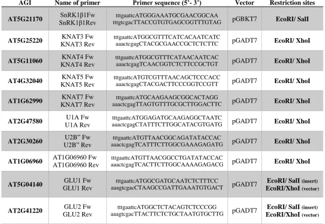
Table I: Sequence of the primers used in this work for PCR amplifications.
The restriction sites are represented in lowercase letters.
2.2. Yeast two-hybrid screen
2.2.1. Transformation of Y2HGold with pGBKT7-SnRK1β1
To identify proteins encoded by the Arabidopsis genome that interact with SnRK1β1, an Y2H screen was performed using a Mate & Plate™ Library - Universal Arabidopsis (Clontech), and a normalized library into pGADT7-RecAB (Clontech) constructed from mRNA isolated from 11 Arabidopsis tissues, mixed in equal quantities and transformed into yeast strain Y187. The bait construction BD-SnRK1β1 was used to transform Y2HGold yeast cells (Clontech), using the Yeastmaker Yeast Transformation System 2 (Clontech) ac-cording to the manufacturer's instructions. Briefly, 100ng of pGBKT7- SnRK1β1 were com-bined with 50µg of Yeastmaker Carrier DNA (Clontech), 50μl of yeast competent cells and 500μl of PEG/LiAc (40% PEG 3350, 0.1M lithium acetate in TE buffer) in a sterile tube (1.5 ml). After were added and mixed to join incubation at 30°C for 30 min, 20μl of DMSO were added and the mix was incubated at 42° for 20 min. After that, the tube was centrifuged (2000rpm, 5min) to obtain the pellet and the supernatant was removed. The pellet yeast cells
AGI Name of primer Primer sequence (5’- 3’) Vector Restriction sites AT5G21170 SnRK1β1Fw
SnRK1β1Rev
tttgaattcATGGGAAATGCGAACGGCAA
tttgtcgacTTACCGTGTGAGCGGTTTGTAG pGBKT7 EcoRI/ SalI
AT5G25220 KNAT3 Fw KNAT3 Rev
tttgaattcATGGCGTTTCATCACAATCATC
aaactcgagCTACGCGAACCGCTCTCTTC pGADT7 EcoRI/ XhoI
AT5G11060 KNAT4 Fw KNAT4 Rev
tttgaattcATGGCGTTTCATAACAATCAC
aaactcgagTCAACGGTCTCTTCCGCTGT pGADT7 EcoRI/ XhoI
AT4G32040 KNAT5 Fw KNAT5 Rev
tttgaattcATGTCGTTTAACAGCTCCCACC
aaactcgagCTACGACTTCCCGGTCCGTT pGADT7 EcoRI/ XhoI
AT1G62990 KNAT7 Fw KNAT7 Rev
tttgaattcATGCAAGAAGCGGCACTAGG
aaactcgagTTAGTGTTTGCGCTTGGACTTC pGADT7 EcoRI/ XhoI
AT2G47580 U1A Fw U1A Rev
tttgaattcATGGAGATGCAAGAGGCTAATC
aaactcgagCTATTTCTTGGCATACGTGATG pGADT7 EcoRI/ XhoI
AT2G30260 U2B” Fw U2B” Rev
tttgaattcATGTTAACGGCAGATATACCAC
aaactcgagTCATTTCTTGGCGAAAGAGATG pGADT7 EcoRI/ XhoI
AT1G06960 AT1G06960 Fw AT1G06960 Rev
tttgaattcATGTTAACGGCCTGATATACCAC
aaactcgagTCACTTCTTGGCAAAAGAGACG pGADT7 EcoRI/ XhoI
AT5G04140 GLU1 Fw GLU1 Rev
tttgaattcATGGCGATGCAATCTCTTTCC
aaagtcgacCTAAGCCGATTGAAATGTGACT pGADT7
EcoRI/ SalI (insert)
EcoRI/XhoI (vector)
AT2G41220 GLU2 Fw GLU2 Rev
tttgaattcATGGCTCTACAGTCTCCCGG
aaagtcgacTTACTTCTCTGCTAATGTGCTTG pGADT7
EcoRI/ SalI (insert)
were resuspended in 100μl and plated onto a 100mm plate with SD -Trp (SD-W) agar me-dium.
2.2.2. Mating and selection of clones
The screening of the Y2H library was performed using the Matchmaker® Gold Yeast Two-Hybrid System (Clontech) according to the manufacturer’s instructions. Briefly, 45ml of 2X YPDA (supplemented with 50μg/ml kanamycin) were inoculated with 5ml of a bait culture (with a cell density higher than 108 cells/ml) and an aliquot (1 ml) of the Y2H library, and incubated for 24h at 30°C with slowly shaking (40 rpm). After incubation, the culture was centrifuged (1000g for 10min). The supernatant was removed and the cell pellet was resuspended in 10ml of water. The cells were spread onto 150 mm diameter plates (200μl per plate) containing SD medium lacking leucine and tryptophan (SD-L-W) supplemented with X-α-GAL (10 mg/L) and Aureobasidin A (AbA) (200 µg/L) and the plates were incu-bated for 5 days at 30ºC. To control the mating efficiency and the number of diploids formed 100 µl of successive dilutions 10-1, 10-2, 10-3 and 10-4 of the resuspended cells were spread onto SD-L, SD-W and SD-L-W (SD-2) media plates and incubated for 3 days at 30ºC (Table II).
The blue colonies that grew on SD-L-W supplemented with X-α-GAL and AbA were patched out onto higher stringency SD medium lacking leucine, tryptophan, adenine and histidine (SD-L-W-A-H), also supplemented with X-α-GAL and AbA. Those colonies that continued to report blue colour were used for further analysis and identification of proteins that possibly interact with SnRK1β1.
Materials and Methods
22
Table II: Measurement of the viability of the Prey Library, the Bait and diploids expressing inter-acting proteins.
After proceed the mating between the two strains used, 100μl were plated in different media with four dilutions (10-1; 10-2; 10-3; 10-4).
Strain Plate on SD Minimal Agar
Medium Selects for
Y2HGold [pGBKT7-53 + SnRK1β1]
x
Y187[pGADT7-T + li-brary] with normalized
library SD -L pGADT7-T SD -W pGBKT7-53 SD -L-W Diploids containing pGBKT7-53 and pGADT7-T SD -L/-W/ X-α-Gal/AbA
Diploids that have also activated AbA resistance and α-galacto-sidase through proteprotein
in-teractions
2.2.3. Determination of transformation and mating efficiency
The total volume obtained after 20hr of mating (pelleted cells plus 10ml of medium) was measured to determine the mating efficiency and the number of colonies diploids. The number of screened clones (diploids) was calculated considering the total volume used, the plating volume, the number of cfu/ml of diploids and the dilution considered (Appendix 1). The mating efficiency was calculate using the ratio between number of cfu/ml of diploids and No. of cfu/ml grew in SD-Leu (SD-L) agar plate.
2.2.4. Yeast plasmid DNA extraction
The blue colonies that grown in SD -L-W-A-H (SD-4) medium supplemented with X-α-Gal and AbA, were used to inoculate 2ml of SD -L-W liquid medium that was incubated for 48h at 30ºC with shaking. DNA extraction was performed using a modified smash and grab method (Hoffman and Winston, 1987). After incubation cells were pelleted by centrif-ugation (1 min, 10000g) and resuspended in 200μl of lysis buffer (10mM Tris, pH 7.5; 1mM EDTA; 100mM NaCl; 1% of SDS; 2% of 100XTriton). After adding 200μl of glass beads and 200μl of phenol-chloroform (50:50) the mix was vortexed for 4 min. Then, after 200μl of TE was added and the mix was vortexed again for 1 minute. After centrifugation at RT for 5 min (13000g) the aqueous phase was transferred to another centrifuge tube and the DNA was precipitated adding 1/10 volume of NaAc 3M, pH=5.2 plus 3 volumes of 100% ethanol and incubating at -20°C for 30 min. Next, the mix was centrifuged at 4°C for 20 min
(13000g) and the supernatant was discarded. The pellet was washed with 750μl of 70% Eth-anol and dried. Finally the DNA was resuspended in 10μl of sterile water.
The cDNA cloned in each of the rescued plasmids was amplified by PCR using the
primers AAATATTCGATGATGAAGATACCCCACCAAACCCA and
TTTAG-TGAACTTGCGGGGTTTTTCAGTATCTACGAT, using the OneTaq® DNA Polymerase according to the manufacturer's instructions in a 50μl of reaction volume. The PCR reaction was performed using 35 cycles with denaturation at 94ºC for 30 sec an annealing/extension at 68°C for 4 min. The amplification product was analyzed by agarose gel electrophoresis. Sequencing of the PCR products was performed using the BigDye Terminator v1.1 Cycle Sequencing Kit (Thermo Fisher) according to the manufacturer's instructions.
2.3. Binary interactions
To study the possible interaction between SnRK1β1 and specific proteins, the Y2H technique was used. The cotransformation of yeast was done mainly according to the lithium acetate protocol (Gietz et al., 1997). Briefly, 25μl of competent cells Y2HGold were mixed with 1μg of each plasmid construction (bait and prey) and 700μl of PEG/LiAc. The mix was incubated at 30°C for 30 min and after further incubated 20min at 42ºC. Next, the cells were pelleted by centrifugation for 5 min (2000g) and the supernatant was discarded. The cells pellet was resuspended in 100μl of sterile water and spread into SD -L-W agar plate that were incubated for 2 days at 30°C. Next, 3 colonies were randomly picked and used to in-oculate 2ml of SD -L-W liquid medium that was incubated at 30ºC until the culture was saturated (48h). Finally, the interaction was determined by growth assay on SD -L-W-A-H agar medium, 3µl of 10-1, 10-2 and 10-3 dilutions of the saturated cultures were spotted onto the plates. As control the growth on SD -L-W medium was also tested.
2.4. Auxiliaries software’s
2.4.1. The Arabidopsis Information Resource (TAIR)
Identification of the sequences obtained was performed using the online BLAST pro-gram of the Arabidopsis Information Resource (TAIR)
(https://www.arabidop-Materials and Methods
24
2.4.2. The MIPS Functional Catalogue Database (FunCatDB)
Functional characterization of the identified genes was done using the online MIPS Functional Catalogue Database (FunCatDB) (http://mips.helmholtz-muenchen.de/fun-catDB/). Additionally this program provides statistics of the functional distribution of a given set of genes (Ruepp et al., 2004). In this case a set of 64 proteins was submitted and compare with the whole genome of Arabidopsis thaliana (Taxonomy ID: 3702)
2.4.3. The Arabidopsis Protein Phosphorylation Site Database (PhosPhAt
4.0)
The information about the experimentally-identified phosphorylation sites of pro-teins were obtained using the PhosPhAt 4.0 database (http://phosphat.uni-hohen-heim.de/phosphat.html). (Heazlewood et al., 2008; Durek et al., 2010; Zulawski et al., 2013)
3. Results
3.1. Identification of SnRK1β1 protein interactors
As previously mentioned in the introduction, the protein-protein interaction is one of the most common processes involved in the response to a certain stimuli (Baena-Gonzalez et al., 2007; Baena-Gonzalez and Sheen, 2008). Besides the importance of α-subunits in the AMPK/SNF1/SnRK1 complex, the regulatory subunits (β- and γ-subunits) are also very im-portant to activate the complex, to select the downstream targets, and to establish relevant interactions that are important for the regulation of gene expression during stress (Lovas et al., 2003; Gissot et al., 2006; Pierre et al., 2007; Li et al., 2009; Baena-Gonzalez, 2010; Ramon et al., 2013). Despite that, the knowledge about the molecular components involved in the stress signalling pathway is scarce. In order to identify new proteins that possibly interact with SnRK1β1 and, in this way, are involved in the stress-response, an Y2H screen was performed using SnRK1β1 as bait.
The mating efficiency was 4.4%, within the expected limits according to the manu-facturer of the kit used to perform the screen (2% to 5%). The number of diploid clones screened was equal to 4 million, four times the minimal number of clones recommended by the manufacturer of the kit used (see Appendix 1).
The blue colonies that grew in (SD -L-W) agar plates (150 mm plates) supplemented with X-α-Gal and AbA were patched out onto a higher stringency medium also supple-mented with X-α-Gal and AbA (SD -L-W-A-H agar plates). Of the 297 yeast colonies that continued to grow and presented blue color in this new medium, 72 were randomly selected to be further analyzed. The remaining colonies were used to inoculate SD-L-W liquid me-dium. After incubation for 2 days at 30ºC, glycerol was added (25% final concentration) and they were stored at -80 °C.
The analysis of the 72 selected colonies allowed the identification of 64 different proteins that possibly interact with SnRK1β1 (Table III). Two of these proteins, SnRK1.2 (KIN11) (Baena-Gonzalez et al., 2007; Fragoso et al., 2009; Williams et al., 2014) and Hsp70 (Slocombe et al., 2004) have previously been identified as SnRK1β1 interactors,
sug-Results
28
3.1.1. Functional categorization
A functional categorization of the 64 proteins identified was performed using the MIPS FunCat (Ruepp et al., 2004). A large number of these proteins are involved in metab-olism (33 proteins), namely of sugars, phosphates and fatty acids, in energy processes (14 proteins) like glycolysis, gluconeogenesis and oxidation of fatty acids, and in nutrient star-vation response (4 proteins) (table IV and Table III in Appendix 2). Some other of the iden-tified proteins are related to binding functions (27 proteins), cellular transport (11 proteins) and cell defense (13 proteins) (table IV and Table III in Appendix 2), being that all these proteins may be included in different categories. Taken together, these results are clearly in line with the importance of SnRK1 complex in the response to energy stress and the re-establishment of energy homeostasis.
Results
30
3.2. Yeast-two Hybrid System
Considering their relevance in stress response and/or their relationship with the en-ergy stress response, three different proteins were chosen to further analysis: the Spliceoso-mal protein U1A (At2g47580); KNOTTED1-like homeobox gene 5, KNAT5 (At4g32040); and Glutamate Synthase 1, GLU1 (At5g04140). The U1A and KNAT5 proteins are involved in transcription and DNA processing (Forment et al., 2002; Scofield et al., 2008), while GLU1 is related to metabolism having a crucial role in nitrogen, glutamate and amino acids metabolisms and is also related to stress response (Kissen et al., 2010; Hirel et al., 2011; Aliyev, 2012; Kangasjarvi et al., 2012). The reconfirmation of the interaction of those pro-tein with SnRK1β1 was performed using the Y2H system. Additionally, the interaction of
FUNCTIONAL CATEGORY Rel. SET (%) Rel. GENOME (%) P-VALUE
01 METABOLISM 39,2 21,2 1,24E-04
01.04 phosphate metabolism 11,9 4,2 2,76E-03
01.05 C-compound and carbohydrate metabolism 20,2 8,21 4,20E-04
01.05.02 sugar, glucoside, polyol and carboxylate metabolism 10,7 1,91 3,38E-05
01.05.02.07 sugar, glucoside, polyol and carboxylate catabolism 8,33 1,04 2,96E-05
01.05.25 regulation of C-compound and carbohydrate metabolism 3,57 0,65 1,80E-02
01.06.05 fatty acid metabolism 7,14 0,61 1,39E-05
01.07 metabolism of vitamins, cofactors, and prosthetic groups 4,76 1,6 4,64E-02
01.07.01 biosynthesis of vitamins, cofactors, and prosthetic groups 3,57 0,95 4,63E-02
02 ENERGY 16,6 4,44 1,89E-05
02.01 glycolysis and gluconeogenesis 4,76 0,88 6,62E-03
02.07 pentose-phosphate pathway 4,76 0,29 1,12E-04
02.10 tricarboxylic-acid pathway (citrate cycle, Krebs cycle, TCA cycle) 2,38 0,34 3,47E-02
02.25 oxidation of fatty acids 7,14 0,21 3,14E-08
14.07.03 modification by phosphorylation, dephosphorylation, autophosphorylation 5,95 2,04 2,90E-02
16 PROTEIN WITH BINDING FUNCTION OR COFACTOR REQUIREMENT 32,1 22,2 2,35E-02
18.01.01 regulation by modification 5,95 1,29 4,82E-03
18.02.09 regulator of transcription factor 3,57 0,93 4,42E-02
20.03.01.01 ion channels 2,38 0,38 4,26E-02
20.09.07.27 vesicle fusion 2,38 0,28 2,35E-02
20.09.16 cellular export and secretion 3,57 0,95 4,63E-02
20.09.16.09 vesicular cellular export 2,38 0,25 1,91E-02
20.09.18.09 vesicular cellular import 3,57 0,73 2,38E-02
20.09.18.09.01 endocytosis 3,57 0,73 2,38E-02
30.01.05 enzyme mediated signal transduction 7,14 2,45 1,74E-02
30.01.09 second messenger mediated signal transduction 4,76 0,91 7,42E-03
30.01.09.07 cAMP/cGMP mediated signal transduction 3,57 0,04 5,47E-06
32.01 stress response 15,4 6,31 2,27E-03
32.01.11 nutrient starvation response 4,76 0,12 3,87E-06
32.07.03 detoxification by modification 2,38 0,34 3,47E-02
32.07.07.07 superoxide metabolism 2,38 0,08 2,60E-03
34.07 cell adhesion 3,57 0,21 8,07E-04
34.07.02 cell-matrix adhesion 3,57 0,02 8,51E-07
40.01.05 growth regulators / regulation of cell size 3,57 0,55 1,15E-02
40.20 cell aging 5,95 0,35 1,20E-05
43 CELL TYPE DIFFERENTIATION 7,14 1,74 3,58E-03
43.01 fungal/microorganismic cell type differentiation 7,14 1,74 3,58E-03
43.01.03 fungal and other eukaryotic cell type differentiation 7,14 1,74 3,58E-03
43.01.03.05 budding, cell polarity and filament formation 5,95 0,86 8,15E-04
Table IV: The main overrepresented FunCat categories with a P-value < 0.05. The Categories in bold are the main category, the others are sub-categories.
other proteins of the same families with SnRK1β1, was also tested using the same method: U2 Small Nuclear Ribonucleoprotein B, U2B” (AT2G30260) and a RNA binding protein (AT1G06960); KNAT3 (AT5G25220), KNAT4 (AT5G11060) and KNAT7 (AT1G62990); and GLU2 (AT2G41220) respectively. All these proteins coding sequence were expressed as a fusion with the GAL4 activation domain (AD) using the pGADT7 vector while the coding sequence of SnRK1β1 was expressed in a fusion with GAL4 DNA-binding domain (BD) using the pGBKT7 vector.
As illustrated in figure 4, the Y2H assay shown that U1A interacts with SnRK1β1 corroborating the observations in Y2H Screen. The other two proteins tested, which are sim-ilar to U1A, also interact with the SnRK1β1 in the Y2H system, suggesting that they might be a target of SnRK1 (figure 4). pGBKT7 SnRK1β1 SnRK1β1 pGBKT7 pGBKT7 SnRK1β1
Bait (BD)
-L -W +A +H -L -W -A -HPrey (AD)
U1A U1A U2B” U2B” AT1G06960 AT1G06960
Figure 4: Protein-protein interaction between SnRK1β1 and U1A, U2B” and AT1G06960 (protein with RNA binding motif).
Interaction was determined by growth assay on SD -4 (-L -W -A –H) agar medium. 3μl of three serial dilutions (1/10, 1/100, 1/1000) of each saturated culture were spotted onto the plates. Cells transformed with the empty vector (pGBKT7) were used as a control.
Results
32
All KNAT proteins tested were able to interact with β-subunit in the Y2H system (Figure 5). Interestingly, the interaction seems to be stronger in the case of KNAT3 and KNAT7 compare to the interaction with KNAT5 (the protein identified in the screen) and KNAT4 evidencing the weaker interaction with SnRK1β1.
Finally, GLU1 and GLU2 also interact with SnRK1β1 in the Y2H system and GLU2 appears to have a stronger interaction with the bait in comparison with the GLU1 (figure 6).
All controls using the empty pGBKT7 and each of the prey construction were nega-tive, confirming that none of the proteins was able by itself to activate de Y2H reporter genes. pGBKT7 KNAT5 KNAT5 SnRK1β1 pGBKT7 KNAT3 KNAT3 SnRK1β1 pGBKT7 KNAT4 KNAT4 SnRK1β1 SnRK1β1 KNAT7 KNAT7 pGBKT7
Bait (BD)
-L -W +A +H -L -W -A -HPrey (AD)
Figure 5: Protein-protein interaction between SnRK1β1 and KNATs.
Interaction was determined by growth assay on SD -4 (-L -W -A -H) agar medium. 3μl of three serial dilutions (1/10, 1/100, 1/1000) of each saturated culture were spotted onto the plates. Cells trans-formed with the empty vector (pGBKT7) were used as a control
Since the β subunits probably facilitate the association of the complex with down-stream targets, after the interactions between the proteins previously mentioned and SnRK1β1 were verified, the Y2H system was also used to test the interaction between these proteins and de catalytic subunit. As shown in figure 7, none of the proteins mentioned pre-viously seem to interact with SnRK1.1, suggesting that the interaction of SnRK1β1 with these targets is independent of the presence of SnRK1.1. However, after a longer incubation (7 days at room temperature), some of these proteins, particularly the KNATs, showed evi-dences of a weak interaction with the α-subunit (SnRK1.1) (figure 8).
SnRK1β1 pGBKT7 SnRK1β1
pGBKT7
Bait (BD)
-L -W +A +H -L -W -A -HPrey (AD)
GLU1 GLU1 GLU2 GLU2
Figure 6: Protein-protein interaction between SnRK1β1 and GLUs.
Interaction was determined by growth assay on SD -4 (-L -W -A -H) agar medium. 3μl of three serial dilutions (1/10, 1/100, 1/1000) of each saturated culture were spotted onto the plates. Cells transformed with the empty vector (pGBKT7) were used as a control.
Results
34 SnRK1.1 KNAT5 SnRK1.1 GLU2 SnRK1.1 U2B” SnRK1.1 KNAT3 SnRK1.1 U1A SnRK1.1 AT1G06960 SnRK1.1 KNAT7 SnRK1.1 GLU1 SnRK1.1 KNAT4Bait (BD)
-L -W +A +H -L -W -A -HPrey (AD)
SnRK1.1 pGADT7
Figure 7: Protein-protein interaction between SnRK1.1 as a bait with all proteins stud-ied.
Interaction was determined by growth assay on SD -4 (-L -W -A –H) agar medium. 3μl of three serial dilutions (1/10, 1/100, 1/1000) of each saturated culture were spotted onto the plates. Cells transformed with the empty vector (pGADT7) were used as a control.
GLU2 U2B” KNAT3 U1A AT1G06960 KNAT7 GLU1 KNAT4 KNAT5 SnRK1.1 SnRK1.1 SnRK1.1 SnRK1.1 SnRK1.1 SnRK1.1 SnRK1.1 SnRK1.1 SnRK1.1
Bait (BD)
-L -W +A +H -L -W - A -HPrey (AD)
Figure 8: Protein-protein interaction between SnRK1.1 with all proteins studied under a longer incubation period.
Interaction was determined by growth assay on SD -4 (-L -W -A –H) agar medium. 3μl of three serial dilutions (1/10, 1/100, 1/1000) of each saturated culture were spotted onto the plates. The cells growth was verified after 7 days at room temperature.
4. Discussion and conclusions
4.1. Identification of SnRK1β1 protein interactors
The way plants respond to biotic and abiotic stresses has been studied for several years (Halford and Hardie, 1998; Serrano et al., 1999; Xiong et al., 2002; Schwachtje et al., 2006; Baena-Gonzalez et al., 2007; Polge and Thomas, 2007; Tuteja, 2007). This study is critical to improve crop production and to minimize the adverse effects of stress on plant growth and development (Serrano et al., 1999; Tuteja, 2007).
The SnRK1 complex has been defined as a central integrator of the stress re-sponse. Under stress conditions, SnRK1 activation induces a reprogramming of the tran-scriptome, by switching on/off specific genes, with a direct impact in the metabolic path-ways, energy cycles and carbon sources, that ultimately result in the re-establishment of energy homeostasis and allows the plants to tolerate these adverse conditions (Baena-Gonzalez et al., 2007). Several SnRK1 related genes involved in stress response have already been identified. However, the mechanisms involved are largely unknown and probably most of molecular components remain to be identified (Baena-Gonzalez and Sheen, 2008; Akpinar et al., 2012; Cho et al., 2012; Nietzsche et al., 2014).
In this study, as a primary approach, an Y2H screen was performed using SnRK1β1 subunit. It has been suggested that the beta subunits are involved in the recog-nition of the SnRK1 substrates (Hedbacker and Carlson, 2008; Li et al., 2009) and so the identification of downstream targets of SnRK1 could be expected. Of the 297 yeast colo-nies that grew in selective media, 72 were analyzed leading to the identification of 64 proteins that putatively interact with SnRK1. Noteworthy, functional characterization of these proteins shown that more than half of them are related to metabolism, namely of sugars, phosphates and fatty acids. These proteins are also involved in energy processes like glycolysis and gluconeogenesis, Krebs cycle and oxidation of fatty acids (table IV and table III in Appendix 2). These results are consistent with the important role played by SnRK1 in controlling metabolism under stress (Lovas et al., 2003; McKibbin et al., 2006; Pierre et al., 2007; Polge and Thomas, 2007; Baena-Gonzalez and Sheen, 2008; Li et al., 2009; Cho et al., 2012; Crozet et al., 2014) and suggests that some of these proteins are probably downstream targets of SnRK1. These results also support, at least partially,