RESUMO.- [Potencial de diferenciação in vitro de cé-lulas provenientes da geleia de Wharton de cordões umbilicais ovinos isolados em diferentes meios de cul-tivo.] A geleia de Wharton do cordão umbilical (GWCU) de mamíferos é uma fonte promissora de células multipo-tentes, sendo vantajosa por aspectos éticos, facilidade de coleta e não causar teratomas em ensaios pré-clínicos. Em ovinos, células multipotentes já foram isoladas de vários tecidos, no entanto, não existem relatos do isolamento a partir do cordão umbilical nesta espécie. O objetivo do pre-sente estudo foi investigar o melhor meio para o transporte do cordão umbilical, isolar e manter as células da GWCU
Potential for
in vitro
mesoderm differentiation of Wharton’s
jelly cells from ovine umbilical cord isolated in different
culture media
1Ronaldo P. Dias2*, Maria F.S. Teixeira2,3, Edmara C. Costa4, Anderson C. Farias2,
Dalva A.A. Azevedo2, Tereza D.F. Aguiar2 and Mariana A. Pinheiro5
ABSTRACT.- Dias R.P., Teixeira M.F.S., Costa E.C., Farias A.C., Azevedo D.A.A., Aguiar T.D.F. & Pi-nheiro M.A. 2016. Potential for in vitro mesoderm differentiation of Wharton’s jelly cells from ovine umbilical cord isolated in different culture media.Pesquisa Veterinária Bra-sileira 36(Supl.1):79-88. Programa de Pós-Graduação em Ciências Veterinárias, Laboratório de Virologia, Faculdade de Veterinária, Universidade Estadual do Ceará, Av. Silas Munguba, 1700, Itaperi, Fortaleza, CE 60740-000, Brasil. E-mail: [email protected]
The mammalian Wharton’s jelly of umbilical cord (WJUC) is a promising source of mul-tipotent cells, providing advantages due to ethical implications, ease of collection and the absence of teratomas in pre-clinical trials. Ovine multipotent cells have already been isola-ted from various tissues, however there are no reports using umbilical cords in this species. This study aimed to investigate the best medium to transport the umbilical cord, to isolate and maintain ovine WJUC cells and to compare in vitro growth and mesodermal differen-tiation potential. Eight ovine umbilical cords were obtained during parturition, sectioned and transported in six different media: MEM, low glucose DMEM, M199, RPMI 1640, PBS and saline. For each transportation medium, four culture media were used and the tis-sue was explanted in 24-well plates and cultured in MEM, low glucose DMEM, M199 and RPMI 1640, all with 10% FBS. Every experiment was conducted with low-passage (P2), investigating MTT viability during four days and adipogenic, chondrogenic and osteogene-sis differentiation was induced in vitro. The most effective transport medium (p<0.1) was low glucose DMEM. There was no bacterial or fungal contamination from collection. Cells from Wharton’s jelly of ovine umbilical cords collected at natural birth possess fibroblastic morphology and the capacity for in vitro differentiation into adipogenic, chondrogenic and osteogenic cell lines. MTT tests and in vitro differentiation experiments revealed that cell culture medium modulates the behavior of cells and is an important factor for proliferation and maintenance of multipotency. Low glucose DMEM was the most suitable medium for the isolation of cells from Wharton’s jelly of ovine umbilical cord.
INDEX TERMS: Sheep umbilical cord matrix, multipotent cells, adipogenesis, chondrogenesis, osteogenesis.
1 Received on November 14, 2015.
Accepted for publication on April 11, 2016.
2 Programa de Pós-Graduação em Ciências Veterinárias, Laboratório
de Virologia, Faculdade de Veterinária, Universidade Estadual do Ceará (UECE), Avenida Silas Munguba 1700, Itaperi, Fortaleza, CE 60740-000, Brazil. Pesquisa de doutorado com apoio da CAPES. *Corresponding au-thor: [email protected]
3 Bolsista de Produtividade em Pesquisa do CNPq.
4 Universidade da Integração Internacional da Lusofonia Afro-Brasileira,
Avenida da Abolição 3, Centro, Redenção, CE 62790-000, Brazil.
5 Bolsista de Iniciação Científica, Fundação Cearense de Apoio ao Desen
-volvimento Científico e Tecnológico (Funcap), Faculdade de Veterinária,
ovino em diferentes meios e comparar a proliferação e o potencial de diferenciação mesodermal in vitro. Oito cor-dões umbilicais foram obtidos, por ocasião do parto natu-ral, seccionados e transportados em seis diferentes meios que consistiram em MEM, DMEM baixa glicose, M199, RPMI 1640, PBS e soro fisiológico. Para cada meio de transporte foram utilizados quatro meios de cultivo, sendo o tecido explantado em placas de 24 poços e cultivados em MEM, DMEM baixa glicose, M199 e RPMI 1640, todos com 10% SFB. Todo o experimento foi realizado em baixa passagem (P2) investigando viabilidade pelo MTT por quatro dias além da indução à diferenciação adipogênica, condrogêni-ca e osteogênicondrogêni-ca in vitro.O meio de transporte mais efetivo (P<0,10) foi o DMEM baixa glicose. Não houve contamina-ções bacterianas ou fúngicas decorrentes da coleta. Células oriundas da geleia de Wharton do cordão umbilical ovino colhido por ocasião do parto natural possuem morfologia fibroblastóide e capacidade de diferenciação in vitro nas linhagens adipogênica, condrogênica e osteogênica.Os en-saios de MTT e diferenciação in vitro, revelaram que o meio
de cultura celular modula o comportamento destas células, sendo um fator importante tanto para a proliferação como para a manutenção da multipotência, destacando o DMEM baixa glicose como o meio mais adequado para o transpor-te e isolamento de células da geleia de Wharton do cordão umbilical ovino.
TERMOS DE INDEXAÇÃO: Matriz do cordão umbilical ovino, célu-las multipotentes, adipogênese, condrogênese, osteogênese.
INTRODUCTION
In recent years, fetal membranes such as the placenta, pla-cental membranes, umbilical cord, and amniotic fluid have been recognized as an unexplored resource for the field of regenerative medicine. These tissues have been shown to be a rich source of cells with elevated multipotency and potent immunosuppressive properties that demonstrate how these cells are exciting promising new tool for the tre-atment of diseases (Murphy & Atala 2013). In addition, the absence of ethical conflicts implied in collection and of te -ratogenic effects involved in the application are considera-ble advantages when compared to embryogenic stem cells and pluripotent-induced cells (Condic & Rao 2010).
Fetal membranes are a rich, non-invasive and easy to collect source of these cells, which present great capacity to proliferate and differentiate (De Vita et al. 2012). Umbi-lical cord is one example of tissue with promising results in research to provide new tools to treating several disea-ses. This fetal membrane is covered by amniotic epithelium that protects a gelatinous and elastic matrix composed by mucopolysaccharides (hyaluronic acid and chondroitin sulfate) referred as Wharton’s jelly (WJ), named after Tho-mas Wharton in 1856 (Forraz & McGuckin 2010).
Umbilical cord is routinely discarded after parturition and cell isolation is facilitated due to the location outside the fetal body. This excludes the necessity of invasive col-lection procedures, which often occurs when adult cells are isolated from other sources, possibly causing discomfort and risk to patients. Furthermore, the relatively large
vo-lume of umbilical cord and easy manipulation theoretically enhances the number of cells that may be extracted. This favors a substantial number of cells in several passages, avoiding long culture procedures and extended ex vivo ex-pansion (Iacono et al. 2015), which becomes a viable solu-tion for obtaining cells to veterinary practice.
The ability of multipotent cells to differentiate into a variety of connective tissue cell types has rendered them an ideal candidate cell source for clinical tissue regenera-tion strategies (Baksh et al. 2004). WJ cells are immuno--privileged, immunosuppressive, have a multipotent diffe-rentiation capacity and are readily available as a cell source (Taghizadeh et al. 2011). In addition, the capacity of human cells derived from Wharton’s jelly to proliferate and diffe-rentiate have been demonstrated and are similar to bone marrow cells (Baksh et al. 2007).
In humans, a simple method for obtaining cells from Wharton’s jelly of umbilical cord (WJUC) have been des-cribed without enzymatic digestion, but simply culturing the tissue explants (Hendijani et al. 2014). With goats, the beneficial effect of cells from WJUC in cutaneous cicatriza -tion have already been demonstrated (Azari et al. 2011). In another study, caprine cells from WJUC also enhanced the healing potential through xenotransplant in cutaneous wounds of rabbits, demonstrating improvement in the for-mation of epithelium and collagen with vascularization (Pratheesh et al. 2014).
With sheep, cells isolated from different sources have demonstrated in vitro capacity to differentiate into osteo-blasts, adipocytes and chondrocytes, such as periodontal ligament (Gronthos et al. 2006), bone marrow (Mccarty et al, 2009), adipose tissue, umbilical cord blood (Fadel et al, 2011), peripheral blood (Lyahyai et al. 2012), amniotic li-quid (Colosimo et al. 2013) and dermal skin (Jahroomishi-razi et al. 2015). However, there are no studies reporting the isolation and in vitro mesoderm differentiation of cells from Wharton’s jelly of ovine umbilical cord.
Sheep are an ideal model for studies in bone tissue en-gineering (Guo et al. 2004), and have also been proposed as an animal model for several applications in biomedical research with illnesses such as respiratory diseases (Sche-erlinck 2008), cardiomyopathies (Psaltis et al. 2010, Sill et al. 2011) and neurological disorders (Fauza et al. 2008). In addition, the use of these animals in orthopedic investiga-tions have improved due to their similarities with humans in weight, size, common structures and regenerative bone/ cartilage processes, leading to a potential in regenerative processes that have also been studied.
An important factor that must be considered in the culture of primary cells is the medium used. Studies have shown that it interferes in the differentiation potential of isolated cells (Wu et al. 2009, Lima et al. 2012, Ribeiro et al. 2013). In order to achieve success in tissues or organs engineering, it is imperative to provide a culture microen-vironment to the cells that supports their proliferation and maintains their differentiation capacity.
aspects in different culture media and their influence in the capacity for in vitro mesoderm differentiation.
MATERIALS AND METHODS
Collection and transportation of sheep umbilical cords. Ovine umbilical cords (n=8) were collected during parturition of healthy mixed breed sheep from two farms located in Guaiuba city territory, in Ceará State. Before rupture, each umbilical cord was pinched with sterile hemostatic scissors and cut in a section of 6 to 10cm. Then, the entire fragment was washed with sterile PBS ad-ded 2% of penicillin-streptomycin (P/S) (GibcoTM, Cat Nº
15140-122, 10,000 U/ml) for disinfection and blood removal. Each cord was immediately sectioned on a sterile petri dish in six fragments of 1cm2 , which were then placed in falcon tubes (TPP, Cat Nº.
91015), each containing a different transport solution. Media used for transportation were: MEM (GibcoTM, Cat Nº. 61100-061), low
glucose DMEM (GibcoTM, Cat Nº 31600-034), Medium 199 (M199)
(GibcoTM, Cat Nº 11150-067), RPMI 1640 (GibcoTM, Cat Nº.
31800-022), phosphate buffered saline (PBS) and saline 0.9% (S); to all of which 4% P/S and 1% amphotericin B were added. Tubes were placed in isothermal box containing recyclable ice (4oC) and sent
to laboratory of Virology from State University of Ceará for the re-maining procedure. This experiment was performed according to the current law and ethical principles for animal experimentation approved by the Ethics Committee for the Use of Animals of the State University of Ceará protocol number 12776979-0.
Transport media selection. In a 24-well plate (Kasvi, Cat. K12-024), fibroblast development was assessed in each cord frag -ment transported in each of the six different media. Every frag-ment from each cord originated explants, which were submitted to culture with four different cellular culture media, displaced in simple arrange (A6x4). Culture media used were: MEM, low glu-cose DMEM, M199 and RPMI 1640, and to all of these 10% fetal bovine serum (FBS) (GibcoTM, Cat Nº 10270-098), 2% P/S and 1%
amphotericin B were added. In each well, three explants were placed, cultivated for 30minutes for plate adherence in incubator at 37ºC with humid atmosphere and 5% CO2, then 0.5mL of cul-ture medium was added. Plates were observed daily in inverted microscope for fibroblast visualization around the explants. At day four, when the first cells were observed, scores were attribu -ted according to the presence and quantity of fibroblasts around the explants: zero cells (score 0), one to five cells (score 1), six to twenty cells (score 2) and uncountable number of cells (score 3). Results were then compared and the best transport medium for cord tissue was selected.
Cord cell culture in different media. After confluence, cells were trypsinized in 24-well plates (Kasvi, Cat. K12-024) and ex-panded in culture flasks of 25cm2 (Kasvi, Cat. K11-2050), each
containing one culture medium, MEM, low glucose DMEM, M199 or RPMI 1640, added 10% FBS, 2% P/S and 1% amphotericin B during the entire experiment. Cell counting in all experiments with plates was performed with Neubauer chambers.
MTT assay for cell viability evaluation (Thiazolyl Blue Tetrazolium Bromide). Growth of second passage (P2) ovine umbilical cord cells was assessed with a method that measures the activity of mitochondrial dehydrogenase enzyme, which when active is capable of metabolizing the MTT reagent resulting in a colored compound named formazan. Therefore, 100µL aliquots of cell suspension in culture medium (MEM, low glucose DMEM, M199 or RPMI 1640) added 10% FBS, 2% P/S and 1% amphote-ricin B, in a concentration of 1x10⁵ cells/mL were placed in
96-well microplates (Kasvi, Cat. K12-096). For each culture medium, four repetitions in triplicate were used, resulting in 12-wells/ day. After every 24h of incubation, the culture medium in the well
was replaced by 100µL of the same medium added MTT (Sigma, Cat. M2003) in a 0.5mg/mL concentration. Microplates were in-cubated for 4h at 37°C to allow MTT permeation and formation of formazan crystals. Then, media containing MTT were removed and formazan crystals were solubilized with 100µL of DMSO un-der stirring for 5 minutes. Spectrophotometry analysis was per-formed with microplate reader (Metertech model ∑ 960) with an absorbance of 600nm. Readings were performed with 24, 48, 72 and 96 hours of culture.
Differentiation in mesoderm cell line. Wharton’s jelly cells cultured in second passage in four distinct media were used to identify their potential for in vitro differentiation into osteocytes, adipocytes and chondrocytes.
Osteogenic differentiation. Osteogenesis was performed using a differentiation kit (StemPro Osteogenesis Kit, Gibco, Cat. Nº A10072-01). Cells were trypsinized and placed in 24-well pla-tes (Kasvi, Cat. K12-024) in a 5x10³ cells/cm2 density with culture
medium added 10% FBS, antibiotics and antifungal, all incubated in humid atmosphere with 5% CO2 at 37°C.
After 24 hours, each medium was replaced by the osteoge-nesis-inducing medium in the wells of the differentiation expe-riment, while control wells remained with maintenance media MEM, low glucose DMEM, M199 and RPMI 1640. Differentiation as well as maintenance media were replaced every two to three days. After 21 days, cells were fixated in 4% buffered formalin for 30 minutes and then washed with distilled water. Alizarin Red S 2% solution (pH 4.2) (Sigma-Aldrich, Cat. A5533) was added to each well for three minutes and then three washings were perfor-med with distilled water.
Adipogenic differentiation. Adipogenesis was performed using a differentiation kit (StemPro Adipogenesis Kit, Gibco, Cat. Nº A10070-01). Cells were trypsinized and placed in 24-well pla-tes (Kasvi, Cat. K12-024) in a 1x10⁴ cells/cm2 density in culture
media added 10% FBS, antibiotics and antifungal in humid atmos-phere with 5% CO2 at 37° C.
After 24 hours, media were replaced by adipogenesis-indu-cing medium in the wells of the differentiation experiment, while control remained with maintenance media. Differentiation and maintenance media were replaced every two to three days.
After 14 days, cells were fixated with 10% buffered formalin for 10 minutes, followed by two washings with PBS. Oil Red O so-lution (Sigma, Cat. O1391) was added to the cells for 15 minutes and then washed twice with distilled water. Counter coloration was performed with Mayer’s hematoxylin (Sigma, Cat. MHS16) for three minutes.
Chondrogenic differentiation. Chondrogenesis was indu-ced using micromass technique in 24-well plates (Kasvi, Cat. K12-024) with a chondrogenesis-inducing kit (StemPro Chon-drogenesis Kit, Gibco, Cat Nº A10071-01). Cells were trypsini-zed and a suspension of 1,6x10⁷ cells/mL was prepared. Three
micromass cultures were prepared in each well, using three 5µl drops of the cell suspension. After two hours, chondrogenesis--inducing medium was added to wells and replaced every two to three days. Control cells were submitted to the same protocol, however after two hours the same maintenance media in which they were cultured were added. After 16 days, media were re-moved from the wells, which were washed with PBS and micro-masses were fixated in 4% formalin for 30 minutes. Following, washing with PBS was performed and Alcian Blue solution (Sig-ma, Cat. B8438) in 3% acetic acid was added for 30 minutes for coloration. Then, three washings in wells were performed with 3% acetic acid and then distilled water was added to neutralize the acidity.
whi-ch were expressed as scores and the difference between means was determined by Kruskal-Wallis test. Significance level was set at p<0.10. ANOVA parametric test for inequality of popula-tion means, Bartlett’s test for inequality of populapopula-tion variances and Mann-Whitney/Wilcoxon two-sample test were applied for multiple comparisons. In order to detect statistical differences in MTT assay, a p<0.05 significance level was considered by Epi Info 7 processing.
RESULTS
Transport and isolation
Cells were isolated from Wharton’s jelly of ovine um-bilical cord at natural birth using an explant culture pro-tocol without enzymatic digestion. All samples presented adhesion to the plastic surface of the culture plates and fibroblastic morphology. For each transport medium, four culture media were tested. Cells initiated migration from
the edge of the tissue fragments of Wharton’s jelly and first fibroblastic cells were observed after four days of culture. Scores of 0-3 were assigned to each treatment (Table 1) and the best results were observed in the wells with ex-plants transported with low glucose DMEM (p<0.10) with an intermediate classification (19.6), followed by RPMI 1640 (17.0), MEM (12.2), M199 (11.5), SF (7.6) and PBS (7.0). Cells obtained from transportation with low glucose DMEM were selected to continue the experiment. After 16 days, single layers cultured in four different media achie-ved 100% confluence (Fig.1) and then were transferred to tissue culture flasks.
MTT essay for cell viability
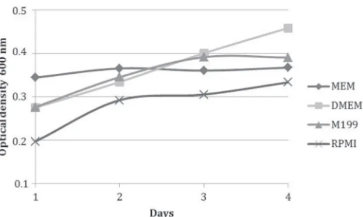
Culture with MEM provided more viable cells after trypsinization (day 1) with an average optical density of 0.344, followed by M199 with 0.276, low glucose DMEM with 0.275 and RPMI 1640 with 0.197 (Fig.2). Growth was analyzed every 24 hours, and low glucose DMEM maintai-ned growth, with the lowest decrease in optical density percentage during the four days, followed by RPMI 1640, M199 and MEM (Table 2). Optical density was significantly different (p<0.05) among media in all days of the experi-ment.
Adipogenic differentiation
After 14 days of adipogenic induction, cells isolated in MEM, low glucose DMEM and M199 presented adipogenic differentiation capacity, however cells isolated in RPMI
Fig.1. (A) Morphology of ovine WJUC cells at day 16 of culture in MEM, (B) low glucose DMEM, (C) M199 and (D) RPMI 1640. P0. 100x, Bars: 100µm.
Fig.2. Viability assessment with MTT test of ovine WJUC cells cul-tured in MEM, low glucose DMEM, M199 and RPMI 1640 du-ring four days.
Table 2. Growth analyzed by absorbance percentage of MTT test results performed in fibroblastic cells from ovine WJUC cells cultured in MEM, low glucose DMEM, M199 and RPMI
1640 during four days
Day MEM Low Glucose DMEM M199 RPMI
ABS Growth ABS Growth ABS Growth ABS Growth
means % means % means % means %
01 0.344 - 0.275 - 0.276 0.197
02 0.365 5.87a 0.334 21.37a 0.345 25.00a 0.262 33.08a
03 0.360 -1.35b 0.400 19.67b 0.391 13.14b 0.305 16.34b
04 0.367 1.99c 0.458 14.36b 0.390 -0.33c 0.333 9.18c
ABS = Absorbance. Distinct letters in sequence within columns mark sta-tistical difference. (p<0.05).
Table 1. Means and total score classification assigned to fibroblastic cell quantities around Wharton’s jelly explants
from ovine umbilical cords at day four of culture with samples transported in MEM, low glucose DMEM, RPMI
1640, M199, PBS and S
Transport media Culture media Classification Individual classification TOTAL MEANS
MEM DMEM M199 RPMI
MEM 8 11.5 21.5 8 49 12.2
DMEM 14 19.5 21.5 23.5 78.5* 19.6*
RPMI 11.5 16.5 23.5 16.5 68 17.0
M199 16.7 5 19.5 5 46.2 11.5
PBS 8 16.5 1 2.5 28 7.0
S 11.5 11.5 5 2.5 30.5 7.6
Fig.3. Adipogenic differentiation of ovine WJUC cells confirmed by the visualization of intracytoplasmic lipid drops stained in red (arro -ws). (A) Isolated cells MEM, (B) low glucose DMEM, (C) M199 and (D) RPMI exposed to adipogenesis-inducing medium. (E) Control wells with ovine WJUC cells not induced to differentiation and cultured in MEM, (F) low glucose DMEM, (G) M199 (G) and (H) RPMI 1640, P2, day 14. Stained with Oil Red O and counterstained with hematoxylin. 400x, Bars: 25µm.
Fig.5. Chondrogenic differentiation of ovine WJUC cells confirmed by the visualization of a blue coloration, which indicates proteoglycan syn -thesis by chondrocytes. (A) Chondrogenic islands formed by cells isolated in MEM, (B) low glucose DMEM, (C) M199 and (D) RPMI 1640, and exposed to chondrogenesis-inducing medium. P2, day 16. 400x. Bars: 25µm. (E) Control wells with ovine WJUC cells not induced to di-fferentiation and cultured in MEM, (F) low glucose DMEM, (G) M199 and (H) RPMI 1640. P2, day 16. 100x, Alcian Blue stain, Bars: 100µm.
1640 did not. Differences were observed in the adipogenic differentiation capacity of cells due to the formation of in-tracytoplasmic lipid drops, which were stained by Oil Red O solution. Cells isolated in low glucose DMEM presented the highest concentration of these drops, followed by M199 and MEM isolated cells. All adipogenesis-induced cells presented morphological alterations of polygonal shape (Fig.3A-D) and control group cells remained with fusiform fibroblastic morphology (Fig.3E-G). However, cells from RPMI 1640 control presented an irregular and less stre-tched morphology (Fig.3H). Adipogenic differentiation did not occur in any of the control wells.
Chondrogenic differentiation
In 13 hours after the addition of chondrogenesis-indu-cing medium, drops in cells isolated in low glucose DMEM aggregated into micromasses, followed by MEM isolated cells in 19 hours and M199 along with RPMI 1640 only in 34 hours (Fig.4A-D).
Drops in cells from control wells also agglutinated, ho-wever a single layer of fibroblasts grew from them, while micromass cells detached along the experiment (Fig.4E-H). After 16 days of differentiation, Alcian blue coloration was performed directly onto the plates, which allowed the visu-alization of intense blue coloration indicative of proteogly-can synthesis by chondrocytes in cells isolated and main-tained in MEM, low glucose DMEM, M199 and RPMI 1640 (Fig.5A-D). In control wells, the blue color was discreet or absent (Fig.5E-H).
Osteogenic differentiation
After 21 days of osteogenesis induction, all isolated cells presented the capacity to differentiate (Fig.6A-D), however a superior quantity of extracellular calcium deposition was observed, stained red in cells isolated and maintained with low glucose DMEM, indicating that these cells differentia-ted into functional osteoblasts in higher proportion. Cells isolated in MEM presented the lowest quantities of calcium deposition among treatments. Cells isolated in RPMI 1640 showed a dispersed calcium deposition in comparison to other groups (Fig.6D). There was no calcium deposition in control groups (Fig.6E-G), except in cells cultured in RPMI 1640, which presented extracellular calcium deposition (Fig.6H).
DISCUSSION
Multipotent cells have a lot of potential for several cell--dependent therapies and the current isolation techniques provide low quantities of these cells, which may be impro-ved with in vitro expansion. However, the potential to ex-pand is limited by in vitro aging, which leads to a loss of multipotency and replicative senescence. Stress induced by culture conditions is probably one of the main factors that affect these phenomena, which may be reduced through improvement of culture techniques (Stolzing et al. 2006). This study describes collection, isolation and in vitro me-sodermal differentiation of cells from ovine Wharton’s jelly maintained in different culture media that have different concentrations of amino acids, vitamins, ions and calories.
Caloric restriction is the only method established for slowing down aging and extending cellular lifespan and it has been proven that reducing glucose concentrations lead to an apoptosis reduction and improvement of cellu-lar proliferation (Stolzing et al. 2006). In addition, calcium and phosphate concentrations also influence growth and differentiation, and optimal concentrations for calcium ion of 1.8mM and for phosphate ion 0.09mM have been estab-lished. Changes in these concentrations may lead to inhibi-tion in growth and/or differentiainhibi-tion capacity in these cells (Liu et al. 2009).
Several culture media have been used in studies with cells from Wharton’s jelly of mammalian umbilical cords, among which are α-MEM, MSCBM (Passeri et al. 2009), DMEM/TCM199 (Iacono et al. 2012), high glucose DMEM (Hendijani et al. 2014) and DMEM (Sreekumar et al. 2014). The media used in the present study, which are MEM, low glucose DMEM, M199 and RPMI 1640, are usually used due to their capacity of supporting growth of different types of cells. However, there are differences in the compositions of these culture media, which may influence proliferation, vi -ability and in vitro cellular differentiation (Wu et al. 2009). Choosing an adequate culture medium is an important step for success in cell culture (Wu et al. 2009), therefore, this study aimed to investigate which medium is ideal for
in vitro culture of ovine umbilical cord. MEM is the most commonly used medium among others in cellular culture and is considered to have a low concentration of phosphate (1mM) and glucose (1g/L) and high calcium concentration (1.8mM) (Lopez-Cazaux et al. 2006). Low glucose DMEM is unique among other media due to the elevated amino acid and vitamins concentrations, which are four times higher than MEM, while possessing the same levels of glucose, cal-cium and phosphate. M199 has the same levels of glucose, calcium and phosphate then MEM and low glucose DMEM; however, it possesses additional exclusive components, which are adenine, adenosine, hypoxanthine, thymine and additional vitamins (Morgan & Morton 1957). RPMI 1640 medium contains biotin, vitamin B12 and PABA, which are not found in other media, and has nutritional values sim-ilar to MEM, but different ionic concentrations. This me-dium has a low calcium concentration (0.8mM) and high phosphate concentration (5mM), however with twice the glucose concentration (2g/L) when compared to MEM, low glucose DMEM and M199 (Lopez-Cazaux et al. 2006).
Iacono et al. (2012) reported problems with bacterial and yeast contamination in the culture of equine Wharton’s jelly cells by the microbiota in the birth canal and environ-mental conditions of collection, even with immersion of umbilical cords for 10 minutes in 70% alcohol. Passeri et al. (2009) tested three transportation and isolation protocols of equine umbilical cords to avoid fungal and bacterial con-taminations, and the best results were obtained with im-mersion of the umbilical cords overnight in the transport medium containing 5% P/S and 2% amphotericin B.
in transport solutions containing 4% P/S and 1% ampho-tericin B.
The long distances between the locations in which an-imals are housed and the laboratory demand ideal trans-port conditions. Therefore, the study was performed to assess the effects of transport media in the development of first cells with in vitro culture as a strategy to improve collections, and low glucose DMEM was significantly su -perior (p<0.10). Explants from umbilical cords that were transported in low glucose DMEM produced more prima-ry cells after four days of culture when compared to oth-er media used in transportation, which woth-ere RPMI 1640, MEM, M199, PBS and S, as shown in Table 1. Sigalas et al. (2004) reported that the culture medium DMEM preserved better the cells from human periodontal ligament stored at room-temperature and in ice for one hour, after which su-perior viability was observed in 0, 24 and 48 hours of cul-ture and these results corroborate with the present study.
The isolation method used in this study was efficient, simple and consistent, producing great quantities of ho-mogeneous fibroblastic cell populations in the four media studied, without visible morphological differences (Fig.1). However, in second passage, morphological differences were observed. Cells cultured in low glucose DMEM pre-sented a narrow and stretched aspect, while cells cul-tured in MEM and M199 revealed a discreet cytoplasm enlargement close to the nucleus position, and cells iso-lated in RPMI 1640 presented a disorganized morphology, with a round and irregular conformation when confluent (Fig.3E-H). These findings corroborate with other studies that demonstrated that the number of passages (Maciel et al. 2014) and the culture medium (Fong et al. 2007, Ribeiro et al. 2013) influence the morphology of cells during cul -ture.
No difference in the duration necessary to obtain con-fluence was observed and in day 16, primary cells (P0) of Wharton’s jelly cultured in MEM, low glucose DMEM, M199 and RPMI 1640 achieved 100% of confluence, when were trypsinized and transferred to culture flasks of 25cm2.
Metabolic assessment by MTT test demonstrated that cells presented a higher proliferation (p<0.05) when cul-tured in low glucose DMEM, followed by RPMI 1640, M199 and MEM, due to the minor decrease in growth percentage during culture, maintaining 14.36% of growth at day four, while MEM, M199 and RPMI 1640 grew 1.99%, -0.33% and 9.18%, respectively at the same day (Table 2). However, culture with medium RPMI 1640 revealed that after tryp-sinizations, many cells remained in suspension, indicating cellular death, which was confirmed with a minor absor -bance in MTT test with 24 hours. In this day, optical densi-ty obtained with RPMI 1640 medium was the lowest, with an average of 0.197 when compared to low glucose DMEM, which was 0.275, while M199 was 0.276 and the higher was MEM with 0.344, which presented the best support to trypsinization process (Fig.2 and Table 2).
Multipotent cells tend to decrease their multipotency over culture duration (Liu et al. 2004). Therefore, a dif-ferentiation assay with three mesodermal cell lines was performed in low passage (P2) in order to solely verify the
effects of culture media on cell isolation and maintenance of multipotency.
In adipogenesis induction, only the cells cultured in RPMI 1640 did not differentiate, not presenting intracyto-plasmic lipid drops stained red by Oil Red O (Fig.3D). Low glucose DMEM and M199 allowed cells to respond better to this induction, with a discreet but visible superiority when compared to MEM (Fig.3B,C). These data corroborate with the methodology proposed by Carswell et al. (2012), which indicate both DMEM and M199 as the ideal media for iso-lation and culture of adipocytes. Janderová et al. (2003) report a superior differentiation in human mesenchymal stem cells into adipocytes provided by M199 added with adipogenic factors, when compared to high glucose DMEM. There was no adipogenic differentiation in any of the neg-ative controls.
The best method for in vitro chondrogenic studies is micromass culture (Zhang et al. 2010). Although there are no studies correlating the formation speed of cellular ag-gregates with a chondrogenic differentiation potential of multipotent cells, in this study there was difference in this behavior specifically concerning time. After 13 hours with the chondrogenesis-inducing medium, cells isolated in low glucose DMEM coalesced forming micromasses, followed by MEM cells, after 19 hours, and M199 and RPMI 1640 cells after 34 hours.
Miura e Shiota (2000) studying micromasses in vitro
demonstrated that cells from bone marrow of rat fetuses presented different cellular motility during migration for chondrogenic islands formation, depending if they were cultured in liquid medium or gel (agarose or collagen), re-porting the occurrence of chondrogenic islands in 24 hours of culture. In addition, Awad et al. (2000), observed that contraction kinetics vary according to the initial cellular density of the culture, demonstrating a shorter time to the formation of chondrogenic aggregates, when cells are seed-ed in high density.
Chondrogenic aggregates are a consequence of the sep-aration of chondrogenic cells from a heterogeneous pop-ulation (Cottrill et al. 1987a). The presence of myofibro -blasts may affect in vitro morphogenesis of cartilage and micromass behavior reflects the interactions with a differ -ent cell population (Cottrill et al. 1987b). This possible cel-lular heterogeneity is reinforced by the fact that tissue from Wharton’s jelly of umbilical cords have stromal cells that, during pregnancy, acquire specific characteristics of myo -fibroblasts with the objective of protecting umbilical veins from compressions, but also serving as a niche for mesen-chymal stem cells (Kobayashi et al. 1998).
Therefore, different times in which cells coalesce may have resulted from a cellular heterogeneity, in a greater or minor proportion, caused by different culture medium compositions in which the ovine WJUC cells were isolat-ed and maintainisolat-ed. At the end of 16 days, positive intense marking with Alcian blue was observed confirming chon -drogenesis in all four treatments (Fig.5A-D).
which cells are isolated and maintained affects, maintain-ing or not, the capacity for in vitro osteogenic differentia-tion. The visualization in red of the extracellular calcium by alizarin red demonstrated the superiority of low glucose DMEM in the isolation and maintenance of the osteogenic differentiation capacity of ovine WJUC cells.
The data from this study corroborate with the findings of Wu et al. 2009, which reported in canine periosteum cells the superiority of low glucose DMEM for cellular dif-ferentiation when compared to RPMI 1640. In addition, these authors reported that mineralized nodules from cul-ture with RPMI 1640 were distributed in a more dispersed pattern. This behavior was also demonstrated in this study, in which extracellular calcium deposition occurred in a less concentrated and more dispersed manner on the single lay-er of cells isolated in this medium (Fig.6D).
Osteogenic differentiation in cells isolated and main-tained with M199 presented inferior quantities of extra-cellular calcium deposition when compared to DMEM and RPMI, followed by MEM maintained cells, which demon-strated the least effective capacity of osteoblast differ-entiation among the four tested media (Fig.6A-D). These findings disagree with the results from Lopez-Cazaux et al. (2006), who reported the superiority of MEM in com-parison with RPMI in proliferation and differentiation into odontoblast-like cells from human dental pulp. Possibly, cells from different origins have distinct nutritional re-quirements, which reflect in a behavioral variation when using the same culture medium.
In the control group negative for osteogenic differen-tiation, extracellular calcium deposition did not occur, except in cells cultured in RPMI, which presented posi-tive red markings (Fig.6H), however these cells were not exposed to the osteogenesis-inducing medium. Sponta-neous mineralization in vitro and without induction with β-glycerophosphate or dexamethasone have been reported in cellular culture of human dental pulp (Alliot-Licht et al. 2001) and canine periosteum (Wu et al. 2009), both cul-tured in RPMI 1640 medium. The hypothesis created by one of the authors for this phenomenon is the presence of pericytes or myofibroblasts that incorporate calcium phosphate from nodule formation that appear when cells remain in a long period at 100% of confluence. Also, spon -taneous mineralization is cell-density dependent and our study demonstrated that culture medium is an important factor as well, since there was no calcium deposition in the control group of other treatments.
Low glucose DMEM is a medium with caloric restric-tion, only 1g/L of glucose, and ideal concentrations of phos-phate and calcium for multipotent cells culture, however it is also unique for possessing four times the quantity of ami-no acids and vitamins. When compared to MEM and M199, which has the same glucose, phosphate and calcium con-centrations, low glucose DMEM was superior in this study, demonstrating the positive effect caused by the superior concentration of amino acids and vitamins in maintaining cellular multipotency . RPMI 1640 induced a spontaneous osteogenic differentiation, which probably hindered the adipogenic differentiation in cells isolated in this medium,
and therefore does not preserve multipotency of cells from ovine Wharton’s jelly.
Based on our findings, cells from Wharton’s jelly of ovi -ne umbilical cord present different potentials for in vitro
differentiation depending on the culture medium used in isolation and maintenance. Low glucose DMEM was the most adequate medium for culture, providing the most in-tense cellular proliferation and best results in adipogenic, chondrogenic and osteogenic in vitro differentiations.
CONCLUSIONS
Cells from Wharton’s jelly of ovine umbilical cords col-lected at natural birth possess fibroblastic morphology and the capacity for in vitro differentiation into adipogenic, chondrogenic and osteogenic cell lines.
Cellular culture medium modulates behavior of these cells and is a major factor for proliferation and mainte-nance of multipotency, for which low glucose DMEM is the most adequate for transportation and culture of cells from Wharton’s jelly of ovine umbilical cords.
Acknowledgments- The authors would like to thank CNPq (Universal
proc. 487425/2012-0) and AUXPE-PROEX 533/2014 for the financial su -pport, and to CAPES for granting the doctoral scholarship.
Conflict of interest statement.- The authors have no competing
inte-rests.
REFERENCES
Alliot-Licht B., Hurtrel D. & Gregoire M. 2001. Characterization of a-smooth muscle actin positive cells in mineralized human dental pulp cultures. Arch. Oral Biol. 46:221-228.
Awad H.A., Butler D.L., Harris M.T., Ibrahim R.E., Wu Y., Young R.G., Kadiya-la S. & Boivin G.P. 2000. In vitro characterization of mesenchymal stem cell-seeded collagen scaffolds for tendon repair: effects of initial seeding density on contraction kinetics. J. Biomed. Mater Res. 51:233-240. Azari O., Babaei H., Derakhshanfar A., Nematollahi-Mahani S.N.,
Poursa-hebi R. & Moshrefi M. 2011. Effects of transplanted mesenchymal stem
cells isolated from Wharton’s jelly of caprine umbilical cord on cutane-ous wound healing; histopathological evaluation. Vet. Res. Commun. 35(4):211-22.
Baksh D., Yao R. & Tuan R.S. 2007. Comparison of proliferative and mul-tilineage differentiation potential of human mesenchymal stem cells derived from umbilical cord and bone marrow. Stem Cells 25(6):1384-1392.
Baksh D., Song L. & Tuan R.S. 2004. Adult mesenchymal stem cells: char-acterization, differentiation, and application in cell and gene therapy. J. Cell Mol. Med. 8(03):301-316.
Carswell K.A., Lee M. & Fried S.K. 2012. Culture of isolated human adipo-cytes and isolated adipose tissue. Methods Mol. Biol. 806:203-214. Colosimo A., Russo V., Mauro A., Curini V., Marchisio M., Bernabò N., Alfonsi
M., Mattioli M. & Barboni B. 2013. Prolonged in vitro expansion partially affects phenotypic features and osteogenic potential of ovine amniotic
fluid-derived mesenchymal stromal cells. Cytotherapy 15(8):930-950.
Condic M.L. & Rao M. 2010. Alternative sources of pluripotent stem cells:
Ethical and scientific issues revisited. Stem Cells Dev. 19(8):1121-1129.
Cottrill C.P., Archer C.W., Hornbruch A. & Wolpert L. 1987a. The differen-tiation of normal and muscle-free distal chick limb bud mesenchyme in micromass culture. Dev. Biol. 119:143-156.
Cottrill C.P., Archer C.W. & Wolpert L. 1987b. Cell sorting and chondro-genic aggregate formation in micromass culture. Dev. Biol. 122:503-515. De Vita B., Campos L.L., Listoni A.J., Maia L., Freitas N.P.P., Alvarenga F.L.
tronco mesenquimais para a medicina veterinária equina. Vet. Zootec. 19(1):8-22.
Fadel L., Viana B.R., Feitosa M.L., Ercolin A.C., Roballo K.C., Casals J.B., Pie-ri N.C., Meirelles F.V., Martins D.S., Miglino M.A. & Ambrosio C.E. 2011. Protocols for obtainment and isolation of two mesenchymal stem cell sources in sheep. Acta Cir. Bras. 26:267-273.
Fauza D.O., Jennings R.W., Teng Y.D. & Snyder E.Y. 2008. Neural stem cell delivery to the spinal cord in an ovine model of fetal surgery for spina
bifida. Surgery 144:367-373.
Fong C.Y., Richards M., Manasi N., Biswas A. & Bongso A. 2007. Compara-tive growth behaviour and characterization of stem cells from human Wharton’s jelly. Reprod. BioMed. Online 15(6):708-718.
Forraz N. & McGguckin C.P. 2010. The umbilical cord: a rich and ethical stem cell source to advance regenerative medicine. Cell Prolif. 44(1):60-69. Guo X., Wang C., Duan C., Descamps M., Zhao Q., Dong L., Lu S., Anselme
K., Lu J. & Song Y.Q. 2004. Repair of osteochondral defects with autol-ogous chondrocytes seeded onto bioceramic scaffold in sheep. Tissue Eng. 10:1830-1840.
Gronthos S., Mrozik K., Shi S. & Bartold P.M. 2006. Ovine periodontal lig-ament stem cells: Isolation, characterization, and differentiation poten-tial. Calcif. Tissue Int. 79:310-317.
Hendijani F., Sadeghi-Aliabadi H. & Javanmard S.H. 2014. Comparison of human mesenchymal stem cells isolated by explant culture method from entire umbilical cord and Wharton’s jelly matrix. Cell Tissue Bank 15:555-565.
Iacono E., Brunori L., Pirrone A., Pagliaro P.P., Ricci F., Tazzari P.L. & Merlo B. 2012. Isolation, characterization and differentiation of mesenchymal
stem cells from amniotic fluid, umbilical cord blood and Wharton’s jelly
in the horse. Reproduction 143:455-468.
Iacono E., Rossi B. & Merlo B. 2015. Stem cells from foetal adnexa and fluid
in domestic animals: An update on their features and clinical applica-tion. Reprod. Dom. Anim. 50:353-364.
Jahroomishirazi R., Bader A., Ebert S., Schmidt C., Sedaghati B., Schulz-Sieg-mund M. & Zscharnack M. 2015. Isolation and characterization of cd271+ stem cells derived from sheep dermal skin. Cells Tissues Organs
200(2):141-152.
Janderová L., McNeil M., Murrell A.N., Mynatt R.L. & Smith S.R. 2003. Hu-man mesenchymal stem cells as an in vitro model for huHu-man adipogen-esis. Obes Res. 11:65-74.
Kobayashi K., Kubota T. & Aso T. 1998. Study on myofibroblast differentia -tion in the stromal cells of Wharton’s jelly Expression and localiza-tion of a-smooth muscle actin. Early Human Develop. 51:223-233.
Lima S.A.F., Wodewotzky T., Lima-Neto J.F., Beltrão-Braga P.C.B. & Alvaren-ga F.C.L. 2012. Diferenciação in vitro de células-tronco mesenquimais da medula óssea de cães em precursores osteogênicos. Pesq. Vet. Bras. 32(5):463-469.
Liu L., DiGirolamo C.M., Navarro P.A.A.S., Blasco M.A. & Keefea D.L. 2004.
Telomerase deficiency impairs differentiation of mesenchymal stem
cells. Exp. Cell Res. 294(1):1-8.
Liu Y.K., Lu Q.Z., Pei R., Ji H.J., Zhou G.S., Zhao X.L., Tang R.K. & Zhang M. 2009. The effect of extracellular calcium and inorganic phosphate on the growth and osteogenic differentiation of mesenchymal stem cells in vitro: implication for bone tissue engineering. Biomed. Mater. 4:1-8.
Lopez-Cazaux S., Bluteau G., Magne D., Lieubeau B., Guicheux J. & Al-liot-Licht B. 2006. Culture medium modulates the behaviour of human dental pulp derived cells: technical note. Euro. Cells and Mater. 11:35-42.
Lyahyai J., Mediano D.R., Ranera B., Sanz A., Remacha A.R., Bolea R., Zara-goza P., Rodellar C. & Martín-Burriel I. 2012. Isolation and characteri-zation of ovine mesenchymal stem cells derived from peripheral blood. BMC Vet. Res. 8:169.
Maciel B.B., Rebelatto C.L.K., Brofman P.R.S., Brito H.F.V., Patricio L.F.L., Cruz M.A. & Locatelli-Dittrich R. 2014. Morphology and morphometry of feline bone marrow-derived mesenchymal stem cells in culture. Pesq. Vet. Bras. 34(11):1127-1134.
Miura T. & Shiota K. 2000. Extracellular matrix environment influences
chondrogenic pattern formation in limb bud micromass culture:
Experi-mental verification of theoretical models. Anat. Rec. 258:100-107.
McCarty R.C., Gronthos S., Zannettino A.C., Foster B.K. & Xian C.J. 2009. characterisation and developmental potential of ovine bone marrow de-rived mesenchymal stem cells. J. Cell. Physiol. 219: 324-333.
Morgan J.F. & Morton H.J. 1957. The nutrition of animal tissues cultivated in vitro. J. Biophysic. Biochem. Cytol. 3(2):141-150.
Murphy S.V. & Atala A. 2013. Amniotic fluid and placental membranes:
unexpected sources of highly multipotent cells. Semin. Reprod. Med. 31(1):62-68.
Passeri S., Nocchi F., Lamanna R., Lapi S., Miragliotta V., Giannessi E., Abra-mo F., Stornelli M.R., Matarazzo M., Plenteda D., Urciuoli P., Scatena F. & Coli A. 2009. Isolation and expansion of equine umbilical cord-derived matrix cells (EUCMCs). Cell Biol. Int. 33:100-105.
Pratheesh M.D., Gade N.E., Dubey P.K., Nath A., Sivanarayanan T.B., Madhu D.N., Sharma B., Amarpal A., Saikumar G. & Sharma G.T. 2014. Molecular characterization and xenogenic application of Wharton’s jelly derived caprine mesenchymal stem cells. Vet. Res. Commun. 38:139-148. Psaltis P.J., Carbone A., Nelson A.J., Lau D.H., Jantzen T., Manavis J.,
Wil-liams K., Itescu S., Sanders P., Gronthos S., Zannettino A.C. & Worthley S.G. 2010. Reparative effects of allogeneic mesenchymal precursor cells delivered transendocardially in experimental nonischemic cardiomyop-athy. JACC Cardiovasc. Interv. 3:974-983.
Ribeiro G., Massoco C.O. & Lacerda Neto J.C. 2013. Culture of equine bone marrow mononuclear fraction and adipose tissue-derived stromal vas-cular fraction cells in different media. Pesq. Vet. Bras. 33(1):20-24. Scheerlinck J.P., Snibson K.J., Bowles V.M. & Sutton P. 2008. Biomedical
ap-plications of sheep models: from asthma to vaccines. Trends Biotechnol. 26:259-266.
Sigalas E., Regan J.D., Kramer P.R., Witherspoon D.E. & Opperman L.A. 2004. Survival of human periodontal ligament cells in media proposed for transport od avulsed teeth. Dent. Traumatol. 20:21-28.
Sill B., Roy N., Hammer P.E., Triedman J.K., Sigg D.C., Kelly M.F., Nedder A., Dunning P.S. & Cowan D.B. 2011. Development of an ovine model of pe-diatric complete heart block. J. Surg. Res. 166:103-108.
Sreekumar T.R., Ansari M.M., Chandra V. & Sharma G.T. 2014. Isolation and characterization of buffalo Wharton’s jelly derived mesenchymal stem cells. J. Stem Cell Res. 4:207.
Stolzing A., Coleman N. & Scutt A. 2006. Glucoinduced replicative se-nescence in mesenchymal stem cells. Rejuv. Res. 9(1):31-35.
Taghizadeh R.R., Cetrulo K.J. & Cetrulo C.L. 2011. Wharton’s Jelly stem cells: Future clinical applications. Placenta 32:311-315.
Wu X., Lin M., Li Y., Zhao X. & Yan F. 2009. Effects of DMEM and RPMI 1640 on the biological behavior of dog periosteum-derived cells. Cytotechnol-ogy 59:103-111.