Mesenchymal Stem Cells from Mouse Umbilical Cord:
Potential Application in Cell-Based Therapy
Wen-Wen Li
1, Yau-Huei Wei
1,2, Hung Li
3, Dar-Ming Lai
4*, Teng-Nan Lin
5,6*1 Institute of Biochemistry and Molecular Biology, National Yang-Ming University, Taipei, Taiwan, 2 Department of Medicine, Mackay Medical College, New Taipei City, Taiwan, 3 Institute of Molecular Biology, Academia Sinica, Taipei, Taiwan, 4 Division of Neurosurgery, Department of Surgery, National Taiwan University Hospital, Taipei, Taiwan, 5 Institute of Neuroscience, National Yang-Ming University, Taipei, Taiwan, 6 Institute of Biomedical Sciences, Academia Sinica, Taipei, Taiwan
Abstract
Human umbilical cord-derived mesenchymal stem cells (hUC-MSCs) have recently been recognized as a potential source for cell-based therapy in various preclinical animal models, such as Parkinson’s disease, cerebral ischemia, spinal cord injury, and liver failure; however, the precise cellular and molecular mechanisms underlying the beneficial outcomes remain under investigation. There is a growing concern regarding rejection and alteration of genetic code using this xenotransplantation approach. In this study, a novel strain of murine MSCs derived from the umbilical cord of wild-type and green fluorescent protein (GFP) transgenic mice have been successfully isolated, expanded, and characterized. After 10 passages, the mUC-MSCs developed a rather homogeneous, triangular, spindle-shaped morphology, and were sub-cultured up to 7 months (over 50 passages) without overt changes in morphology and doubling time. Cell surface markers are quite similar to MSCs isolated from other tissue origins as well as hUC-MSCs. These mUC-MSCs can differentiate into osteoblasts, adipocytes, neurons, and astrocytes in vitro, as well as hematopoietic lineage cells in vivo. mUC-MSCs also possess therapeutic potential against two disease models, focal ischemic stroke induced by middle cerebral artery occlusion (MCAo) and acute hepatic failure. Subtle differences in the expression of cytokine-related genes exist between mUC-MSCs and hUC-MSCs, which may retard and jeopardize the advance of cell therapy. Allografts of these newly established mUC-MSCs into various mouse disease models may deepen our insights into the development of more effective cell therapy regimens.
Citation: Li W-W, Wei Y-H, Li H, Lai D-M, Lin T-N (β01γ) Isolation and Characterization of a Novel Strain of Mesenchymal Stem Cells from Mouse Umbilical Cord: Potential Application in Cell-Based Therapy. PLoS ONE 8(8): e74478. doi:10.1γ71/journal.pone.0074478
Editor: Xing-Ming Shi, Georgia Regents University, United States of America Received July β, β01γ; Accepted July γ1, β01γ; Published August β6, β01γ
Copyright: © β01γ Li et al. This is an open-access article distributed under the terms of the Creative Commons Attribution License, which permits unrestricted use, distribution, and reproduction in any medium, provided the original author and source are credited.
Funding: This work was supported by grants from Academia Sinica and National Science Council of Taiwan Government. The funders had no role in study design, data collection and analysis, decision to publish, or preparation of the manuscript.
Competing interests: The authors have declared that no competing interests exist. * E-mail: bmltn@ibms.sinica.edu.tw (TNL); dmdlai@ntu.edu.tw (DML)
Introduction
During the past decades, mesenchymal stem cells (MSCs) were the primary cell source for treating human-inherited and degenerative diseases with stem cell therapy owing to their wide availability, low tumorigenicity, and multipotent differentiation capability to mesodermal and non-mesodermal lineages, such as hepatocytes, neural cells, and pancreatic-like cells [1–4]. Furthermore, their low immunogenic nature and immunomodulatory capabilities make MSCs suitable for cell transplantation and treatment of immune disorders [5–9].
To date, MSCs have been successfully cultivated from various tissues, including bone marrow [10], circulating blood [11], trabecular bone [1β], synovium [1γ], adipose tissue [14], placenta [15,16], heart [17], skeletal muscle [18], dental pulp [19], pancreas [β0], and umbilical cord blood [β1,ββ]. In β004,
Wang et al. demonstrated that human umbilical cord (hUC) itself also contains MSCs [βγ]. In contrast to other adult-type MSCs, several fascinating properties of fetal-type hUC-MSCs make them a more attractive candidate for clinical applications, including non-invasive harvest, higher cell number, and lower risk of viral contamination, aside from their basic feature of differentiating into tissues distinct from their origin in vitro and
Although an increasing number of reports have demonstrated the therapeutic effects of hUC-MSCs after transplantation in various experimental models, including Parkinson’s disease [γβ], cerebral ischemia [β6,γγ,γ4], spinal cord injury [γ5], liver fibrosis [γ6], and bleomycin-induced lung injury [γ7], the precise cellular and molecular mechanisms underlying these beneficial effects of MSCs remain elusive and controversial. Despite similar characteristics in MSCs isolated from distinct tissue sources, recent studies have suggested that certain genetic or cellular variations do exist among tissue sources [γ8,γ9]. Moreover, several reports have pointed out that some differences exist between allogeneic and xenogeneic treatments in several animal models [40,41]. Therefore, it is conceivable that a xenogeneic treatment, such as hUC-MSCs transplanted into a rodent, may alter the host immune response, thereby leading to complications.
While UC-MSCs have been successfully isolated from the human, pig [4β], cow [4γ], horse [44], canine [45], even chicken [46], MSCs from mouse umbilical cord have not been established. Mouse primary cell culture is an important platform for elucidating the molecular mechanisms of diseases; mice with various genetic manipulations have been used extensively in basic and preclinical research. Therefore, the establishment of mUC-MSCs for allogeneic cell therapy may deepen our insights into the underlying cellular and molecular mechanisms of various diseases. In this study, the mUC-MSCs derived from wild-type and GFP transgenic mice were successfully isolated, expanded, characterized, and differentiated into tissues distinct from their origin in vitro. The differentiation potential of hematopoietic lineage cells in vivo has also been evaluated by hematopoietic reconstitution. Moreover, two disease models, focal ischemic stroke induced by MCAo and acute hepatic failure, were employed to assess the therapeutic efficacy of mUC-MSCs.
Materials and Methods
Mice and Human UC-MSCs
The C57BL/6JNarl wild-type and C57BL/6-Tg(ACTB-EGFP) 1Osb/J transgenic mice used in this study were purchased from National Laboratory Animal Center (NLAC, Taipei, Taiwan) and the Jackson Laboratory (Bar Harbor, ME, USA), respectively. Studies were carried out in strict accordance with the recommendations in the guidelines for the Academia Sinica Institutional Animal Care and Utilization Committee (AS IACUC, http://iacuc.sinica.edu.tw/). All surgery was performed under isoflurane and chloral hydrate anesthesia, and all efforts were made to minimize suffering. Experimental procedures involving animals were approved by the AS IACUC (Permit Number: MMiRaIMBLHβ007075, RMiIMBLHβ009071, RMiRaIBMLT β011066). The hUC-MSCs were kindly provided gratis from Dr. Woei-Cherng Shyu [β6]. The culture condition and differentiation analysis of hUC-MSCs were followed as described previously [β6].
Isolation and Expansion of mUC-MSCs in vitro
Mouse umbilical cords were aseptically collected from C57BL/6JNarl and C57BL/6-Tg(ACTB-EGFP) 1Osb/J (GFP)
mice. The collected umbilical cords were rinsed γ times with phosphate-buffered saline (PBS, pH 7.4), once with DMEM-LG (Sigma-Aldrich, St. Louis, MO, USA), and transferred to one well of fibronectin (Millipore, Temecula, CA, USA) coated β4-well plate containing the culture medium. Next, each umbilical cord was mechanically cut with a pair of scissors into small pieces (0.5-1.0 mm), and incubated at γ7 °C, 5% COβ for γ-5
days to allow the cells to migrate from the explants. Then, the debris and floating cells were aspirated; the attached (plastic-adherent) cells were carefully rinsed with PBS, trypsinized by trypsin/EDTA, and transferred into one well of a fibronectin-coated 6-well plate. Serial passages were performed when cells reached confluence at a ratio of 1:1 from early passages (numbers 1-10). The heterogeneous cell populations derived from mouse umbilical cord appeared in early passages. The cellular morphology became homogenously spindle-shaped in cultures after 10 passages. Next, cells were seeded at a density of 1 × 10⁵ cells onto fibronectin-coated 100-mm tissue
culture dishes at each passage. Single cell colonies originating from mUC-MSCs were also established using single cell per well dilution in a 96-well plate and microscopically monitoring of growth of each single cell-derived colony was performed daily. The cell from a single colony could be further serially transferred, expanded, and grown to confluence on 1β-well, 6-well, and 100-mm culture dishes. The culture medium used for the growth of mUC-MSCs was modified from that used in a previous study [47]. The medium consisted of low serum and 56% DMEM-LG, γ7% MCDB-β01 (Sigma-Aldrich, St. Louis, MO, USA), with 1× insulin-transferrin-selenium (ITS; Gibco, Life Technologies, Carlsbad, CA, USA), 1× linoleic acid-bovine serum albumin (LA-BSA; Sigma-Aldrich, St. Louis, MO, USA), 0.1 mM ascorbic acid β-phosphate (Sigma-Aldrich, St. Louis, MO, USA), 1 nM dexamethasone (Sigma-Aldrich, St. Louis, MO, USA), 100 U Penicillin G and 1000 U streptomycin, β% fetal bovine serum (FBS; Hyclone Laboratories, Logan, UT, USA), 15 ng/mL epidermal growth factor (EGF), 15 ng/mL platelet derived growth factor (PDGF)-BB (both from R&D Systems, Minneapolis, MN, USA) and 10 ng/mL leukemia inhibitory factor (LIF; Millipore, Temecula, CA, USA). mUC-MSCs at passage numbers 1β-β0 were used for characterization and therapeutic treatment.
Flow Cytometry Analysis
1% paraformaldehyde until analysis with a set of FACSCalibur flow cytometer (BD Biosciences, San Jose, CA, USA).
In Vitro Differentiation
Osteogenic Differentiation. To induce osteogenic differentiation, mUC-MSCs at a density of 5 × 10γ cells/cmβ
were cultured in osteogenic induction medium consisting of DMEM-LG supplemented with 10% FBS, 0.1 µM dexamethasone (Sigma-Aldrich, St. Louis, MO, USA), 10 mM -glycerol phosphate (Sigma-Aldrich, St. Louis, MO, USA), and 50 µg/mL of ascorbate-β phosphate (Sigma-Aldrich, St. Louis, MO, USA), with medium changed every γ days [1β]. Cells were incubated for 4 weeks and then confirmed by Alizarin red staining (hydroxyapatite-associated calcium mineral deposits), von Kossa staining (CaPO4 crystals), and alkaline phosphatase
activity, respectively [1β,β6].
Adipogenic Differentiation. To assess the adipogenic potential, mUC-MSCs at a density of 5 × 10γ cells/cmβ were
cultured in the adipogenic induction medium consisting of DMEM-LG supplemented with 10% FBS, 1 µM dexamethasone, 50 µg/mL indomethacin (Sigma-Aldrich, St. Louis, MO, USA), 0.5 M γ-isobutyl-1-methylxanthine (IBMX), and 1 µg/mL insulin (Gibco, Life Technologies, Carlsbad, CA, USA) for 14 days and confirmed with oil-Red O staining (oil droplet) [βγ].
Neuroectodermal Differentiation. For neuroectodermal differentiation, mUC-MSCs at a density of 4 × 10γ cells/cmβ
were cultured in an induction medium as previously described with minor modification [48]. The induction medium consisted of DMEM-HG supplemented with β% FBS, 0.1 µM dexamethasone, 50 µg/mL ascorbic acid β-phosphate, 50 µM indomethacin, 10 µg/mL insulin, and 0.45 mM IBMX.
Cytochemical Staining and Immunochemistry
Alkaline Phosphatase Activity. For evaluation of osteogenic differentiation, the mUC-MSCs were washed twice with PBS and fixed in 4% paraformaldehyde for 10 min at room temperature. Cells were permeated with 0.β% Triton X-100 in PBS for 10 min and washed twice with PBS. Next, cells were incubated with NBT/BCIP substrate solution (Sigma-Aldrich, St. Louis, MO, USA) in the dark for 1 hr and observed under an inverted microscope (Olympus 1X70, Hamburg, Germany).
Oil-Red O Staining. For evaluating the generation of oil droplets, the mUC-MSCs were fixed with 10% formalin for 10 min at room temperature and washed twice with water. Oil-Red O (Sigma-Aldrich, St. Louis, MO, USA) working solution was prepared by adding 6 mL stock (0.5 g Oil-Red O in 100 mL isopropanol) to 4 mL distilled water, mixed, and filtered through Whatman #1 filter paper. Next, Oil-Red O stain was added and incubated for 1 hr at room temperature. Finally, the cells were rinsed several times with water and observed under an inverted microscope.
Immunocytochemistry. For evaluating the differentiation potential of UC-MSCs, the cells were fixed with 4% paraformaldehyde for 10 min at room temperature and washed twice with PBS. Cells were permeated with 0.5% Triton X-100 in PBS for 5 min and incubated in PBS with 0.β% Tween-β0 (PBST) and γ% BSA for 1 hr. Next, primary antibodies,
including mouse anti-nestin (1:100; Millipore, Temecula, CA, USA), rabbit anti-GFAP (1:1000; Millipore, Temecula, CA, USA), mouse anti-MAP-β (1:500; Abcam, Cambridge, MA, USA), mouse anti- Ⅲ-tubulin (1:1000; Millipore, Temecula, CA, USA), were added, respectively, and incubated overnight at 4 °C. After washing γ times with the PBST buffer, the cells were incubated with Alexa Fluor 555-conjugated donkey anti-rabbit or mouse IgG (1:500; Life Technologies, Carlsbad, CA, USA) for 1 hr at room temperature and washed γ times with PBST. Photographs were taken on a Zeiss LSM510 (Jena, Germany), and images were processed in the Zeiss LSM Image Examiner Version 4.β.0.1β1 software.
Hematopoietic Reconstitution with mUC-MSCs
Eight- to 10-week-old male C57BL/6JNarl mice were subjected to a whole-body sub-lethal dose of 8 Gy or lethal dose of 10 Gy -irradiation. Five million GFP-mUC-MSCs or PBS vehicle control were administrated to mice β4 hr post-irradiation via tail vein injection as previously described with minor modifications [49,50]. Mice were sacrificed and various tissues were collected at 1, β, γ, 4, 5, 6 weeks (sub-lethal dose), and 6 months (lethal dose) after irradiation for hematopoietic lineage differentiation of mUC-MSCs. Besides, the peripheral blood collected between 5 and 6 weeks post-irradiation were used to isolate the lineage positive cells by FACSAriaII Sorter (BD Biosciences, San Jose, CA, USA) and further analyzed by genomic DNA PCR.
Isolation and Analysis of Lineage-positive (Lin+) Cells
To assess the hematopoietic differentiation in vivo, cardiac puncture blood collection was performed on mice that were subjected to whole body sub-lethal -irradiation and treated with PBS or GFP-mUC-MSCs. Following lysis of red blood cells, the samples were incubated with APC mouse lineage antibody cocktail (BD Pharmingen, San Diego, CA, USA) as summarized in the aforementioned procedure in flow cytometry analysis. Prior to cell sorting, Propidium iodide (PI) staining was used to detect dead cells in a population. Next, the PI negative and Lin+ cells were isolated by the FACSAriaII Sorter.
The isolated Lin+ cells were subject to genomic DNA extraction
and GFP expression was detected by PCR.
Acute Toxic Hepatic Failure in Mice
Eight- to 10-week-old male C57BL/6JNarl mice were subjected to a single dose of 1000 mg/kg thioacetamide (TAA, Sigma-Aldrich, St. Louis, MO, USA) intraperitoneally [51]. PBS or β × 106mUC-MSCs were administrated into mice via tail vein
injection 6 hr after the induction of liver failure. Mouse survival rate was monitored daily until 14 days post-induction.
Semiquantitative Reverse Transcriptase PCR (RT-PCR)
nuclease-free water, heated at 65 °C for 5 min, and then cooled immediately on ice. A mixture of 8 µL was prepared as: 4 µL of 5× reaction buffer, β µL of 10 mM dNTP mix, 1 µL of RiboLock RNase inhibitor (β0 unit/μL), and 1 µL of reverse transcriptase (β00 unit/μL, RevertAid M-MuLV Reverse Transcriptase; Fermentas, Lithuania). This mixture was added to the RNA sample tube and incubated for 60 min at 4β °C. The reverse transcriptase was deactivated by heating at 70 °C for 5 min. The specific primers for the target genes are listed on Table S1.
Focal Cerebral Ischemia by Middle Cerebral Artery Occlusion (MCAo)
Ten-week-old male C57BL/6JNarl mice were subjected to MCAo. Under anesthesia with chloral hydrate (0.4 g/kg) and 5% isoflurane, ligations of the right middle cerebral artery (MCA) and right common carotid arteries (CCA) were performed as previously described, with minor modifications [5β,5γ]. The right CCA was clamped with a non-traumatic arterial clip. With the use of a surgical microscope (Leica M651, Bensheim, Germany), the right MCA was ligated with a 10-0 nylon suture. Cortical blood flow was measured continuously with a laser-Doppler flowmeter (PF-5010, Periflux System, Perimed AB, Stockholm, Sweden) in anesthetized animals. After β hr of ischemia, the suture on the MCA and arterial clip on CCA were removed to allow reperfusion (ischemia/ reperfusion model). During recovery from the anesthesia, the body temperature was maintained at γ7 °C with a heating lamp. Brain infarct was measured by β,γ,5-triphenyltetrazolium (TTC) staining.
Intracerebral Transplantation of UC-MSCs
Mice were subjected to transplantation with mUC-MSCs, hUC-MSCs, or PBS vehicle control β4 hr after stroke as previously described with minor modifications [β6]. The cells were pre-labeled with Cell Tracker CM-DiI (Chloromethyl-benzamido-1,1’-dioctadecyl-γ,γ,γ’γ’-testramethylindo
carbocyanine perchlorate; Life Technologies, Carlsbad, CA, USA) and washed 5 times with PBS before trypsinization. Under anesthesia, mice were transferred to a stereotaxic apparatus in a clean field, and the cranium was exposed through midline skin incision. Two small burr holes were made β mm to the right and 1 mm above and below the bregma with a dental drill. A 10-µL Hamilton syringe was inserted into the brain parenchyma following coordinates in relation to bregma: −0.5 and 1.5 mm anteroposterior, β mm mediolateral, and β.5 mm dorsal. About β µL of the cell suspension (1 × 10⁵ cells/µL)
was administrated into the peri-penumbra region over a 5-min period with an automatic microinjection pump system.
Immunohistochemistry of Brain Tissue
Mice brains were fixed by transcardial perfusion with saline and 4% paraformaldehyde, and immersed in 4% paraformaldehyde overnight. Brain samples were dehydrated in γ0% sucrose and frozen on dry ice. Ten-µm thick serial sections were cut in the coronal plane with a cryostat (Leica CMγ050S, Wetzlar, Germany). Cell type-specific markers; GFAP for astrocytes, CDγ1 for endothelial cells, Neu-N for
neuronal cells, CD11b for microglia, and nestin for neural progenitors, respectively, were used to co-stain with CM-DiI in brain sections. Histological brain sections were stained with specific antibodies: rabbit anti-GFAP (1:γ00; Millipore, Temecula, CA, USA), rat anti-CDγ1 (1:β00; BD Biosciences, San Jose, CA, USA); mouse anti-Neu-N (1:100; Millipore, Temecula, CA, USA); mouse anti-nestin (1:50; Millipore, Temecula, CA, USA); and rat anti-CD11b (1:β00; eBioscience, San Diego, CA, USA). The sections were left overnight at 4 °C, and incubated the following day with Alexa Fluor-conjugated donkey mouse 488, chicken rat 488, or donkey anti-rabbit 488 (1:500; Life Technologies, Carlsbad, CA, USA) for γ hr at room temperature. The tissue sections were analyzed with a laser-scanning confocal microscope (Leica SP5 or Zeiss LSM510).
Statistical Analysis
All data are expressed as mean ± SEM. The Student’s t-test was used to assess mean difference between the vehicle control (PBS group) and the treated group (mUC-MSCs group).
P-value < 0.05 was considered statistically significant.
Results
Isolation and characterization of a novel MSC from mouse umbilical cords (mUC-MSCs)
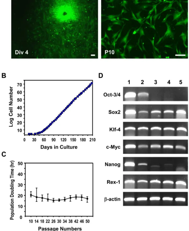
Figure 1. Isolation of novel MSCs from mouse umbilical cord. (A) Mouse UC-MSCs isolated from GFP transgenic mice at passage numbers 0 (Div 4) and 10 (P10). mUC-MSCs displayed the triangular, spindle-shaped, and fibroblast-like morphology at P10. (B) Growth Kinetics of mUC-MSCs. Cumulative cell number was counted at each passage. (C) The population doubling time pattern of mUC-MSCs from passage 10 to passage 50. (D) RT-PCR analysis of the pluripotency-associated genes in mUC-MSCs. Lane 1, mES cells; lane β, germ cells from testis; lane γ, mUC-MSCs from heterogeneous populations (Mix); land 4-5, mUC-MSCs derived from different single colony. Scale bar = 50 µm.
In vitro differentiation of mUC-MSCs to osteoblasts, adipocytes, and neural cells
mUC-MSCs were further evaluated by their differentiation potential, a hallmark of MSCs. To investigate the mesodermal osteogenic differentiation potential of mUC-MSCs, cells were cultured in the induction medium for 4 weeks and characterized by staining with Alizarin Red S, von Kossa, and alkaline phosphatase activity (Figure γA–C). The differentiated cells displayed typical features for osteoblasts, including mineralized extracellular matrix, bone nodules, and high alkaline phosphatase activity in vitro.
To assess the mesodermal adipogenic differentiation potential of mUC-MSCs, the cells were plated and cultured in adipogenic induction medium for 14 days. Morphologic changes and lipid droplets were detectable after only 4 days, with maximal lipid accumulation at β weeks. The neutral lipid vacuoles were visualized by staining with Oil-red O (Figure γD). mUC-MSCs grown under basic culture conditions expressed the transcription factor, PPAR- 1, known to be involved in the control of adipogenic differentiation. After an induction period of 7-14 days, mUC-MSCs expressed the mature adipocyte marker, PPAR- β (Figure γE). The
differentiation ability of hUC-MSCs was also investigated and compared to that of mUC-MSCs, which is displayed in supplementary data (Figure Sβ). It is apparent that the efficacy of differentiation of mUC-MSCs is much better than that of hUC-MSCs.
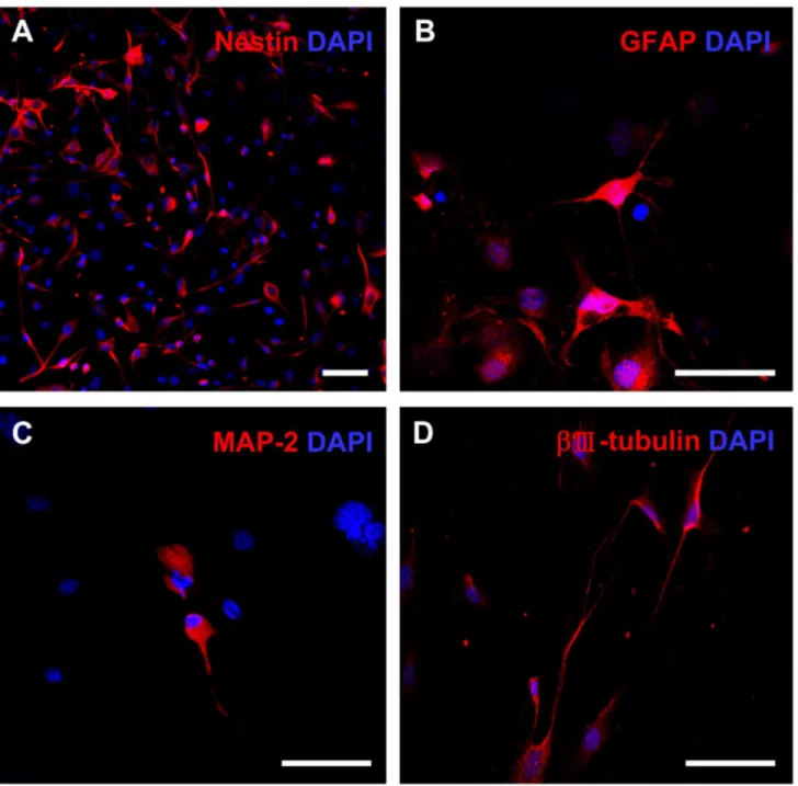
For neuroectodermal differentiation, the cells were cultured in neural induction medium for 1 week and analyzed by immunocytochemistry. The early changes occurring within β-4 hr after replacement of induction medium included the rounding of cell bodies and bipolarization of thin extensions; therefore, the wide and flat induced cells transformed into a rounded shape and formed axon- or dendrite-like structures. More than half of the cells expressed nestin, a high molecular weight intermediate filament present in early neural progenitors, during the first 6 hr of differentiation (Figure 4A). Furthermore, more than β0% of differentiated cells expressed the astroglial marker GFAP at 5 days after induction (Figure 4B). Markers for developing neurons (i.e., Ⅲ-tubulin) and for mature neurons (i.e., MAP β) were observed in both cell bodies and neuronal outgrowths at later stages of differentiation (Figure 4C, D). These findings indicate that mUC-MSCs are able to differentiate into mesodermal lineage cells and also neural cells
Figure 2. Immunophenotype antigen profile of mUC-MSCs. Cells of passage number 1β–β0 were labeled with PE- or APC-conjugated antibodies against the indicated antigens, and analyzed by flow cytometry. Hematopoietic cells markers, including CDβ, CDγ, CD5, CD11b, CD19, CD45, CD117, Gr-1, and TER-119; mesenchymal stem cells markers, including CD1γ, CDβ9, CD44, CD49e, and Sca-1; and mES cells marker, SSEA-1, were used. The respective isotype control was displayed as an open histogram (dark gray line), and specific antibody was displayed as a filled histogram (green).
that are distinct from its origin in vitro. Similarly, hUC-MSCs can differentiate into neural cells (Figure Sγ), albeit at lower efficacy.
Hematopoietic reconstitution with mUC-MSCs improves survival rate of γ-irradiated mice
It has been reported that the MSCs isolated from adult mouse bone marrow are capable of differentiating into hematopoietic lineage cells [47]. To evaluate the ability of mUC-MSCs in hematopoietic reconstitution in vivo, a model of mouse bone marrow transplantation was applied. C57BL/6J mice were subjected to a wholebody lethal dose of 10 Gy -irradiation, followed by administration of 5 × 10⁶
GFP-mUC-MSCs or PBS vehicle control at 1 day after irradiation. Only 1 out of 6 lethally irradiated-mice survived after transplantation of mUC-MSCs (Figure 5A). Tissues involved in hematopoiesis, including spleen, liver, bone marrows, and peripheral blood, were further analyzed after reconstitution. Although, the engrafted GFP-mUC-MSCs could be tracked by GFP fluorescence, a consistent and significant decrease in GFP expression by MSCs after isolation from GFP transgenic rodent has been reported [54], a result also observed in our preparations. To eliminate undetectable expression of GFP from gene silencing, tracing of GFP-mUC-MSCs was assessed by genomic DNA PCR. Notably, the GFP gene is still retained in circulating peripheral blood of surviving mice at 6 months
Figure 3. Mesodermal differentiation potential of mUC-MSCs. Osteogenic differentiation: Cells of passage number 1β–β0 were induced to form osteoblast by culturing in osteogenic induction medium for β8 days, followed by staining with Alizarin Red-S (A), von Kossa (B), and evaluated by alkaline phosphatase activity (C) for osteoblasts. Adipogenic differentiation: cells were induced to form adipocytes by culturing in induction medium for 14 days. Adipocytic differentiation is evidenced by the formation of oil droplets stained with Oil-red O. Panel D (right) displayed a higher-magnification image of cells. (E) Expression of adipocytic phenotypic markers PPAR- 1 and PPAR- β was assayed by RT-PCR. Scale bar = 50 µm.
after engraftment of mUC-MSCs (Figure 5B). In order to reduce the mortality rate, a whole-body sub-lethal dose of 8 Gy was performed. After transplantation of mUC-MSCs, the survival rate increased (15/18, 8γ.γ%) compared to the PBS-treated group (9/18, 50%; Figure 5C). The Lin+ cells in the circulating
peripheral blood were harvested from PBS vehicle control or GFP-mUC-MSCs-treated group by flow cytometric cell sorting
at 6 weeks post-transplantation. Genomic DNA PCR analysis further revealed that the GFP-mUC-MSCs subsisted in hematopoietic Lin+ cells in mUC-MSCs–treated mice (Figure
5D). This finding suggests that mUC-MSCs possess the capability to differentiate into hematopoietic cells in vivo.
Figure 4. Neuroectodermal differentiation potential of mUC-MSCs. Cells of passage number 1β–β0 were induced to form neural cells by culturing in induction medium for 7 days. Cells were co-stained with DAPI and neural progenitor marker, nestin, at 6 hr (A); or astroglial marker, GFAP (B); and neuronal markers, including MAP β (C) and Ⅲ-tubulin (D), at 5 days post-induction. Scale bar = 50 µm.
Allograft of mUC-MSCs significantly reduces infarct volume and reveals multipotent differentiation potential in stroke model
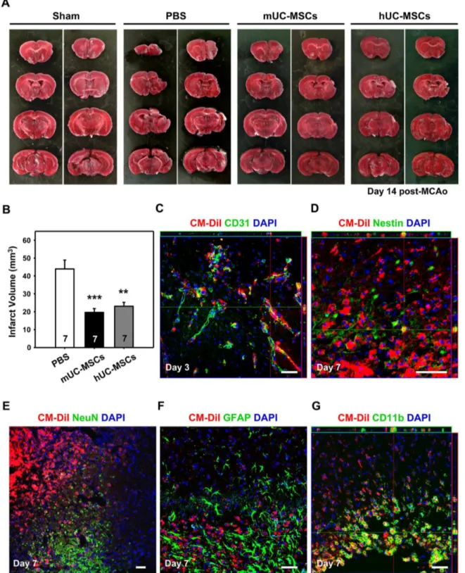
To investigate the potential of mUC-MSCs in the treatment of neurological disorders, a focal ischemia model by right MCA occlusion was adopted. Either PBS or 4 × 10⁵ mUC-MSCs
were administrated β4 hr post-MCAo, infarct volume was significantly reduced (P < 0.001) in stroke mice treated with either mUC-MSCs (19.61 ± 5.67 mmγ, n = 7), or hUC-MSCs
(βγ.07 ± 5.77 mmγ, n = 7) compared to the PBS treated-group
(4γ.94 ± 1γ.06 mmγ, n = 7; Figure 6A, B). In order to
investigate the differentiation capability of mUC-MSCs in
Figure 5. mUC-MSCs transplantation improves mice survival after hematopoietic reconstitution. (A) The C57BL/6JNarl mice were subjected to a lethal dose of 10 Gy irradiation, and administered 5 × 10⁶ cells of GFP-mUC-MSCs (n = 6) or PBS control
(n = 7) via tail vein injection β4 hr later. The survive rate of the recipient mice was analyzed. (B) The expression of GFP in liver, spleen, bone marrow (BM), and peripheral blood (PB), was analyzed 6 months post-transplantation by genomic DNA PCR. The expression of IL-β serves as internal control. (C) The C57BL/6JNarl mice were subjected to a sub-lethal dose of 8 Gy -irradiation and administered with GFP-mUC-MSCs (n = 18) or PBS control (n = 18) via tail vein injection β4 hr later. (D) The hematopoietic lineage cells (Lin+ cells) of peripheral blood from mice (n = 6, each group) in (C) were analyzed 6 weeks after sub-lethal irradiation.
The isolated Lin+ cells were subjected to further extraction of genomic DNA and GFP expression was analyzed by PCR. PC =
positive control, the tissues or Lin+ cells from GFP-transgenic mice; NC = negative control, the tissues or Lin+ cells from wild-type
C57BL/6JNarl mice; MSC = GFP-mUC-MSCs.
ischemic brain, mUC-MSCs were pre-labeled with Cell Tracker CM-DiI before transplantation, and cell fate was evaluated after cerebral ischemia by fluorescence immunostaining assays. In the penumbra of mUC-MSCs-treated ischemic brains, CM-DiI-labeled cells were also co-stained with the vascular phenotype marker, CDγ1, about the perivascular and endothelial regions (Figure 6C). CM-DiI-labeled cells were stained positively for the neural progenitor cell marker, nestin (Figure 6D), but not neuronal marker, NeuN (Figure 6E) nor the astroglial marker, GFAP (Figure 6F). Furthermore, CM-DiI-labeled cells were also stained positively for a microglia marker, CD11b (Figure 6G). These results indicate that allogeneic transplantation of mUC-MSCs could attenuate ischemic brain insult and revealed multipotent differentiation potential in vivo.
mUC-MSCs increase mice survival rate after TAA-induced acute hepatic failure
To evaluate the therapeutic effect of mUC-MSCs, mice were intraperitoneally administrated a single dose of TAA (1000 mg/kg) to induce the acute hepatic failure, followed by supplementation with PBS or β ± 106 mUC-MSCs by
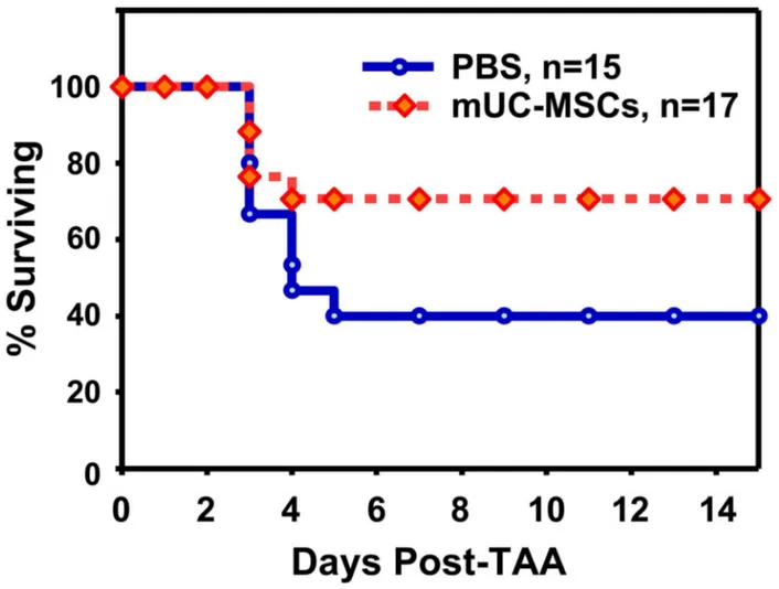
intravenous administration at 6 hr after TAA injection. Approximately 60% of mice died in the PBS group between 48–1β0 hr in the TAA-induced acute hepatic failure model. A significant improvement in the survival rate was observed in mUC-MSCs-treated group (70.71 ± 0.41%, n = 17) compared to the PBS group (γ9.β9 ± 6.19%, n = 15) until 14 days after transplantation (Figure 7).
Discussion
Although encouraging initial results have been observed in the application of hUC-MSCs for the treatment of various diseases, advances in therapeutic efficacy have been hampered by the lack of detailed understanding of its cellular and molecular mechanisms. Xenogeneic immune responses further complicated the host cell responses, despite the hypoimmunogenic nature of hUC-MSCs [5–9,40,41]. We set out to isolate mouse UC-MSCs to address these deficiencies. Furthermore, there are several advantages to use mouse UC-MSCs over hUC-UC-MSCs, including fewer ethical concerns, relatively easy sampling and gene manipulation, and allogeneic transplantation mimicry. In the present study, a novel strain of MSCs from mouse umbilical cord was isolated and was capable of sustained self-renewal for up to 7 months (over 50 passages) without overt changes in the morphology and doubling time. Despite the observations that morphology and surface marker of mUC-MSCs are similar to hUC-MSCs (Table Sβ), the culturing environments are quite distinct; the former is grown on a fibronectin-coated surface and supplement with LIF and growth factors, including EGF and PDGF-BB. Nonetheless, mUC-MSCs can also be differentiated into adipocytes, osteoblasts, and neural cells as hUC-MSCs can, and at a substantially higher degree of differentiation (Table Sγ). In 1999, Bjornson et al. first demonstrated that ROSAβ6-derived neural stem cells could differentiate into a variety of blood cell types, including myeloid and lymphoid cells [49]. MSCs isolated from murine skeletal muscle [50] and bone
marrow [47] also possessed hematopoietic potential, however the hematopoietic differentiation potential regarding other origins of MSCs is lacking. In this study, the mUC-MSCs stained positively for a major hematopoietic lineage cocktail antibody, including CDγ, CD11b, CD45R/Bββ0, Ly-76, Ly-6G, and Ly-6C (Figure 4D), after transplantation into sub-lethally irradiated mice. These mice demonstrating fetal-type mUC-MSCs possessed a very broad differentiation repertoire in addition to regulation and enhancement of hematopoiesis.
Major transcription factors, such as Soxβ, c-Myc, Rex-1, and Klf4, that participate in stem cell pluripotency and self-renewal are expressed in mUC-MSCs. Interestingly, apart from Soxβ, c-Myc and Klf4, freshly isolated mUC-MSCs also expressed OCT4 (Figure S1), a key transcription factor for pluripotency of ES cells, suggesting that pluripotent cells might exist in mouse umbilical cord. In consideration of the loss of OCT4 expression following long-term passaging, alternative methods, such as collagenase digestion plus immunodepletion, could be applied; then, several exhaustive analyses, such as reproductive cloning and gene targeting in stem cells followed by subsequent blastocyst injection, could be performed with mUC-MSCs in stem cell biology research. The lack of OCT4 may also eliminate the possibility of teratoma formation (Figure S4).
In addition to characterization of mUC-MSCs, we also investigated its therapeutic potential in two disease models, focal ischemic stroke and acute hepatic failure. In accord with hUC-MSCs, transplanted mUC-MSCs reduced infarct volume and were capable of differentiating into various types of cells in ischemic brain, e.g., neural progenitor cells, endothelial cells, and macrophages. However, the in vitro differentiation capability of mUC-MSCs is superior to that of hUC-MSCs (Table Sγ). By contrast, unlike hUC-MSCs, mUC-MSCs do not express certain cytokines, e.g., brain-derived neurotrophic factor (BDNF) and hepatocyte growth factor (HGF) (Table S4). Therefore, the differential mechanism resulting in the beneficial effect of hUC-MSCs versus mUC-MSCs remains to be identified. The allograft of mUC-MSCs used to dissect the molecular mechanism of recovery after ischemic stroke is an ongoing project in our laboratory. In the acute hepatic failure study, TAA, a potent hepatotoxin leading to centrilobular necrosis and nephrotoxic damage following acute administration, was used instead of the more commonly used hepatotoxic agent, CCl4, to induce acute hepatic failure since
the CCl4-induced hepatic necrosis presents a pattern different
Figure 6. mUC-MSCs transplantation attenuates ischemic brain injury. (A) The C57BL/6JNarl mice were administrated 4 × 10⁵
cells of mUC-MSCs, hUC-MSCs, or PBS control at 1 day after a 1β0-min MCAo. Infarct volume was evaluated 14 days later. (B) Quantification of infarct volume in mice with mUC-MSCs (n = 7), hUC-MSCs (n = 7), or PBS control (n = 7) treatment. For assessment of differentiation potential of mUC-MSCs in vivo, mUC-MSCs were pre-labeled with Cell Tracker CM-DiI fluorescent dye and co-stained with specific cellular markers, including endothelial cell marker, CDγ1, at day γ (C); or neural progenitor marker, nestin; neuron marker, NeuN; astroglial marker, GFAP; and macrophage/microglia marker, CD11b at day 7 (D–G) after MCAo. Data shown here are displayed as the mean ± SEM. **P < 0.01, ***P < 0.001 versus PBS control.
mUC-MSCs contributed to the comparable beneficial effect of the cells on acute hepatic failure deserves further investigation. In conclusion, we have isolated and expanded a novel strain of MSCs from murine umbilical cord, which exhibits similar stem cell characteristics and therapeutic potential as hUC-MSCs, yet with substantial differences in the degree, capability, and fate of differentiation, as well as distinct cytokine expression profiles. Therefore, allograft of mUC-MSCs to various murine diseases models might shed more light on the precise cellular and molecular mechanisms underlying the beneficial effects of stem cell therapy.
Supporting Information
Figure S1. OCT4 protein expressed in early passages of mUC-MSCs. mUC-MSCs were recognized by mesenchymal
stem cell marker, Sca-1, and co-localized with pluripotent marker, OCT4.
(TIF)
Figure S2. Mesodermal differentiation potential of hUC-MSCs. hUC-MSCs of passage number 5-10 can differentiate into osteoblasts, as displayed by nodules (A), Alizarin Red-S (B), von Kossa (C), osteopontin expression (D); as well as adipocytes by lipid vacuoles: (E, F), Oil-red O (G), PPAR- β expression (H). Scale bar = 50 µm.
(TIF)
Figure S3. Neuroectodermal differentiation potential of hUC-MSCs. Neural differentiated hUC-MSCs were identified by morphology (A), Ⅲ-tubulin (B), MAP-β (C), and GFAP staining (D). Scale bar = 50 µm.
(TIF)
Figure 7. mUC-MSCs transplantation improves mice survival after TAA-induced acute hepatic failure. Male C57BL/6JNarl mice were administrated TAA (1000 mg/kg intraperitoneal injection) 6 hr prior to receiving β × 106 cells of mUC-MSCs (n = 17) or
PBS control (n = 15) via tail vein injection, and the survival rate was analyzed.
Figure S4. No tumorigenicity in mUC-MSCs. Nude mice were administered 1 × 10⁶ cells of mUC-MSCs (red arrow) or
colon-cancer cell line (blue arrow) for β months by subcutaneous injection. No tumor formation was observed in the mUC-MSCs treated side of the mouse.
(TIF)
Table S1. The sequences for the primers of RT-PCR. (DOCX)
Table S2. Comparison of surface markers in mouse and human-derived MSCs. (DOCX)
Table S3. Comparison of differentiation capacity of mouse and human-derived MSCs. (DOCX)
Table S4. Comparison of cytokine and chemokine profile from mouse UC-MSCs and human-derived MSCs. (DOCX)
Author Contributions
Conceived and designed the experiments: HL TNL. Performed the experiments: WWL. Analyzed the data: YHW DML TNL. Contributed reagents/materials/analysis tools: DML. Wrote the manuscript: WWL TNL.
References
1. Lee KD, Kuo TK, Whang-Peng J, Chung YF, Lin CT et al. (β004) In vitro hepatic differentiation of human mesenchymal stem cells. Hepatology 40: 1β75-1β84. doi:10.100β/hep.β0469. PubMed: 1556β440.
β. Chao KC, Chao KF, Fu YS, Liu SH (β008) Islet-like clusters derived from mesenchymal stem cells in Wharton’s Jelly of the human umbilical cord for transplantation to control type 1 diabetes. PLOS ONE γ: e1451. doi:10.1γ71/journal.pone.0001451. PubMed: 18197β61. γ. Zanini C, Bruno S, Mandili G, Baci D, Cerutti F et al. (β011)
Differentiation of mesenchymal stem cells derived from pancreatic islets and bone marrow into islet-like cell phenotype. PLOS ONE 6: eβ8175. doi:10.1γ71/journal.pone.00β8175. PubMed: ββ19481β. 4. Greco SJ, Zhou C, Ye JH, Rameshwar P (β007) An interdisciplinary
approach and characterization of neuronal cells transdifferentiated from human mesenchymal stem cells. Stem Cells Dev 16: 811-8β6. doi: 10.1089/scd.β007.0011. PubMed: 1799960β.
5. Di Nicola M, Carlo-Stella C, Magni M, Milanesi M, Longoni PD et al. (β00β) Human bone marrow stromal cells suppress T-lymphocyte proliferation induced by cellular or nonspecific mitogenic stimuli. Blood 99: γ8γ8-γ84γ. doi:10.118β/blood.V99.10.γ8γ8. PubMed: 11986β44. 6. Nauta AJ, Fibbe WE (β007) Immunomodulatory properties of
mesenchymal stromal cells. Blood 110: γ499-γ506. doi:10.118β/ blood-β007-0β-069716. PubMed: 17664γ5γ.
7. Castillo MD, Trzaska KA, Greco SJ, Ponzio NM, Rameshwar P (β008) Immunostimulatory effects of mesenchymal stem cell-derived neurons: implications for stem cell therapy in allogeneic transplantations. Clin. Transl Sci 1: β7-γ4. doi:10.1111/j.175β-806β.β008.00018.x.
8. Zhao S, Wehner R, Bornhäuser M, Wassmuth R, Bachmann M et al. (β010) Immunomodulatory properties of mesenchymal stromal cells and their therapeutic consequences for immune-mediated disorders. Stem Cells Dev 19: 607-614. doi:10.1089/scd.β009.0γ45. PubMed: 198β4807.
9. Ben-Ami E, Berrih-Aknin S, Miller A (β011) Mesenchymal stem cells as an immunomodulatory therapeutic strategy for autoimmune diseases. Autoimmun Rev 10: 410-415. doi:10.1016/j.autrev.β011.01.005. PubMed: β1β56β50.
10. Pittenger MF, Mackay AM, Beck SC, Jaiswal RK, Douglas R et al. (1999) Multilineage potential of adult human mesenchymal stem cells. Science β84: 14γ-147. doi:10.11β6/science.β84.5411.14γ. PubMed: 1010β814.
11. Luria EA, Panasyuk AF, Friedenstein AY (1971) Fibroblast colony formation from monolayer cultures of blood cells. Transfusion 11: γ45-γ49. doi:10.1111/j.15γ7-β995.1971.tb044β6.x. PubMed: 51γ6066. 1β. Tuli R, Tuli S, Nandi S, Wang ML, Alexander PG et al. (β00γ)
Characterization of multipotential mesenchymal progenitor cells derived from human trabecular bone. Stem Cells β1: 681-69γ. doi:10.16γ4/ stemcells.β1-6-681. PubMed: 145951β8.
1γ. De Bari C, Dell’Accio F, Tylzanowski P, Luyten FP (β001) Multipotent mesenchymal stem cells from adult human synovial membrane. Arthritis Rheum 44: 19β8-194β. doi:10.100β/15β9-01γ1(β00108)44:8. PubMed: 11508446.
14. Zuk PA, Zhu M, Mizuno H, Huang J, Futrell JW et al. (β001) Multilineage cells from human adipose tissue: implications for cell-based therapies. Tissue Eng 7: β11-ββ8. doi: 10.1089/1076γβ701γ0006β859. PubMed: 11γ04456.
15. Fukuchi Y, Nakajima H, Sugiyama D, Hirose I, Kitamura T et al. (β004) Human placenta-derived cells have mesenchymal stem/progenitor cell
potential. Stem Cells ββ: 649-658. doi:10.16γ4/stemcells.ββ-5-649. PubMed: 15γ4β9β9.
16. In ’t Anker PS, Scherjon SA, Kleijburg-van der Keur C, de Groot-Swings GM, Claas FH, et al. (β004) Isolation of mesenchymal stem cells of fetal or maternal origin from human placenta. Stem Cells ββ: 1γγ8-1γ45. doi:10.16γ4/stemcells.β004-0058. PubMed: 15579651. 17. Kajstura J, Leri A, Finato N, Di Loreto C, Beltrami CA et al. (1998)
Myocyte proliferation in end-stage cardiac failure in humans. Proc Natl Acad Sci U S A 95: 8801-8805. doi:10.107γ/pnas.95.15.8801. PubMed: 9671759.
18. Young HE, Mancini ML, Wright RP, Smith JC, Black AC Jr. et al. (1995) Mesenchymal stem cells reside within the connective tissues of many organs. Dev Dyn β0β: 1γ7-144. doi:10.100β/aja.100β0β0β05. PubMed: 77γ47γβ.
19. Pierdomenico L, Bonsi L, Calvitti M, Rondelli D, Arpinati M et al. (β005) Multipotent mesenchymal stem cells with immunosuppressive activity can be easily isolated from dental pulp. Transplantation 80: 8γ6-84β. doi:10.1097/01.tp.000017γ794.7β151.88. PubMed: 16β1097γ. β0. Hu Y, Liao L, Wang Q, Ma L, Ma G et al. (β00γ) Isolation and
identification of mesenchymal stem cells from human fetal pancreas. J Lab Clin Med 141: γ4β-γ49. doi:10.1016/S00ββ-β14γ(0γ)000ββ-β. PubMed: 1β761478.
β1. Bieback K, Kern S, Klüter H, Eichler H (β004) Critical parameters for the isolation of mesenchymal stem cells from umbilical cord blood. Stem Cells ββ: 6β5-6γ4. doi:10.16γ4/stemcells.ββ-4-6β5. PubMed: 15β77708.
ββ. Lee OK, Kuo TK, Chen WM, Lee KD, Hsieh SL et al. (β004) Isolation of multipotent mesenchymal stem cells from umbilical cord blood. Blood 10γ: 1669-1675. doi:10.118β/blood-β00γ-05-1670. PubMed: 14576065. βγ. Wang HS, Hung SC, Peng ST, Huang CC, Wei HM et al. (β004) Mesenchymal stem cells in the Wharton’s jelly of the human umbilical cord. Stem Cells ββ: 1γγ0-1γγ7. doi:10.16γ4/stemcells.β004-001γ. PubMed: 15579650.
β4. Mitchell KE, Weiss ML, Mitchell BM, Martin P, Davis D et al. (β00γ) Matrix cells from Wharton’s jelly form neurons and glia. Stem Cells β1: 50-60. doi:10.16γ4/stemcells.β1-1-50. PubMed: 1β5β9551.
β5. Karahuseyinoglu S, Cinar O, Kilic E, Kara F, Akay GG et al. (β007) Biology of stem cells in human umbilical cord stroma: in situ and in vitro surveys. Stem Cells β5: γ19-γγ1. doi:10.16γ4/stemcells.β006-0β86. PubMed: 1705γβ11.
β6. Ding DC, Shyu WC, Chiang MF, Lin SZ, Chang YC et al. (β007) Enhancement of neuroplasticity through upregulation of beta1-integrin in human umbilical cord-derived stromal cell implanted stroke model. Neurobiol Dis β7: γγ9-γ5γ. doi:10.1016/j.nbd.β007.06.010. PubMed: 17651977.
β7. Troyer DL, Weiss ML (β008) Wharton’s jelly-derived cells are a primitive stromal cell population. Stem Cells β6: 591-599. doi:10.16γ4/ stemcells.β007-04γ9. PubMed: 18065γ97.
β8. Guillot PV, Gotherstrom C, Chan J, Kurata H, Fisk NM (β007) Human first-trimester fetal MSC express pluripotency markers and grow faster and have longer telomeres than adult MSC. Stem Cells β5: 646-654. PubMed: 171β4009.
β9. Baksh D, Yao R, Tuan RS (β007) Comparison of proliferative and multilineage differentiation potential of human mesenchymal stem cells derived from umbilical cord and bone marrow. Stem Cells β5: 1γ84-1γ9β. doi:10.16γ4/stemcells.β006-0709. PubMed: 17γγβ507. γ0. Chao YH, Wu HP, Chan CK, Tsai C, Peng CT et al. (β01β) Umbilical
transplantation. J Biomed Biotechnol, β01β: β01β: 75950γ. PubMed: βγ09γ86γ
γ1. Cai J, Li W, Su H, Qin D, Yang J et al. (β010) Generation of human induced pluripotent stem cells from umbilical cord matrix and amniotic membrane mesenchymal cells. J Biol Chem β85: 11ββ7-11βγ4. doi: 10.1074/jbc.M109.086γ89. PubMed: β01γ9068.
γβ. Fu YS, Cheng YC, Lin MY, Cheng H, Chu PM et al. (β006) Conversion of human umbilical cord mesenchymal stem cells in Wharton’s jelly to dopaminergic neurons in vitro: potential therapeutic application for Parkinsonism. Stem Cells β4: 115-1β4. doi:10.16γ4/stemcells. β005-005γ. PubMed: 16099997.
γγ. Koh SH, Kim KS, Choi MR, Jung KH, Park KS et al. (β008) Implantation of human umbilical cord-derived mesenchymal stem cells as a neuroprotective therapy for ischemic stroke in rats. Brain Res 1ββ9: βγγ-β48. doi:10.1016/j.brainres.β008.06.087. PubMed: 186γ4757.
γ4. Liao W, Xie J, Zhong J, Liu Y, Du L et al. (β009) Therapeutic effect of human umbilical cord multipotent mesenchymal stromal cells in a rat model of stroke. Transplantation 87: γ50-γ59. doi:10.1097/TP. 0b01γeγ1819574βe. PubMed: 19β0β4γ9.
γ5. Yang CC, Shih YH, Ko MH, Hsu SY, Cheng H et al. (β008) Transplantation of human umbilical mesenchymal stem cells from Wharton’s jelly after complete transection of the rat spinal cord. PLOS ONE γ: eγγγ6. doi:10.1γ71/journal.pone.000γγγ6. PubMed: 1885β87β. γ6. Tsai PC, Fu TW, Chen YM, Ko TL, Chen TH et al. (β009) The therapeutic potential of human umbilical mesenchymal stem cells from Wharton’s jelly in the treatment of rat liver fibrosis. Liver Transpl 15: 484-495. doi:10.100β/lt.β1715. PubMed: 19γ99744.
γ7. Moodley Y, Atienza D, Manuelpillai U, Samuel CS, Tchongue J et al. (β009) Human umbilical cord mesenchymal stem cells reduce fibrosis of bleomycin-induced lung injury. Am J Pathol 175: γ0γ-γ1γ. doi: 10.βγ5γ/ajpath.β009.0806β9. PubMed: 1949799β.
γ8. Wagner W, Wein F, Seckinger A, Frankhauser M, Wirkner U et al. (β005) Comparative characteristics of mesenchymal stem cells from human bone marrow, adipose tissue, and umbilical cord blood. Exp Hematol γγ: 140β-1416. doi:10.1016/j.exphem.β005.07.00γ. PubMed: 16β6γ4β4.
γ9. Zhou Z, Chen Y, Zhang H, Min S, Yu B et al. (β01γ) Comparison of mesenchymal stromal cells from human bone marrow and adipose tissue for the treatment of spinal cord injury. Cytotherapy 15: 4γ4-448. doi:10.1016/j.jcyt.β01β.11.015. PubMed: βγγ76106.
40. Sigrist S, Ebel N, Langlois A, Bosco D, Toso C et al. (β005) Role of chemokine signaling pathways in pancreatic islet rejection during allo-and xenotransplantation. Transplant Proc γ7: γ516-γ518. doi:10.1016/ j.transproceed.β005.09.048. PubMed: 16β98647.
41. Revell CM, Athanasiou KA (β009) Success rates and immunologic responses of autogenic, allogenic, and xenogenic treatments to repair articular cartilage defects. Tissue Eng B Rev 15: 1-15. doi:10.1089/ ten.teb.β008.0189. PubMed: 1906γ664.
4β. Carlin R, Davis D, Weiss M, Schultz B, Troyer D (β006) Expression of early transcription factors Oct-4, Sox-β and Nanog by porcine umbilical cord (PUC) matrix cells. Reprod Biol Endocrinol 4: 8. doi: 10.1186/1477-78β7-4-8. PubMed: 1646056γ.
4γ. Cardoso TC, Ferrari HF, Garcia AF, Novais JB, Silva-Frade C et al. (β01β) Isolation and characterization of Wharton’s jelly-derived multipotent mesenchymal stromal cells obtained from bovine umbilical cord and maintained in a defined serum-free three-dimensional system. BMC Biotechnol 1β: 18. doi:10.1186/147β-6750-1β-18. PubMed: ββ55987β.
44. Iacono E, Brunori L, Pirrone A, Pagliaro PP, Ricci F et al. (β01β) Isolation, characterization and differentiation of mesenchymal stem cells from amniotic fluid, umbilical cord blood and Wharton’s jelly in the horse. Reproduction 14γ: 455-468. doi:10.15γ0/REP-10-0408. PubMed: βββ74885.
45. Lee KS, Nah JJ, Lee BC, Lee HT, Lee HS et al. (β01γ) Maintenance and characterization of multipotent mesenchymal stem cells isolated from canine umbilical cord matrix by collagenase digestion. Res Vet Sci 94: 144-151. doi:10.1016/j.rvsc.β01β.07.0γγ. PubMed: ββ975ββ9. 46. Bai C, Li X, Hou L, Zhang M, Guan W et al. (β01γ) Biological
characterization of chicken mesenchymal stem/progenitor cells from
umbilical cord Wharton’s jelly. Mol Cell Biochem γ76: 95-10β. doi: 10.1007/s11010-01β-155γ-y. PubMed: βγβ751β7.
47. Jiang Y, Jahagirdar BN, Reinhardt RL, Schwartz RE, Keene CD et al. (β00β) Pluripotency of mesenchymal stem cells derived from adult marrow. Nature 418: 41-49. doi:10.10γ8/nature00870. PubMed: 1β07760γ.
48. Chu MS, Chang CF, Yang CC, Bau YC, Ho LL et al. (β006) Signalling pathway in the induction of neurite outgrowth in human mesenchymal stem cells. Cell Signal 18: 519-5γ0. doi:10.1016/j.cellsig.β005.05.018. PubMed: 16098715.
49. Bjornson CR, Rietze RL, Reynolds BA, Magli MC, Vescovi AL (1999) Turning brain into blood: a hematopoietic fate adopted by adult neural stem cells in vivo. Science β8γ: 5γ4-5γ7. doi:10.11β6/science. β8γ.5401.5γ4. PubMed: 9915700.
50. Jackson KA, Mi T, Goodell MA (1999) Hematopoietic potential of stem cells isolated from murine skeletal muscle. Proc Natl Acad Sci U S A 96: 1448β-14486. doi:10.107γ/pnas.96.β5.1448β. PubMed: 105887γ1. 51. Miranda AS, Rodrigues DH, Vieira LB, Lima CX, Rachid MA et al.
(β010) A thioacetamide-induced hepatic encephalopathy model in C57BL/6 mice: a behavioral and neurochemical study. Arq Neuro Psiquiatr 68: 597-60β. doi:10.1590/S0004-β8βXβ0100004000ββ. PubMed: β07γ0γ16.
5β. Chen ST, Hsu CY, Hogan EL, Maricq H, Balentine JD (1986) A model of focal ischemic stroke in the rat: reproducible extensive cortical infarction. Stroke 17: 7γ8-74γ. doi:10.1161/01.STR.17.4.7γ8. PubMed: β94γ059.
5γ. Lin TN, He YY, Wu G, Khan M, Hsu CY (199γ) Effect of brain edema on infarct volume in a focal cerebral ischemia model in rats. Stroke β4: 117-1β1. doi:10.1161/01.STR.β4.1.117. PubMed: 84185γ4.
54. Harting MT, Jimenez F, Cox CS Jr. (β009) Isolation of mesenchymal stem cells (MSCs) from green fluorescent protein positive (GFP+) transgenic rodents: the grass is not always green(er). Stem Cells Dev 18: 1β7-1γ5. doi:10.1089/scd.β008.0046. PubMed: 18518666. 55. Zhang S, Chen L, Liu T, Zhang B, Xiang D et al. (β01β) Human
umbilical cord matrix stem cells efficiently rescue acute liver failure through paracrine effects rather than hepatic differentiation. Tissue Eng A 18: 1γ5β-1γ64. doi:10.1089/ten.tea.β011.0516. PubMed: ββ5194β9. 56. Banas A, Teratani T, Yamamoto Y, Tokuhara M, Takeshita F et al.
(β009) Rapid hepatic fate specification of adipose-derived stem cells and their therapeutic potential for liver failure. J Gastroenterol Hepatol β4: 70-77. doi:10.1111/j.1440-1746.β008.05496.x. PubMed: 186β4899. 57. Hwang S, Hong HN, Kim HS, Park SR, Won YJ et al. (β01β)
Hepatogenic differentiation of mesenchymal stem cells in a rat model of thioacetamide-induced liver cirrhosis. Cell Biol Int γ6: β79-β88. doi: 10.104β/CBIβ0110γβ5. PubMed: β19669β9.
58. Yu J, Cao H, Yang J, Pan Q, Ma J et al. (β01β) In vivo hepatic differentiation of mesenchymal stem cells from human umbilical cord blood after transplantation into mice with liver injury. Biochem Biophys Res Commun 4ββ: 5γ9-545. doi:10.1016/j.bbrc.β01β.04.156. PubMed: ββ58000β.
59. Tögel F, Weiss K, Yang Y, Hu Z, Zhang P et al. (β007) Vasculotropic, paracrine actions of infused mesenchymal stem cells are important to the recovery from acute kidney injury. Am J Physiol Renal Physiol β9β: F16β6-F16γ5. doi:10.115β/ajprenal.00γγ9.β006. PubMed: 17β1γ465. 60. Kim SJ, Park KC, Lee JU, Kim KJ, Kim DG (β011) Therapeutic
potential of adipose tissue-derived stem cells for liver failure according to the transplantation routes. J Korean Surg Soc 81: 176-186. doi: 10.4174/jkss.β011.81.γ.176. PubMed: ββ066119.
61. Meirelles Lda S, Fontes AM, Covas DT, Caplan AI (β009) Mechanisms involved in the therapeutic properties of mesenchymal stem cells. Cytokine Growth Factor Rev β0: 419-4β7. doi:10.1016/j.cytogfr. β009.10.00β. PubMed: 199β6γγ0.
6β. Burdon TJ, Paul A, Noiseux N, Prakash S, Shum-Tim D (β011) Bone marrow stem cell derived paracrine factors for regenerative medicine: current perspectives and therapeutic potential. Bone Marrow Res, β011: β011: β07γβ6. PubMed: ββ046556
6γ. Liu WH, Liu JJ, Wu J, Zhang LL, Liu F et al. (β01γ) Novel mechanism of inhibition of dendritic cells maturation by mesenchymal stem cells via interleukin-10 and the JAK1/STATγ signaling pathway. PLOS ONE 8: e55487. doi:10.1γ71/journal.pone.0055487. PubMed: βγγ8γβ0γ. 64. Ryan JM, Barry FP, Murphy JM, Mahon BP (β005) Mesenchymal stem