Ana Paula Alves Pacheco
Characterization of hairy1 and hairy2 pattern expression
during lung development of Gallus gallus
Tese de Mestrado
Mestrado de Genética Molecular
Trabalho efectuado sob a orientação de:
Professor Doutor Jorge Correia-Pinto
Professor Associado Convidado
Professora Doutora Rute Moura
Professora Auxiliar Convidada
Escola de Ciências da Saúde, da Universidade do Minho, Braga,
Portugal
Junho 2010
Nome: Ana Paula Alves Pacheco
Endereço electrónico: anapacheco@ecsaude.uminho.pt
Título da Tese de Mestrado:
Characterization of hairy1 and hairy2 expression pattern during lung development of Gallus gallus Caracterização do padrão de expressão de hairy1 e hairy2 ao longo do desenvolvimento pulmonar de Gallus gallus
Trabalho efectuado sob a orientação de: Professor Doutor Jorge Correia-Pinto Professora Doutora Rute Moura
Supervisão
Professora Doutora Paula Sampaio
Designação do Mestrado Mestrado em Genética Molecular Ano de Conclusão: 2010
É AUTORIZADA A REPRODUÇÃO INTEGRAL DESTA TESE, APENAS PARA EFEITOS DE INVESTIGAÇÃO, MEDIANTE DECLARAÇÃO ESCRITA AO INTERESSADO, QUE A TAL SE COMPROMETE.
Universidade do Minho, ____/____/____
ACKNOWLEDGMENTS / AGRADECIMENTOS
No decurso da realização desta tese de Mestrado, foram várias as pessoas que desempenharam, directa ou indirectamente, um papel fundamental na sua realização. Gostaria de manifestar aqui os meus sinceros agradecimentos.
Ao Professor Doutor Jorge Correia-Pinto, orientador deste projecto, pela possibilidade que me ofereceu em poder trabalhar e integrar o fantástico Domínio das Ciências Cirúrgicas. Pelo entusiasmo que sempre demonstrou face aos resultados que foram surgindo.
A todos os docentes do Mestrado de Genética Molecular, especialmente à Professora Doutora Paula Sampaio pela supervisão deste trabalho.
À Doutora Rute Moura. Acima de tudo pela dedicação, por tudo aquilo que me ensinou ao longo deste percurso, pela orientação, pela indispensável ajuda em todos os momentos, pelas incansáveis correcções e pela amizade.
À Mónica e à Tatiana pela companhia na ―sala das lupas‖, pela boa disposição e pela amizade. Obrigado pelos conselhos e pelas boas conversas. Tatiana, obrigado pela ajuda na análise estatística. À Sílvia Gonzaga pela amizade e excelente sentido de humor.
Ao Paulo Mota por me ensinar a dissecar os ―pulmõezinhos‖ e pela sua boa disposição tão contagiante.
A todo o grupo do Domínio das Ciências Cirúrgicas. Pela alegria e boa disposição no trabalho e pelo ambiente extraordinário. Desde a experiência e simpatia da Raquel, da Sheeba, passando pelos fantásticos almoços, pelas conversas e pelos bons momentos com as meninas do laboratório ao lado, Sandra, Sara, Olga, Céline, Vera, Helena e ao João Vilaça.
À Professora Doutora Isabel Palmeirim pelo fornecimento dos plasmídeos para a síntese das sondas de hairy1 e hairy2. Pelos comentários construtivos que foi fazendo ao longo deste projecto. A ti por teres estado presente em todos os momentos (principalmente nos menos bons), por todos os conselhos e longas conversas. Obrigado pela tua paciência.
E por último, mas mais importante, à minha família. Aos meus pais por estarem sempre do meu lado e por me apoiarem em todos os momentos. Obrigado por mais esta oportunidade.
ABSTRACT
Although much is known about some aspects of lung development, the mechanisms that regulate the spatiotemporal control of this process are not well defined. The identification of a segmentation clock that ensures the correct spatiotemporal periodicity of somite generation, during chick somitogenesis, provided the first molecular evidence of how embryonic cells count time. The underlying mechanisms of this process involve the oscillation of ―clock-genes‖ like hairy1 and hairy2 (members of hairy-enhancer-of-split –HES- family of transcriptional repressors), which are intimately related to the Notch signaling pathway. More recently, hairy2 expression unveiled a molecular clock operating during limb development.
Lung development share some similarities with somitogenesis and limb development, namely: they all occur along the rostro-caudal axis, the signaling pathways implicated are the same, the formation of consecutive repetitive structures along the anterior-posterior axis is observed, and they occur with exact chronological precision. Considering this parallelism, the characterization of hairy1 and hairy2 during lung branching morphogenesis of Gallus gallus became relevant, since their expression might indicate a possible role in the development of this tissue, although their functions are not yet clarified. Moreover, taking into consideration that FGF and Notch signaling pathways are involved in the molecular clock mechanisms, the link between hairy1 and hairy2, these pathways and branching morphogenesis was assessed by lung explant culture system.
The present work characterizes, for the first time, hairy1 and hairy2 expression pattern in early stages of chick lung development, by in situ hybridization. hairy1 expression was evident mainly in the pulmonary epithelium of the respiratory tract, but also in the mesenchyme. hairy2 expression was evident mainly in the chick pulmonary mesenchyme surrounding the trachea and the trachea bifurcation, and the most distal region of the main bronchus. hairy1 and hairy2 expression was not affected by FGF signaling inhibition; however, treated explants showed atypical lung branching when compared with control ones, which is consistent with the role of FGF signaling in branching morphogenesis. These results indicate that these genes are not downstream targets of FGF signaling pathway in the chick lung. Notch inhibition did not affect hairy1 expression but, on the other hand, hairy2 expression was dramatically reduced when compared with control explants. Moreover, treated explants showed not only an increased number of secondary branches but also an anomalous lung structure in the distal tip of the main bronchus. Notch signaling inhibition appears to affect only hairy2 expression in the lung and seems to interfere with the correct process of lung development.
RESUMO
Hoje em dia muito se sabe sobre desenvolvimento pulmonar. No entanto, os mecanismos que regulam o controlo espacial e temporal deste processo não estão bem definidos. A identificação de um relógio molecular da segmentação responsável pela correcta periodicidade espacial e temporal da formação dos sómitos no modelo da galinha, forneceu a primeira evidência molecular de como de como é feita a contagem do tempo pelas células embrionárias. Os mecanismos subjacentes a este processo envolvem a oscilação de genes como hairy1 e hairy2 (membros da família de repressores de transcrição hairy-enhancer-of-split, HES), que estão intimamente relacionados com a via de sinalização Notch. Mais recentemente, foi descrita a existência de um relógio molecular no desenvolvimento do membro.
O desenvolvimento pulmonar partilha algumas semelhanças com a somitogénese e o desenvolvimento do membro, nomeadamente: ocorre ao longo de um eixo rostro-caudal, as vias de sinalização implicadas são as mesmas, observa-se a formação consecutiva de estruturas repetitivas ao longo de um eixo anterior-posterior, e ocorre com precisão cronológica. Considerando este paralelismo, tornou-se relevante caracterizar o padrão de expressão de hairy1 e hairy2 durante a ramificação pulmonar de Gallus gallus, uma vez que a sua expressão pode indicar um possível papel no desenvolvimento deste tecido. Além disso, tendo em conta que as vias de sinalização Notch e FGF estão envolvidas nos mecanismos do relógio molecular, estas vias conjuntamente com a relação entre hairy1 e hairy2 e a ramificação pulmonar foram avaliadas.
O presente trabalho caracteriza, pela primeira vez, o padrão de expressão de hairy1 e hairy2 em estadios iniciais do desenvolvimento pulmonar de galinha, por hibridização in situ. A expressão de hairy1 foi observada principalmente no epitélio pulmonar do tracto respiratório, mas também no mesênquima. hairy2 é expresso sobretudo no mesênquima pulmonar de galinha que rodeia a traqueia e a bifurcação da traqueia, e na região mais distal do brônquio principal. A expressão destes genes não foi afectada pela inibição da via FGF, indicando que estes genes não são alvos desta via de sinalização neste órgão. No entanto, estes explantes apresentaram uma ramificação pulmonar atípica, o que está de acordo com o papel da via FGF na ramificação pulmonar. A inibição da via Notch não afectou a expressão de hairy1 mas, a expressão de hairy2 foi dramaticamente reduzida. Além disso, os explantes tratados com DAPT apresentaram não só um aumento no número de ramificações secundárias mas também uma estrutura pulmonar anómala. A inibição da via Notch parece afectar a expressão de hairy2 em regiões concretas do pulmão e parece interferir no correcto processo de desenvolvimento pulmonar.
TABLE OF CONTENTS
ACKNOWLEDGMENTS/AGRADECIMENTOS v ABSTRACT vii RESUMO ix TABLE OF CONTENTS xi ABBREVIATIONS xiii LIST OF FIGURES xv 1.INTRODUCTION 1 1.1. Chicken Embryology 31.1.1. Events in Embryonic Development 3
1.1.2. Stages in Chick Embryo Development 4
1.1.3. Chick Model 5
1.2. Segmentation Clock 6
1.2.1. Notch Signaling pathway 9
1.2.1.1.Notch Signaling 10
1.2.1.2.The Notch Cascade 11
1.2.2. FGF Signaling Pathway 12
1.3. Lung Development 14
1.3.1. Branching Morphogenesis 14
1.3.1.1.Notch Signaling 15
1.3.1.2.FGF Signaling 16
1.3.2. Avian Lung Development 16
1.3.2.1. Molecular Aspects of Avian Lung Development 18
1.4. Aims 20
2.MATERIAL AND METHODS 21
2.1. Embryo Dissection 23
2.2. In situ Hybridization 23
2.2.1.1. DNA Extraction 23
2.2.1.2. Plasmid Linearization 25
2.2.1.3. DNA Purification 25
2.2.1.4. Probe Synthesis 26
2.2.2. In situ Hybridization 26
2.3. Chick Lung Explant Culture 28
2.3.1. In vitro Inhibition Studies 29
2.3.1.1. FGF Inhibition 29
2.3.1.2. Notch Inhibition 29
2.3.2. Statistical Analysis 29
3.RESULTS 31
3.1. Characterization of hairy1 and hairy2 expression pattern during chick lung
development 33
3.1.1. hairy1 Expression Pattern 33
3.1.2. hairy2 Expression Pattern 35
3.2. In vitro Inhibition Studies 37
3.2.1. FGF Signaling Inhibition 37
3.2.2. Notch Signaling Inhibition 40
4.DISCUSSION 43
5.CONCLUSIONS 49
ABBREVIATIONS
ADAM desintegrin and metallopeptidase domain AP alkaline phosphatase
BCIP bromo chloro indolyl phosphate bHLH basic helix-loop-helix
BMP bone morphogenetic protein
CHAPS cholamidopropyl dimethylammonio propanesulfonate CSL CBF1 suppressor of hairless Lag-1
DAPT difluorophenacetyl alanyl phenylglycine T-butyl ester DMSO dimethylsulfoxide
DPBS dulbecco´s phosphate buffered saline EDTA ethylen diamin tetra acid
EGF epidermal growth factor
EGTA ethylene glycol tetraacetic acid FGF fibroblast growth factor
FGFR fibroblast growth factor receptor HES hairy and enhancer of split HGF hepatocyte growth factor HH Hamburger and Hamilton HSPG heparin sulfate proteoglycans ISH in situ hybridization
LB Luria broth LN Lin-12-Notch
NBT nitro blue tetrazolium chloride NICD notch intracellular domain NF nuclear factor
OR orange domain
PBS phosphate-buffered saline solution
PBT phosphate-buffered saline solution with Tween PCR polymerase chain reaction
PM plasma membrane PSM presomitic mesoderm RAM RBP-Jk associate molecule
RBP-J recombination signal sequence-binding protein J RT room temperature
RT-PCR real-time PCR SHH sonic hedgehog SSC standard saline citrate St stage
SU5402 2-[(1,2-Dihydro-2-oxo-3H-indol-3-ylidene)methyl]-4-methyl-1H-pyrrole-3-propanoic acid Su(H) supressor of hairless
TACE TNF-converting enzyme Tbx T-box
TAD transactivation domain TGF transforming growth factor WNT wingless wint
LIST OF FIGURES
PAGE
Figure 1.1. Structure of Notch and its ligands 11
Figure 1.2. Notch signaling pathway 12
Figure 1.3. FGF receptors and FGF signaling transduction 13
Figure 1.4. Chick lung development 17
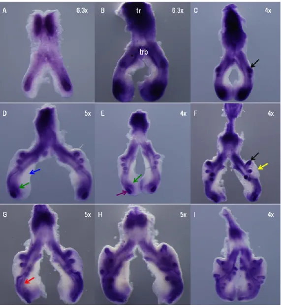
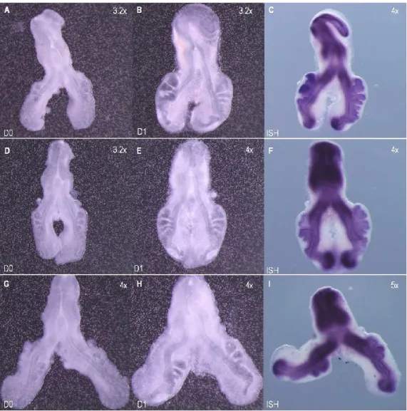
Figure 3.1. hairy1 expression pattern during chick lung development 34
Figure 3.2. hairy1 expression pattern of st b2 lung 34
Figure 3.3. Slide sections of st b1 and b3 lungs probed with hairy1 35 Figure 3.4. hairy2 expression pattern during chick lung development 36
Figure 3.5. hairy2 expression pattern of st b2 lung 36
Figure 3.6. Slide sections of st b0 and b1 lungs probed with hairy2 37 Figure 3.7. In vitro FGF inhibition studies: branching morphogenesis and hairy1 38 Figure 3.8. In vitro FGF inhibition studies: branching morphogenesis and hairy2 39 Figure 3.9. Morphometric analysis of lung explant culture treated with SU5402 39 Figure 3.10. In vitro Notch inhibition studies: branching morphogenesis and hairy1 40 Figure 3.11. In vitro Notch inhibition studies: branching morphogenesis and hairy2 41 Figure 3.12. Morphometric analysis of lung explant culture treated with DAPT 42
1
I
NTRODUCTION
1.1. CHICKEN EMBRYOLOGY
Avian and mammalian embryos are very similar both in its morphological complexity and in the general course of early embryonic development, despite the considerable differences in the adult stage. Avian embryos, however, are easier to obtain and observe: embryo observation and manipulation can be carried out simply by removing the egg‘s shell and embryos can be cultured in an in vitro system. The chicken is now one of the most versatile experimental systems available because of the advances in new technologies, including in vivo electroporation, isolation of embryonic stem cells, and sequencing of the chick genome (Wallis et al., 2004).
1.1.1.EVENTS IN EMBRYONIC DEVELOPMENT
The large yolky egg is fertilized and begins to divide while still in the hen‘s oviduct. Initially, division is confined to a small patch of cytoplasm that contains the nucleus and lies on top of the yolk, leading to the formation of the blastoderm. A layer of cells, called the hypoblast, develops over the yolk to form the floor of the cavity and eventually gives rise to extra-embryonic structures. The embryo is formed from the remaining blastoderm, known as the epiblast. During the 20-hour passage down the oviduct, the egg becomes surrounded by albumen, the shell membranes and the shell. When the egg is laid, some embryonic development has already occurred and usually stops until proper cell environmental conditions are established for incubation to resume. At first all the cells are alike but, as the embryo develops, cell differentiation occurs.
Soon after incubation begins, gastrulation initiates in the epiblast and is marked by the development of a pointed thickened layer of cells visible in the caudal end of the embryo called the primitive streak. This structure is the forerunner of the main body axis of the embryo. Furthermore, a precursor of the digestive tract forms, blood islands appear and somites start to emerge at the anterior end of the embryo.
On the second day of incubation, the blood islands begin linking and form a vascular system, while the heart is being formed elsewhere. Flexion and arching of the embryo begins bringing the two heart rudiments together to form one organ lying ventral to the gut; the heart and vascular systems join and the heart begins to beat. At this stage, ears and lens begin to form.
At the end of the third day of incubation, the head is well developed, the heart is formed, blood vessels are forming and the limbs begin to develop. Blood vessels and blood islands, where blood cells are being formed, have developed in the extra-embryonic tissues. The vessels connect up with
those of the embryo to provide a circulation with a beating heart. At this stage the embryo lies down with its left side on the yolk, the head is strongly flexed so the embryo forms a "C" shape. The embryo gets its nourishment from the yolk through extra-embryonic membranes, which also provide protection. The amnion forms a fluid-filled amniotic sac that provides mechanical protection. A chorion surrounds the whole embryo and lies just beneath the shell, an allantois receives excretory products, and provides the site of oxygen and carbon dioxide exchange, and a yolk sac surrounds the yolk.
Torsion and flexion continue through the fourth day. The digestive and respiratory system develop and the heart continues to enlarge even though it has not been enclosed within the body. It is seen beating if the egg is opened carefully. By the end of the fourth day of incubation, the embryo has all organs needed to sustain life after hatching, and most of the embryo's parts can be identified. The chick embryo cannot, however, be distinguished from that of mammals. In the remaining time before hatching the embryo grows in size, the internal organs develop, wings, legs and beak are formed, and down feathers grow on the wings and body. The chick hatches 21 days after the egg is laid (Wolpert et al., 2007; Gilbert, 2006).
1.1.2.STAGES IN CHICK EMBRYO DEVELOPMENT
The sequence of chick development has been described as an illustrated series of developmental stages by Eyal-Giladi and Kochav (1975) for the first seven hours of development and by Hamburger and Hamilton (1951) for development up to 20 days, allowing standardization between researchers working on chick development. Furthermore the detailed description of the developing anatomy of the chick embryo in ‗The Atlas of Chick Development‘ (Bellairs & Osmond, 2003), which builds on the Hamburger and Hamilton description of chick development, has allowed reliable reporting of manipulated chick embryonic anatomy.
The Hamburger-Hamilton stages (HH) are a series of 46 chronological stages in chick development, starting from laying of the egg and ending with a newly hatched chick. Chick embryos can be staged according to different morphological landmarks. Although most organ systems have a stereotypical appearance at each stage, there are a few which particularly lend themselves to use in staging chick development.
In the very early embryo, the primitive streak is the only visible landmark, and its shape and size is used to stage HH1-6 embryos. Stages 5-8 may be defined by the formation of a head fold, the neural folds, and their fusion to form the neural tube. The expansion of anterior neural tube to form
the brain may also be used to identify later stages. Somitogenesis, the progressive segmentation of the paraxial mesoderm, provides a convenient method for staging embryos between stage 7 and 14. Somites form with surprising regularity every 90 minutes. Stage 10 embryos have 10 somites, and as a rule of thumb, the embryo gains 3 somites during each stage. However, beyond stage 14 (HH14, 22 somites) it becomes increasingly difficult to determine the number of somites accurately. This is due in part to the dispersal of the mesoderm of the anterior most somites, and, in later stages, to the curvature of the tail. Total somite-counts given for the following stages are typical, but sufficiently variable so as not to be diagnostic. For these reasons, other markers (limb-buds, visceral arches) and other externally visible structures are used as identifying criteria from stage 15 onward. Formation of the brachial arches, which will give rise to the structures of the jaw, pharynx and larynx, begins at HH14 and is used as a marker throughout development. The morphology of the limbs, starting with the appearance of wing bud at stage 16, is a useful landmark for staging chick embryos until hatching. Between stages 15 and 35, the appearance of specific structures within the limbs (such as joints and digits), and at later stages the length of the toes are used. The formation and development of the eyelids, primordial feathers and beak are used in a similar way to stage later development (Hamburger & Hamilton, 1951; Bellairs & Osmond, 2003; Gilbert, 2006).
1.1.3.CHICK MODEL
The chick embryo, including its extra-embryonic membranes, has long been used as a developmental model due to its accessibility for surgical manipulations and abundance of data elucidating cellular signaling and interactions during embryonic development (Coleman, 2008). It provides an excellent model system for studying the development of higher vertebrates wherein growth accompanies morphogenesis. This model has several advantages over mammalian systems for in vivo studies, as it is cost-effective, easily manipulated, easily accessible from early stages and throughout organogenesis, and can be used for transgenesis in conjunction with viral vectors or electroporation (Brown et al., 2003). Furthermore, a large database exists on the descriptive aspects of normal and abnormal development of the early avian embryo, and a detailed fate map that shows the locations of progenitor cells prior to gastrulation as well as at later stages, when many different organ rudiments are forming, is available (Darnell & Schoenwolf, 2000).
Chicken eggs can be easily obtained from commercial sources. If eggs are acquired and used within a week, unincubated eggs can be stored in any cool place (16ºC), obviating the need for a special storage facility. At the time the egg is laid, the avian embryo consists of a flat, two-layered blastoderm that lies on the surface of the yolk and, therefore, is readily accessible. Subsequent
development occurs with incubation at 38°C and is rapid. During this period of early development, chick embryos can be easily removed from the shell for culture, or they can be cultured in ovo and follow subsequent development for up to several days. Embryos are semitransparent, allowing the visualization of internal tissues possible under the microscope, and they are big enough to make several types of micromanipulation practical at these early stages (Darnell & Schoenwolf, 2000). Chick embryology studies have demonstrated that it is possible to cleanly separate epithelial and mesenchymal tissues, recombine the two tissues in different orientations or tissues from embryos at different stages of development, and then graft the recombined tissues back into the embryo (Davey & Tickle, 2007).
In the last few years, the classical approaches have been enormously enriched by three major technical advances: the introduction of new methods for gain- and loss-of-function and promoter analysis, the isolation of embryonic stem cells and development of new methods for transgenesis, and the sequencing of the chicken genome and establishment of numerous new electronic resources (Stern, 2005). The recent completion of the sequencing and assembly of the chicken genome represents a major leap forward in avian functional genomics research (Cogburn et al., 2007). Its compact genome and its unique evolutionary position with respect to mammals have greatly facilitated the identification of putative regulatory gene regions, which show high sequence conservation to their mammalian counterparts (Groef et al., 2008).
The special characteristic of chick embryos is that it is possible to bring together well-established embryological manipulations — the classical ‗cut-and-paste‘ experiments, fate mapping and the application of biologically active molecules (by inserting small beads, which act as controlled release carriers, pre-loaded with growth factors, inhibitors, etc) — with more recently developed genetic manipulations, such as mis- and overexpression of genes and increasingly, gene inactivation. Importantly, such genetic manipulation can be precisely targeted in time and in space (Brown et al., 2003).
1.2. SEGMENTATION CLOCK
Somitogenesis, the periodic formation of the vertebrae precursors, is a striking example of a dynamic embryonic process that relies on precise spatial and temporal control of gene expression (Aulehla & Pourquié, 2008). This process comprises the generation and translation of a temporal periodicity into the metameric pattern of somites (Leimeister et al., 2000). Somites constitute the basis of the segmental pattern of the body and give rise to the axial skeleton, the dermis of the back,
and all striated muscles of the adult body. Individual pairs of somites, located symmetrically on either side of the neural tube, emerge from the rostral end of the presomitic mesoderm (PSM), while new mesenchymal cells enter the caudal paraxial mesoderm as a consequence of gastrulation. In the chick embryo, a somite pair is laid down every 90 min in a rostro-caudal progression, and a total of 50 somite pairs are formed during embryogenesis (Palmeirim et al., 1997).
In 1997, Palmeirim and collaborators disclosed a molecular clock underlying vertebrate embryo somitogenesis when analyzing the expression pattern of the hairy1 gene, an avian homolog of the Drosophila hairy segmentation gene, in chick embryos. They demonstrated that hairy1 mRNA is expressed in a highly dynamic manner in the chick PSM appearing as a caudo-rostral wave, which is reiterated during the formation of every somite. The domain of hairy1 expression has the appearance of a wavefront beginning in the broad, caudal PSM, progressing anteriorly and intensifying into the narrow anterior PSM. Finally, in each cycle, expression decays sharply throughout the PSM except for a thin stripe corresponding to the posterior part of the forming somite. This wavefront is not due to cell movements within the PSM, nor to the periodic production of an anterior-to-posterior diffusing signal, but is an autonomous property of the cells in this tissue: cells within the segmental plate cycle autonomously between hairy1 ―on‖ or ―off‖ expression states until they segment and then sustain hairy1 expression in the posterior somite half (Palmeirim et al., 1997; Leimeister et al., 2000).
Analysis of the hairy1 sequence suggests that it belongs to the hairy-enhancer-of-split (Hairy E (Spl)/ HES) family of transcriptional repressor proteins. All HES members share conserved functional motifs: the basic domain (b) required for DNA-binding, a helix-loop-helix dimerization domain (HLH), an orange domain (OR) which confers specificity among family members and the C-terminal WRPW tetrapeptide required for co-factor binding. The evidence to date suggests that HES proteins can be involved in an array of repression mechanisms via the recruitment of different protein partners, forming complexes with different specificities, and regulate a wide variety of developmental processes such as negative control of differentiation, anterior-posterior segmentation in both invertebrates and vertebrates (probably by distinct mechanisms), and sex determination in flies. In many, but not all of these processes, HES proteins function as effectors of the Notch signaling pathway (Kageyama et al., 2007).
Latter, a second chicken hairy-related gene, hairy2, has been identified and was shown to cycle in synchrony with hairy1 across the PSM (Leimeister et al., 2000). Like hairy1, hairy2 is expressed as a wavefront, which sweeps across the PSM during the formation of each somite. Moreover, the
propagation of the hairy2 wavefront is similar to that of hairy1. It does not rely on cell movement or on a propagatory signal traveling through the PSM, suggesting that this dynamic regulation of hairy2 expression constitutes an intrinsic property of the PSM (Jouve et al., 2000). When Jouve and collaborators compared the expression profiles of hairy1 and hairy2, they realized that both genes exhibit similar anterior expression borders in the PSM but become expressed in complementary compartments in the rostral-most PSM. Furthermore, hairy1 and hairy2 expressions cycle together in the PSM, they share the same expression domain, except that hairy2 extends more caudally in the PSM. At the end of one cycle, the expression domains diverge in the rostral PSM, where hairy2 is located in the anterior part of the prospective somite, whereas hairy1 is found in the posterior part (Jouve et al., 2000).
The discovery of oscillating expression of hairy1 in the PSM of chick embryos provided the first molecular evidence for the existence of a segmentation clock that ensures the correct spatiotemporal periodicity of somite generation. Meanwhile, several other genes have also been reported to have a dynamic expression at the level of the PSM in a caudal to rostral direction, such as hairy2 in the chick, lunatic fringe (Lfng), in the chick and mouse, her1 and her7 in zebrafish, Hes1 and Hes7 in mouse, Hey2 both in chick and mouse, esr9 and esr10 in Xenopus and her7 in medaka (Andrade et al., 2007). Furthermore, Aulehla and colleagues (2003) have shown that a repressor of the Wnt signaling pathway, axin2, is also cycling in the mouse PSM (Aulehla et al., 2003).
All studies concerning embryonic cyclic gene expression during development have focused exclusively on the somitogenesis process. However, time control is present during all embryonic processes, suggesting that the molecular clock may not be an exclusive property of the PSM cells but could also be operating in other developing tissues (Andrade et al., 2005). Hirata and coworkers (2002) showed that serum treatment of a variety cultured cell lines induces cyclic expression of both mRNA and protein of Hes1 (mouse hairy2 homologue), with 2-hour periodicity. Recently, hairy2 expression was studied during chick limb bud development and it was observed that this gene is expressed from limb bud initiation until digit formation. This study provided for the first time evidence of a molecular clock working during chick forelimb autopod outgrowth and patterning. Pascoal et al. (2007) showed that the somitogenesis clock gene hairy2 is expressed cyclically in the distal mesenchyme of chick embryo forelimbs, with a 6-hr periodicity and that this periodicity is correlated with the formation time of an autopod limb element. The results of this work support the hypothesis that the molecular clock is not an exclusive property of PSM tissue rather a more general way to count time during vertebrate development, providing positional information to different types of cells (Pascoal et al., 2007).
Although the nature of the clock is still unknown, its existence is revealed by genes expressed in a cyclic fashion within the PSM and the majority of them is intimately related to the Notch signaling pathway (Aulehla et al., 2003; Andrade et al., 2005). These cyclic genes, as well as other components of the Notch signaling pathway, were shown to be required for the proper somite segmentation in mice and zebrafish by mutant analysis or morpholino oligo (MO)-mediated gene-knockdown experiments (Kawamura et al., 2005). Somitogenesis is defective in animals in which this pathway is disrupted by either activating or inactivating mutations. Notch is linked to the segmentation clock although the exact nature of this relationship is not yet understood. Several observations suggest that Notch signaling is a central component of the oscillatory mechanism: (1) the cycling expression of mouse Hes1 depends on the Notch ligand Delta1 (Dll1); (2) the activity of the Notch-dependent factors Her1, Her7, Hes1 and Hes7 is required for their own cyclic expression; (3) the oscillatory expression pattern of lunatic fringe, required for somite segmentation, is under the direct transcriptional control of Notch signaling and is lost in embryos lacking Dll1 or Hes7 (Pasini et al., 2003). Alternatively, it has been proposed that the main function of Notch signaling is to maintain the synchronization of cyclic gene expression in the PSM (Jiang et al., 2000, Aulehla & Pourquié, 2008).
The FGF signaling pathway has also been implicated in the somitogenesis clock mechanism. Kawamura et al. (2005) focused on a zebrafish hairy/Esp1 gene, her13.2 (Hes6-related hairy/Enhancer of split-related gene), which is expressed in the posterior PSM. They provided evidence that her13.2 links FGF signaling to the Notch-regulated oscillation machinery in zebrafish. Expression of her13.2 is induced by FGF-soaked beads and decreased by an FGF signaling inhibitor. her13.2 is required for periodic repression of the Notch-regulated genes her1 and her7, and for proper somite segmentation. Furthermore, Her13.2 augments autorepression of her1 in association with Her1 protein. Therefore, FGF signaling appears to maintain the oscillation machinery by supplying a binding partner, Her13.2, for Her1. It is likely that FGF signaling is transmitted through several different molecular pathways in the posterior PSM, and that Her13.2 mediates specifically one of the roles of FGF signaling, the regulation of the cyclic genes.
1.2.1.NOTCH SIGNALING PATHWAY
Animal development involves many types of cell communication processes and the molecular bases of some of these processes have been unraveled in recent years. Curiously the same molecular pathways, such as Notch signaling pathway, are used to convey messages between cells in vertebrate and invertebrate animals (Bishop et al., 1999). Notch signaling defines a conserved, pleiotropic cell-interaction pathway that controls cell fate and consequently differentiation, proliferation and apoptotic events throughout embryonic development and homeostasis of adult self-renewing organs (Lake et al., 2009; Borggrefe & Oswald, 2009). The central element of this pathway is the transmembrane Notch receptor, which triggers signaling through interaction with membrane-bound ligands expressed on adjacent cells (Lake et al., 2009).
The Notch gene was first identified in Drosophila melanogaster and encodes a large transmembrane protein that acts as a signaling receptor that is required throughout development to regulate the spatial patterning, timing and outcomes of many different cell fate decisions in both vertebrate and invertebrate species (Baron, 2003; Brennan & Gardner, 2002). In mammals, there are four Notch receptors (Notch1–4) and five transmembrane ligands from the Delta (Delta-like1, Dll1, Delta-like3, Dll3 and Delta-like4, Dll4) and Jagged families (Jag1, 2) (Benedito & Duarte, 2005; Borggrefe & Oswald, 2009). Avian have two Delta-like genes, Dll1 and Dll4, and two Jagged genes, called Serrate1 and Serrate2 in chicken. Both Notch and its ligands are integral membrane proteins and generally transmit signals only between cells in direct contact. Moreover, Notch activation has a direct and immediate effect on gene expression, mediated by the detached intracellular domain of Notch itself, acting as a transcriptional regulator in the nucleus (Borggrefe & Oswald, 2009). Thus Notch signaling can readily throw genetic switches that determine choices of cell fate. Furthermore, activation of Notch in a given cell frequently regulates the production of Notch ligands by that cell. Because the level of Notch activation in the cell depends on the level of ligand expression in its neighbors, and vice-versa, this gives rise to feedback loops that correlate the fates of adjacent cells and control spatial pattern of differentiation (Lewis, 1998).
1.2.1.1. Notch Structure
A prototypical Notch gene encodes a single transmembrane receptor composed in its extracellular region of a conserved array of up to 36 EGF-like repeats, mediating direct contact between ligand and receptor involved in interaction; three juxtamembrane repeats, known as Lin-12-Notch (LN) repeats, which modulate interactions between the extracellular and the
membrane-tethered intracellular domains. The intracellular region of Notch includes seven ankyrin repeats flanked by nuclear localization signals, a proline, glutamine, serine, threonine-rich (PEST) domain and a transactivation domain (figure 1.1) (Fiúza & Arias, 2007).
1.2.1.2. The Notch Cascade
The Notch signaling cascade appears remarkably simple with apparently no second messengers involved, although its role and the activation of downstream genes in a given tissue remain often complex and unpredictable (Borggrefe & Oswald, 2009). Notch signaling is activated upon cell-to-cell contact as a result of interactions between Notch receptors and their ligands (Delta or Jagged). Multiple studies focusing on both Drosophila and mammalian Notch receptors have led to a model for Notch signaling that involves a series of cleavages that eventually leads to the release of the intracellular domain, which carries nuclear localization signals, from the cell surface followed by its translocation to the nucleus where it participates directly in transcriptional events (Lake et al., 2009).
A most important feature of Notch is that it acts, at the same time as a transmembrane receptor and as a transcription factor. At the cell surface, extracellular domain of Notch can interact with the extracellular region of Delta and Jagged homologues expressed in a neighboring cell (Arias et al., 2002). This interaction results in the exposure of an extracellular metalloprotease site (S2 site) which thus becomes susceptible to cleavage by transmembrane proteases of the ADAM family (Mumm et al. 2000). Subsequently, two further intramembranous cleavages occur, named S3/S4, by -secretase activity of a membrane protein complex containing members of the Presenilin family and
Figure 1.1. Structure of Notch and its ligands. Notch
ligands, Delta and Jagged/Serrate, are composed of a cysteine-rich region, called DSL, responsible for the interaction with the Notch receptor and several EGF repeats. Jagged/Serrate also contains an extracellular cystein-rich region. Notch is composed by up to 36 EGF-like repeats. Notch also contains a cysteine-rich region known as Lin-12 repeats in close proximity with heterodimerization domains that bind non-covalently extracellular Notch with membrane-tethered intracellular Notch. In its intracellular part, Notch has a region called RAM followed by repeated structural motifs named Ankyrin repeats (mediate the interaction between Notch and CBF1/Su(H)), a transactivation domain (TAD) and a PEST domain. The PEST domain is involved in the degradation of Notch. PM, plasma membrane. (Adapted from Fiúza & Arias, 2007)
Nicastrin as catalytic components (figure 1.2). -secretase activity can be inhibited with small cell-permeant molecules such as DAPT, leading to a blockade of Notch signaling (Daudet et al., 2007). The intracellular domain of Notch (NICD) is thus finally released and translocates into the nucleus where it interacts with members of the RBP-J (also called CSL, CBF1, Su(H), Lag-1) family of transcription factors, regulating gene expression by acting as a transcriptional co-activator. NICD cannot bind directly to DNA but heterodimerizes with the DNA binding protein RBP-J and activates transcription of genes containing RBP-J binding sites. NICD binding to RBP-J is crucial for the switch from repressed to activated state. NICD first displaces corepressors from RBP-J, resulting in derepression of promoters containing RBP-J binding sites and subsequently recruits a coactivator complex to activate transcription of Notch target genes (Borggrefe & Oswald, 2009).
Although signals mediated through Notch receptors have diverse outcomes, only a fairly limited set of Notch target genes have been identified in various cellular and developmental contexts. Hes is a highly conserved protein family that is regulated by Notch in multiple cell types. In mammals, the best-described Notch target genes are indeed the transcription factors Hes1, Hes5 and Hey1. Hes and Hey proteins are bHLH transcription factors that function as transcriptional repressors. Genetic studies in mice have shown that inactivation of many components of the Notch pathway results in dramatic segmentation defects and a severe impairment of the periodic expression of the cyclic genes (Dubrulle & Pourquié, 2002).
Figure 1.2. Notch signaling
pathway. Notch binding to ligand elicits several steps of cleavage. The first one at the S2 site is mediated by the proteases ADAM10 or by TACE. This catalyzes the processing of Notch in the intramembranous S2 and S3 sites by the -secretase complex. Thus, Notch intracellular domain (NICD) is released and translocates into the nucleus where it dislodges repressors (co-R) associated with the DNA-binding CSL transcription factor. (Adapted from Fiúza & Arias, 2007)
1.2.2.FGFSIGNALING PATHWAY
Fibroblast growth factors (FGF) and their specific cell surface receptors (FGFR) make up a large and complex family of signaling molecules that have been shown to play an important role in a variety of processes of embryonic development and tissue homeostasis (Dailey et al., 2005). The FGF family of ligands consists of 23 members in humans and mice, and 13 in chicken. These molecules signal by activating a smaller family of cell surface receptors encoded in four distinct genes (FGFR1-4) that, through alternative splicing in the extracellular immunoglobulin (Ig)-like domain adjacent to the membrane, can produce numerous FGFR isoforms. As the variable region involves the ligand-binding site, the FGFR isoforms differ in their binding affinity toward the FGF ligands. Experimental studies analyzing receptor specificity of the entire FGF family demonstrate a significant redundancy in FGF-FGFR interactions, with all the major FGFR variants being activated by at least five ligands. For instance, the prevalent isoform of FGFR3 (FGFR3c) appears to be strongly activated by FGF1, 2, 4, 8, 9, and 17-20 in vitro. However, its redundancy appears, in contrast, limited in vivo (Krejci et al., 2009).
FGFRs are single-pass transmembrane proteins with tyrosine kinase activity. Ligand binding to the extracellular domain of the receptor initiates a signal transduction cascade (Ras-MAP kinase, PI3 kinase/Akt), that ultimately results in modification of gene expression (Dailey et al., 2005; Thisse & Thisse, 2005). The FGF-FGFR interaction requires the intervention of heparin or HSPG that bind both the ligand and the receptor at specific domains and stabilize the formation of a receptor dimmer bound to the FGF molecules (figure 1.3). FGF signaling can be blocked by chemical inhibitors such as SU5402, an FGF receptor antagonist, that inhibit the tyrosine kinase activity of all four FGFRs by interacting with the catalytic domain (Firnberg & Neubuser et al., 2002).
Figure 1.3. FGF receptors and FGF sinal
transduction. FGFRs are modular proteins comprising 3 immunoglobulin domains (IgI, IgII and IgIII). FGF ligands linked to heparin sulfate proteoglycan (HSPG) bind to IgII and IgIII of FGFR. This results in the dimerization and the subsequent transactivation by phosphorylation of specific tyrosine residues. The main two transduction pathways involve the phospholipase C-g (PLCg) and the Ras/MAP kinase. (Adapted from Thisse & Thisse, 2005)
1.3. LUNG DEVELOPMENT
Lung development is a highly regulated and orchestrated process directed by mesenchymal-epithelial interactions that control and coordinate the temporal and spatial expression of multiple regulatory factors required for proper lung formation. It is now known that the molecular mechanisms involved in patterning, development and differentiation of the lung are orchestrated by finely integrated and mutually regulated networks of transcriptional factors, growth factors, matrix components and physical forces (Shi et al., 2007).
1.3.1.BRANCHING MORPHOGENESIS
Early during mammalian embryonic life, the foregut endoderm is specified into domains that will give rise to organs, such as the thyroid, lung, liver, and pancreas. Once respiratory cell fate has been established, the tracheal and lung primordia form, and the lung subsequently develops into a tree-like system of epithelial tubules and vascular structures that ultimately becomes the airways and the alveoli (Cardoso & Whitsett, 2008; Muratore et al., 2009). The foregut endoderm differentiates into various epithelial cell types, which line the inner surface of the developing lung and trachea. The lung mesenchyme originates from the lateral plate mesoderm and gives rise to multiple components of the lung, including its connective tissue, endothelial cell precursors, the smooth muscle that surrounds airways and blood vessels, the cartilage of the trachea, the lymphatics, and the mesothelial cells that cover the outer surface of the lung, the pleura (Cardoso & Lu, 2006).
Normal growth, morphogenetic patterning and cellular differentiation in the developing lung depend on interactive signaling between the endodermal epithelium and mesenchyme. For instance, distal mesenchyme induces ectopic budding and branching when grafted adjacent to the tracheal endoderm denuded of mesenchyme (Alescio & Cassini, 1962); conversely, tracheal mesenchyme inhibits branching when grafted next to distal epithelium (Wessels, 1970), showing that crosstalk between the epithelium and the mesenchyme drive the branching process. These intimate interactions are mandatory for formation and completion of lung development and ultimately reflect activation of local gene networks along the proximal-distal axis of the respiratory tract, which coordinates the temporal- spatial appearance of buds and clefts, resulting in the bronchial tree (Lebeche et al., 1999). The presence of extracellular matrix molecules, including collagen, fibronectin, laminin, glycosaminoglycans, and proteoglycans, as well as cell membrane-bound integrins, play an important role in directing lung development by influencing the rates of cellular proliferation and differentiation (Shannon & Deterding, 1997). Mechanical distention exerted on the
lung as well as on specific cell types can also significantly affect gene expression and, ultimately, lung growth and development (Pinkerton & Joad, 2000). A myriad of diffusible factors such as FGF, TGF-, BMP-4, retinoic acid, SHH, HGF hepatocyte growth factor and EGF, with their cognate receptors and intracellular signaling molecules modulate cellular proliferation/differentiation and branching (Muraoka et al., 2000). Expression of each molecule occurs in a temporally, spatially, and cell type-specific manner. Also, the role of each molecule should be deemed not in terms of its isolated function in specific cells but, rather, in the context of epithelial-mesenchymal interactions, a process that is central to lung morphogenesis (Demayo et al., 2002).
1.3.1.1. Notch signaling
Notch has been implicated in several aspects of lung biology, including a role in epithelial growth and differentiation. During development, Notch receptors and ligands have been identified in both epithelial and mesenchymal compartments of the lung, and there is in vitro evidence that Notch may act on epithelial differentiation and branching morphogenesis (Tsao et al., 2008).
Aberrant Notch function is associated with several developmental disorders, neurodegenerative disease and cancers (Baron, 2003). Notch pathway activity in normal fetal lung development has been assessed primarily by Hes1 mRNA and immunohistochemistry. Hes1 mRNA has been detected in early pseudoglandular stage mouse lung starting at E12. In the mouse, Hes1 mRNA expression progressively rises until birth and then remains detectable in adult lung as well. Hes1 immunoreactivity is readily detectable in fetal mouse lung in non-endocrine airway epithelial cells that express Notch1 and Notch3. Very limited data are available for other Notch effectors in lung development. Hes5 has not been detected in whole fetal lung RT-PCR (Ito et al., 2000). HeyL is apparently expressed in lung vasculature (Leimeister et al., 2000a). Hey1 mRNA is prominently expressed in adult lung, Hey2 at lower levels (Steidl et al., 2000). Early lethality of mice with homozygous deletions for Notch1, Notch2, Dll1, and Jagged1 limits the assessment of these genetic alterations in lung development (Collins et al., 2004).
Quantitative expression studies from the developing mouse lung demonstrate a progressive increase in Notch1–Notch4, Dll1, and Jagged1 mRNAs from E11.5 into adulthood. In situ hybridization studies and immunohistochemistry suggest that Notch1 is expressed in the distal lung endoderm at least as early as E11.5 and persists through fetal development (Ito et al., 2000). Notch1 expression in pseudoglandular lung clearly overlaps with areas of the epithelium undergoing active growth and branching morphogenesis. Notch1 is not expressed at high levels in fetal lung
mesenchyme that surrounds the primitive epithelium (Post et al., 2000). In contrast to Notch1, Notch2 and Notch3 are both expressed in lung mesenchyme. Notch2 appears to be lacking in epithelial cells by in situ hybridization, whereas Notch3 mRNA and immunoreactivity can be detected in epithelial cells as well as mesenchyme (Post et al., 2000; Ito et al., 2000).
1.3.1.2. FGF signaling
In the embryonic lung, FGF signaling is an essential component of the regulatory networks between epithelium and mesenchyme and is fundamental at several stages of mammalian lung development (Del Moral et al., 2006). A restricted number of FGF family ligands and all FGFRs are present in the embryonic lung, and their expression is regulated in time and space. Perturbation of the FGF signaling pathway during lung development results in dramatic abnormalities of epithelial branching and differentiation (Warburton et al., 1999).
FGF10 and its receptor, Fgfr2b, are required for epithelial branching. Targeted deletion of either genes prevents branching, causing the trachea to terminate as a blind sac (Colvin et al., 2001). At early stages, FGF10 is expressed at high levels in the distal lung mesenchyme in a pattern that appears to correlate with sites of prospective bud formation, acting as a chemo-attractant of lung epithelium, being able to direct bud outgrowths to proper positions in lung organ cultures (Miura et al., 2009; Lebeche et al., 1999). FGF7 can be detected in the early embryonic lung and, together with other factors, plays a role in epithelial branching in vitro, acting as a proliferative factor for the lung epithelium (Lebeche et al., 1999). FGF9 is expressed in the outermost layer of the lung, the mesothelium, and in the epithelium of the developing bronchi (Colvin et al., 1999; Yin et al., 2008). It has been identified as a key factor that signals to mesenchyme to regulate proliferation, differentiation and the expression of other factors that in turn regulate epithelial development. Classical inactivation of FGF9 leads to a reduction in proliferation of the lung mesenchyme resulting in severely reduced branching of the lung epithelium (Colvin et al., 2001).
1.3.2.AVIAN LUNG DEVELOPMENT
Among the air-breathing vertebrates, the avian respiratory apparatus, the lung-air sac system, is the most structurally complex and functionally efficient. After intricate morphogenesis, elaborate pulmonary vascular and airway (bronchial) architectures are formed (Maina et al., 2006).
Although the anatomy of the avian lung differs from that of the mammalian lung, both develop similarly and have anatomical functional equivalents. The avian lung forms by a series of closed
circular buds arising from the main airway branches, which differs from the dichotomous branching morphogenesis in mammalian lung development. In contrast to the mammalian lung, which terminates in alveoli, the avian lung forms a looping anastomotic network of air-vascular surfaces (parabronchi) that end in terminal air buds and air capillaries (Loscertales et al., 2008).
In the embryo of the domestic fowl, Gallus gallus variant domesticus, the development of the respiratory tract proceeds through three stages: the formation of the respiratory rudiments (between 2 and 4 days of incubation, fig. 1.4.A), the bronchial branching (5 days of incubation, fig. 1.4.B) and the formation of the air sacs (6 days of incubation, fig. 1.4.C and D) (Sakiyama et al., 2000).
The lung buds become evident on day 3.5 of embryogenesis, i.e. about stage 23 of development (Hamburger and Hamilton, 1951). They appear as paired protuberances on the lateroventral aspect of the foregut (primitive pharynx) of the developing embryo. On day 5, after fusing on the ventral midline, the single bud divides into left and right primordial lungs that progressively elongate caudally while separating and shifting towards the dorsolateral aspects of the coelomic cavity.
On day 8, the lungs reach their definitive topographical locations in the coelomic cavity. As they develop and increase in size, they grossly changed from a saccular – to a wedge-shaped form.
Figure 1.4. Chick lung development. (A) Drawing of the lung buds and surrounding organs after 3.5 days
of incubation. Lateral view of the developing lung at day 5 (B), day 6 (C) and day 8 (D) of incubation. (Adapted from Sakiyama et al., 2000).
After rotating through an angle of about 180º along the longitudinal axis, the lungs progressively insert and become firmly affixed to the ribs (Maina et al., 2003; Maina et al., 2006). Starting as a solid cord of epithelial cells that runs in a craniocaudal direction of the developing lung, progressively, the intrapulmonary primary bronchus begins to form. In a craniocaudal sequence, secondary bronchi sprout from the lumen of the intrapulmonary primary bronchus, radiating and extending outwards. On reaching the periphery of the lung, parabronchi (tertiary bronchi) bud from the secondary bronchi and project into the surrounding mesenchymal cell mass. The parabronchi proliferate, anastomose and connect the secondary bronchi. The parabronchial lung is capable of gas exchange by the end of the incubation period. On hatching no new structures appear, rather refinement of the existing structures occurs (Maina et al., 2003a).
1.3.2.1. Molecular aspects of avian lung development
Although the morphogenesis of the mammalian lung is now well understood, there is a glaring dearth of data on the avian lung. To elucidate the molecular mechanism for regulating the region-specific morphogenesis of the chicken respiratory tract, Sakiyama and colleagues analyzed the spatiotemporal expression patterns of the Hoxb genes, Bmp-2, Bmp-4, Wnt-5a, and Wnt-11 in the developing respiratory tract and found region-specific expression of these genes in the mesenchymal layer. By tissue recombination experiments, they found that the dorsal and the ventral pulmonary mesenchyme have different inductive capacities toward the tracheal epithelium. These observations suggested the possibility that Hoxb genes are involved in the system specifying regional differences in morphogenesis and cytodifferentiation of respiratory tract. In addition, they argue that it is possible that BMPs and WNTs mediate region-specific epithelial-mesenchymal interaction in this system (Sakiyama et al., 2000).
Muraoka et al. (2000) suggested that nuclear transcription factor kB (NF-kB) may be required to mediate epithelial-mesenchymal interactions in the embryonic chick lung since NF-kB activity in the lung mesenchyme inhibits branching of underlying epithelium (Muraoka et al., 2000). More recently Sakiyama et al. (2003) investigated the role of the Tbx4–Fgf10 system on the separation of the lung bud from the oesophagus in the chicken embryo showing that Tbx4 governs initial endodermal bud formation, respiratory endoderm formation, and septation of the respiratory tract and the esophagus. Moreover, the presence of a feedback loop between Tbx4 and Fgf10 in the regulation of lung development was demonstrated. In the mammalian developing lung, fgf10 expression in the distal mesenchyme at sites where prospective epithelial buds will appear, and its ability to induce epithelial expansion and budding in organ cultures have led to the hypothesis that FGF10 governs the
directional outgrowth of lung buds during branching morphogenesis (Bellusci et al., 1997).
In the chick lung, the branched structure is formed dorsally while the cyst structure (air sac) is formed ventrally during development. Sakiyama et al. (2000) carried out tissue recombination experiments, which showed that the cyst-branch difference in this system is caused by region-specific mesenchymal properties. The results of Miura and coworkers (2009) suggested that the regional cystic-branched difference within the developing chick lung results from a difference in the rate of diffusion of morphogen between the ventral and dorsal regions due to differential levels of HSPG and a different mesenchymal structure (Miura et al., 2009).
1.4. AIMS
It is well known that hairy1 and hairy2 are involved in segmentation events that require tight spatial and temporal regulation, such as somitogenesis and limb development. These processes share some similarities with lung development, namely: they all occur along the rostro-caudal axis, the signaling pathways implicated are the same (FGF, Wnt, Notch), the formation of consecutive repetitive structures along the anterior-posterior axis is observed (somites, limb elements and secondary bronchi in somitogenesis, limb and lung development, respectively), and they occur with exact chronological precision. Considering this parallelism, the characterization of hairy1 and hairy2 during lung branching morphogenesis became relevant, since their expression might indicate a possible role in the development of this tissue, although their functions are not yet clarified.
The purpose of this work was to study the role of hairy1 and hairy2 during embryonic lung development of Gallus gallus.
Thus, the specific aims of this work were:
1. To characterize the expression pattern of hairy1 and hairy2 throughout different stages of lung development by in situ hybridization.
2. To evaluate the effect of Notch and FGF signaling pathway inhibition with DAPT and SU5402, respectively, using in vitro explant culture system:
In hairy1 and hairy2 expression pattern, assessed by in situ hybridization; In lung branching morphology.
2
M
ATERIAL AND
M
ETHODS
2.1. EMBRYO DISSECTION
Fertilized chick (Gallus gallus) eggs, obtained from commercial sources, were incubated for 4-6 days in a humidified atmosphere at 37ºC. After incubation period, eggs shells were cracked open, and embryos removed and immersed in phosphate-buffered saline (PBS). The embryos were then transferred to dissection dishes immersed in PBS and staged according to the developmental table of Hamburger and Hamilton (1951).The attached yolk sac was cut and the embryos killed by decapitation. Embryonic chick lungs were carefully dissected under a dissection microscope (Olympus SZX16, Japan) and were then classified in b1, b2, b3, and so on, taking into account the number of secondary buds formed, 1, 2 or 3 respectively. Lungs were then processed either for ISH or for in vitro culture.
o PBS: 137mM NaCl, 10mM Phosphate, 2.7mM KCl, pH 7.4.
2.2.
I
NS
ITUHYBRIDIZATION
In order to understand how gene expression is guiding development, it is essential to know exactly where and when particular genes are active. Genes are switched on and off during development and gene expression patterns are continuously changing (Wolpert et al., 2007). One of the techniques that show where a gene is being expressed both within whole intact embryos and in sections is ISH.
In situ hybridization, as the name suggests, is a method of localizing and detecting specific RNA or DNA sequences in morphologically preserved tissues or cells preparations by hybridizing the complementary strand of a nucleotide probe to the sequence of interest. This technique involves: generation of a nucleic acid probe, labeled to enable subsequent detection; preparation of fixated tissues; pre-treatment of tissues to increase accessibility of target nucleic acid; hybridization of labeled probe to tissues; washing under conditions that remove non-hybridized probe; detection of the labeled probe, revealing the location of the target cellular nucleic acid (Wilkinson, 1999).
2.2.1.PROBES
2.2.1.1. DNA Extraction
DNA extraction was performed using the GenEluteTM Plasmid Miniprep kit (Sigma, USA). This kit offers a simple, rapid, and cost-effective method for isolating plasmid DNA from recombinant Escherichia coli cultures. Glycerol stocks of plasmids containing the coding sequence of Gallus
gallus hairy1 and hairy2 were kindly provided by Prof. Isabel Palmeirim, Life and Health Sciences Research Institute, School of Health Sciences, University of Minho. An overnight culture of recombinant E. coli containing hairy1 and hairy2 plasmid was harvested by centrifugation and subjected to a modified alkaline-SDS lysis procedure followed by adsorption of the DNA onto silica in the presence of high salts. Contaminants were then removed by a spin-wash step. Finally, the bound DNA was eluted in water and stored at -20ºC.
DNA extraction was performed according to the protocol described below.
1. An overnight culture grown in 5 ml of LB medium was harvested by centrifugation. Two ml of culture volume were transferred to a microcentrifuge tube, cells were centrifuged at 6000 rpm for 1 minute and the supernatant was discarded.
o LB medium: 1% w/v bacto-tryptone; 0.5% w/v bacto-yeast extract; 1% w/v NaCl
2. The bacterial pellet was resuspended in 200 L of Resuspension Solution, and then vigorously vortexed.
3. The resuspended cells were lysed by adding 200 L of Lysis Solution and the contents were immediately mixed by gentle inversion.
4. Following cell lysis, the cell debris was precipitated with 350 L of Neutralization Solution. 5. After gently inversion, the cell debris was centrifuged at 13000 rpm for 10 minutes.
6. Meanwhile, a GenElute Miniprep Binding Column was inserted into a microcentrifuge tube and 750 L of Column Preparation Solution was added. After centrifugation at 13000rpm for 1 minute flow-trough was discarded.
7. The cleared lysate from step 6 was transferred to the prepared column and centrifuged for 1 minute at 13000 rpm.
8. Flow-through was discarded and 750 L of the Wash Solution was added to the column to remove residual salt and other contaminants.
9. Column was centrifuged twice, 13000 rpm for 1 minute, to remove all the ethanol.
10. Finally, the column was transferred to a fresh collection tube and the purified plasmid DNA was eluted by the addition of 50 L water and stored at –20ºC.
2.2.1.2. Plasmid Linearization
hairy1 plasmid, containing an 850 bp fragment of the hairy1 coding sequence cloned in pBluescript KS-vector, was linearized using HindIII (Fermentas, Canada). hairy2 plasmid containing a 350 bp of the hairy2 cDNA cloned in pGEMT was linearized with NcoI (Fermentas) according to the manufacturer´s instructions. Both enzymes were selected to give a 5´overhang, since 3´overhangs are reported to lead to synthesis of abnormal long transcripts. This linearization step was controlled by agarose gel electrophoresis, since circular molecules that are left in the digestion mixture affect the transcription efficiency.
2.2.1.3. DNA Purification
Purification of enzymatic digestion was performed by the QIAquick® PCR purification kit (Qiagen, USA). This system combines the convenience of spin-column technology with the selective binding properties of a uniquely designed silica membrane. DNA adsorbs to the silica membrane in the presence of high concentrations of salt while contaminants pass through the column. Impurities are efficiently washed away, and the pure DNA is eluted with water. The DNA was stored at -20ºC.
DNA purification was performed according to the protocol described below.
1. Five volumes of Buffer PBI were added to 1 volume of the digestion and mixed.
2. The sample was applied to a QIAquick spin column, previously placed in a collection tube, and centrifuged for 1 minute at 13000 rpm.
3. The flow-through was discarded and 750 L of Buffer PE was added, following a centrifugation (1 minute at 13000 rpm).
4. An additional centrifugation step, 1 minute at 13000 rpm, was performed so as to remove any residual Buffer PE, which may interfere with subsequent enzymatic reactions.
5. The QIAquick column was placed in a clean microcentrifuge tube and the DNA was eluted by the addition of 30 L water to the center of the QIAquick membrane. After column was centrifuged (1 min, 13000 rpm). The DNA was stored at -20ºC.
2.2.1.4. Probe Synthesis
T7 RNA polymerase (Promega, USA) was used to synthesize the antisense digoxigenin-labeled hairy1 and hairy2. The in vitro transcription reaction was performed using DIG RNA Labeling Mix (Roche Applied Sciences, Germany) according to the manufacturer´s instructions. The DIG labeling method is based on a steroid isolated from plants, which are the only natural sources of this molecule. Digoxigenin is linked to uridine nucleotides that are incorporated into the RNA probe by RNA polymerase. Hybridized DIG-labeled probes are then detected with high affinity anti-digoxigenin antibody conjugated with alkaline phosphatase (AP), in a colorimetric reaction with two AP substrates: NBT and BCIP (see ISH detection).
2.2.2. IN SITU HYBRIDIZATION
In situ hybridization was performed according to procedure described by Henrique et al. (1995) with minor modifications:
Pre-Hybridization
Prior to hybridization, samples were subjected to a series of pre-treatments that increase the efficiency of hybridization and/or decrease non-specific background.
1. After dissected, chick lungs were fixed overnight at 4ºC in 4% formaldehyde/2 mM EGTA, rinsed in PBS, dehydrated through a methanol series, and stored in 100% methanol at -20ºC.
2. After a step of rehydration, and in order to increase the accessibility of the target RNA, tissues were treated with proteinase K (Promega) to partially digest cellular proteins. Proteinase K is an endopeptidase which is non-specific and attacks all peptide bonds, is active over wide pH range and not easily inactivated. It is used to remove protein that surrounds the target sequence (Polak & McGee, 1998). Incubation has to be carefully monitored because if the digestion proceeds to far you could end up destroying most of the tissue or cell integrity; in this case, and considering the small size of the tissues proteinase K incubation was only for 2 minutes.
o Proteinase K solution: 10g/ml; 0.1% Tween20; PBS
3. This step is followed by a refixation step, to avoid disintegration of the sample, after washing with PBT.