Expression pattern of
zcchc24
during early
Xenopus
development
MARTA VITORINO
1,2, ELIZABETH CORREIA
1,2, ANA-RITA SERRALHEIRO
2, ANA-CATARINA DE-JESUS
1,2,
JOSÉ M. INÁCIO
1,2and JOSÉ A. BELO*
,1,2,31Regenerative Medicine Program, Departamento de Ciências Biomédicas e Medicina, Universidade do Algarve, 2 IBB-Institute for Biotechnology and Bioengineering, Centro de Biomedicina Molecular e Estrutural, Universidade do Algarve,
Campus de Gambelas, Faro and 3Faculdade de Ciências Médicas, Universidade Nova de Lisboa, Lisboa, Portugal.
ABSTRACT We report the expression pattern of a novel Xenopus laevis gene, zcchc24, which en-codes a protein containing two zinc finger domains from the zf-CCHC and zf-3CxxC superfamilies. This protein shares >84% amino acid identity with its vertebrate homologues. During X. laevis embryonic development, zcchc24 is expressed at gastrula stages in the dorsal mesoderm, includ-ing the cardiac precursors region. Durinclud-ing neurula stages, zcchc24 is expressed as two stripes in the dorsal region, more precisely, in the somitogenic mesoderm until the cardiac mesoderm. At early tailbud stages, zcchc24 continues to be expressed in these regions, but starts to be expressed in the migrating neural crest. Later, this gene is expressed in the head, branchial arches, heart and somites. The zinc finger domains present in Zcchc24 proteinand its dynamic gene expression pat-tern suggest that Zcchc24 might be involved in the regulation of heart, somites and of branchial arch formation/patterning, namely in the regulation of apoptosis.
KEY WORDS:
zcchc24, zinc finger, heart development, somitogenic mesoderm, neural crest
The circulatory system is the first one to become functional during vertebrate embryo development, and is composed by the heart, blood cells and vessels. The formation of the heart is a well-conserved process among vertebrate, however, the molecular mechanisms involved in it are not well defined. To address this limitation, a differential screening for genes expressed in the heart precursor cell lineages of chick embryos was performed in our lab (Bento et al., 2011). From the 777 detected genes, 199 were
classified as upregulated uncharacterized genes. Among them, it was obtained ZCCHC24 (zinc finger domain-containing protein 24),
which predicted amino acid sequence is identical 90.8%, 90.5% and 90.8% to its human, mouse and frog homologs, respectively (Bento et al., 2011).
Bioinformatic analysis showed that Xenopus laeviszcchc24
(Genbank accession no. KF438010) encodes a 239 amino acids protein, with a predicted molecular mass of 26.99 kDa, and with two zinc finger domains (Fig. 1, http://www.ncbi.nlm.nih.gov/Structure/ cdd/wrpsb.cgi). Zinc fingers are relatively small protein domains that bind to zinc atoms and, normally, contain finger-like protrusions that make contact with their target molecules. They were initially identified as DNA-binding motifs but several studies showed that
www.intjdevbiol.com
*Address correspondence to: José António Belo. Faculdade de Ciências Médicas da UNL, Edifício CEDOC II, Rua Câmara Pestana nº6, 6-A e 6-B, 1150-082 Lisboa, Portugal. Tel: +351-218-803-102. Fax +351-218-803-010. E-mail: [email protected]
Accepted: 19 December 2013. Final, author-corrected PDF published online: 30 April 2014.
ISSN: Online 1696-3547, Print 0214-6282 © 2014 UBC Press
Printed in Spain
Abbreviations used in this paper: ZCCHC24, zinc finger domain-containing protein 24.
these domains can bind to DNA, RNA, proteins and lipids (Hall, 2005). Indeed, proteins with zinc finger domains are extremely abundant in eukaryotic genomes but can vary both in structure, as well as in function. More, they are involved in biological func-tions as diverse as cell growth, differentiation, DNA recognition, RNA packaging, transcriptional activation, regulation of apoptosis, protein folding and assembly, and lipid binding (Laity et al., 2001).
Consequently, there are several superfamilies of zinc finger motifs, which are classified according to its sequence and structure (Krishna et al., 2003). Zcchc24 protein contains two different zinc
finger domains: one associated to the zf-CCHC superfamily and the other to the zf-3CxxC superfamily (Fig. 1). A typical example of proteins containing the zf-CCHC domain is the inhibitor of apop-tosis (IAP) that has been reported as a regulator of programmed cell death by inhibition of caspases (Krishna et al., 2003). The
zf-3CxxC domain is present in several proteins with functions related with modifications in either DNA or chromatin, such as histone H3 lysine 36, and demethylases KDM2A and B (Birke et al., 2002,
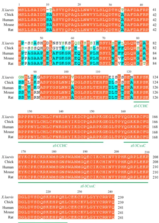
Fig. 1. Sequence alignment of Zcchc24 family members. Comparison of the predicted amino acid sequence of X. laevis Zcchc24 with its chick, human, mouse and rat homologs. X. laevis Zcchc24 (Genbank accession no. KF438010) shares 90.8% of identity with chick ZCCHC24 (XP_421599), 86.3% with human ZCCHC24 (NP_699198), 84.6% with mouse ZCCHC24 (NP_001094903), and 84.2% with ZCCHC24 rat (NP_001101864) homologs. Conserved residues are shaded in orange while identical amino acids among some of the sequences are shaded in light blue. The absence of residues at the corresponding region is indicated by dashes. Both zf-CCHC and zf-3CxxC zinc finger domains are displayed in green.
To analyze the potential function of X. laevis zcchc24 during
early frog embryo development, we examined its expression by whole-mount in situ hybridization (WISH).
zcchc24 transcripts were first detected at midblastula stage,
in both dorsal and ventral marginal zones of the embryos (Fig. 2 A,A’), showing a ring around the marginal zone or presumptive mesoderm. Then, at the onset of gastrulation, zcchc24 expression
was observed in the dorsal mesoderm of both involuting marginal zone (IMZ), a region immediately above the dorsal blastopore lip, and non-involuting marginal zone (NIMZ), which is the region on the top of IMZ (Fig. 2 B,B’).
During gastrulation, and more specifically at stage (st) 11-12,
zcchc24 is expressed in the dorsal mesoderm, in the somitogenic
mesoderm, excluding the dorsal midline (Fig. 2 C C’,E,E’). myoD,
nkx2.5, it is possible to observe that anteriorly zcchc24 expression
is adjacent to the cardiac mesoderm, while more posteriorly, zc-chc24 is expressed in the paraxial mesoderm. (Fig. 3 C,D)
Afterwards, at early tailbud stages, zcchc24 expression is
detected in the unsegmented somitogenic mesoderm and in the somites (Fig. 4 A,B). In contrast, nkx2.5 is predominantly expressed
in the differentiating cardiac muscle (Fig. 4C). In addition, when we compare the expression of zcchc24 with the expression of twi,
a neural crest marker, we observe that zcchc24 transcripts are in
the migrating neural crest cells (Fig. 4 B,D,E).
At later tailbud stages, zcchc24 is expressed in the
differenti-ating cardiac muscle, in the presumptive heart region and in the head, excluding the cleft between the branchial arches and cement gland (Fig. 4 F,G). A transverse section of a st 32 embryo showed whose expression has been described in the
same region, was used as a marker of somi-togenic mesoderm (Fig. 2 D,F). This gene is involved in the formation of somites, and the knock-down of the MyoD disrupts the correct alignment of muscle fibers (Maguire et al.,
2012). At this stage, zcchc24 is also expressed
in two lateral mesoderm stripes around the blastopore that culminate in the dorsal side of the embryo (Fig. 2 C,E). Interestingly, these two lateral mesodermalstripes are correlated with the region in which the heart is originated. As a matter of fact, it has been suggested that, during gastrulation, the precardiac mesoderm migrates in two bilateral heart field located in the anterior lateral mesoderm (Sater and Jacobson, 1989).
From mid (st 11) to the end of gastrulation/ beginning of neurulation (st 13), zcchc24
expression seems to follow the migration of involuting mesoderm along the antero-posterior axis (Fig. 3A). During neurulation,
zcchc24 expression is detected in the anterior
and somitogenic mesoderm (Fig. 3 B-B’’,E), which later will give rise to lateral and neural plate, and the somites, respectively. At these stages, the expression of zcchc24 decreases
B
C
D
E
F
A
A’
B’
C’
E’
that zcchc24 transcripts are clearly detected in heart region, more
precisely, in endocardium, myocardium and pericardium, and in branchial arch mesenchyme (Fig. 4G’). The expression of zcchc24
(Fig. 4G) in the heart is similar to the expression of nkx2.5 in this
region (Fig. 4 H,I). More, when zcchc24 and twi expressions are
compared, it becomes obvious that both genes are expressed in the branchial arches (Fig. 4 J,K).
According to our data, the expression pattern of Xenopuszcchc24
is in part similar to the expression pattern of its chick homolog.
Here, we show that, early in development, Xenopus zcchc24 is
expressed in the dorsal mesoderm, in which the cardiac mesoderm is initially formed, and later is expressed in the head, heart and somites, like its chick homolog at Hamburger and Hamilton stage 10 (HH10; Fig. 5). This suggests that these homologs might have a role in heart formation, both in Xenopus and chick. Moreover,
it demonstrates that zcchc24 expression pattern was conserved
during evolution.
Several zinc finger proteins with a role in heart development Fig. 2. zcchc24 expression from blastula to gastrula stages. Whole mount in situ hybridization using DIG labelled zcchc24 (A-C’, E,E’) or fluorescein labelled myoD(D,F) was performed on embryos from blastula to gastrula stages. (A,A’) At blastula stages, zcchc24 is expressed as a ring around the marginal zone in both dorsal and ventral sides. The embryos display the animal hemisphere to the top. (B,B’) In the beginning of gastrulation (st 10.5), zc-chc24 expression is restricted to the dorsal mesoderm in both involuting and non-involuting marginal zone. (C,C’, E,E’) At late gastrula (st 11 and 12), zcchc24 transcripts are detected in two lateral mesoderm stripes around the blastopore that culminate in the dorsal mesoderm, the somitogenic mesoderm, but is excluded from the midline. (D,F) myoD is expressed in the somitogenic mesoderm. (A’ , B’) Hemisections of (A,B), respectively. (C‘, E’) Transverse sections of st 11 and st 12 embryos, respectively, with dorsal side displayed to the top. (B,C) Vegetal views. (D,E,F) Dorsal views. Dashed lines delimitate the blastopore. An, animal; Vg, vegetal; D, dorsal; V, ventral; dbl, dorsal blastopore lip; sm, somitogenic mesoderm.
Fig. 3. zcchc24 expression during neurula stages. Whole mount in situ hybridization using DIG labelled zcchc24
(A-B’’, E) or fluorescein labelled nkx2.5
(D) and double whole mount in situ hybridization with DIG labelled zcchc24 and fluorescein labelled nkx2.5 (C) were performed on embryos at neurula stages. (A) At late gastrula/early neurula (st 13), zcchc24 is expressed as two stripes in the dorsal side of the embryo along the antero-posterior axis but is excluded from the notochord. (B-B’’, E) During neurula stages, zcchc24 is expressed in anterior and somitogenic mesoderm, decreasing the expression from the posterior to the anterior region. Comparison between the expression of zcchc24 and nkx2.5 (B,C,D) shows that the most anterior expression of zcchc24 is adjacent to the cardiac mesoderm. (A,B, C-E) Embryos in a dorsal view. (B’, B’’) Transversal sections of st 15 embryos with the dorsal region displayed to the top. Red arrows indicate the heart precursor region. A, anterior; P, posterior; n, notochord; sm, somitogenic mesoderm.
B
C
D
E
A
B’
have been described. The GATA zinc finger-containing transcrip-tion factors are a family of proteins that have been implicated in regulation of gene expression in the heart development (Haworth
et al., 2008). It was demonstrated in zebrafish that GATA5 is
necessary for the production of the correct number of myocardial
precursors and for the correct expression of several cardiac genes including nkx2.5. Indeed, the overexpression of GATA5 induces
contractile heart-like tissue (Reiter et al., 1999). GATA 4 is another
GATA family zinc finger that is also implicated in heart and liver development. It was shown that the knock-down of this transcript Fig. 4. Expression pattern of zcchc24
during tailbud stages. Whole mount in situ hybridization using DIG labelled zcchc24 (A, B, F-G’), fluorescein labelled nkx2.5 (C,H) or fluorescein labelled twi(D,J), and double whole mount in situ hybridization using DIG labelled zcchc24 and fluorescein labelled twi
(E,K) or DIG labelled zcchc24 and fluorescein labelled nkx2.5 (I) were performed on embryos at tailbud stages. (A-E) At early tailbud stages, zcchc24 is strongly expressed in the unsegmented somitogenic mesoderm, somites, and migrating neural crest but is absent in the region of differentiating cardiac muscle. (F) Later, at st 28, zc-chc24 transcripts start to be detected in the forming heart, and in the entire head except the region between the branchial arches. (G-K) This expres-sion is maintained throughout tailbud development, being zcchc24 expressed in the somites, branchial arches, head and heart. All the embryos are displayed in a lateral view with the anterior region to the left. (G’) Transversal section of the heart region of a st 32 embryo. The orange arrows indicate the neural crest/ branchial arches, and the red arrows indicate heart-forming region/heart. A, anterior; P, posterior; ec, endocardium; pc, pericardium; mc, myocardium; bam, branchial arch mesenchyme; cg, cement gland.
Fig. 5. In chicken embryos, ZCCHC24 is expressed in the heart and somites. Whole mount in situ hybridization using DIG labelled ZCCHC24 in chick embryos at HH10. (A, A’) At this stage, ZCCHC24 is expressed in the heart (red arrow), somites (yellow arrow) and in head mesenchyme. (A’) Magnification of (A).
G
B
C
D
E
F
H
I
J
K
A
G’
A
A’
affects heart and liver primordia following their specifica-tion (Haworth et al., 2008, Holtzinger and Evans, 2005).
In addition, after heart specification, GATA4 interacts with another GATA family member, GATA6, during its action in the development of heart in mouse, Xenopus and zebrafish
(Holtzinger and Evans, 2005, Peterkin et al., 2003, Zhao et al., 2005). Therefore, since zcchc24 is expressed in
heart precursor cells and later in the heart (Fig. 6), like GATA family genes, it is tempt fate to extrapolate a role for zcchc24 in the formation of the heart.
Nevertheless, zcchc24 is also highly expressed in
Sev-eral zinc fingers proteins like Gli, Gli3 and Gli4 members of the Hedgehog (Shh) signaling pathway have been implicated in somite formation. Gli-type proteins function as transcriptional repressors that respond to Shh signals, and control the expres-sion of Shh-responsive genes such as myf5, the muscle master
regulator (Hui and Angers, 2011). Therefore, we think that Zcchc24 putative role in the regulation of the proper somite segmentation could not be excluded.
The expression of zcchc24 in the migrating neural crest and
later in the branchial arches suggests that zcchc24 might have a
function during the development and/or migration of this tissue. Curiously, several zinc finger proteins of the Snail family have been associated to the neural crest formation (del Barrio and Nieto, 2002, Nieto et al., 1994). For example, it was reported that slug and snail, two members of this family, are important for
neural crest specification and migration. In Xenopus embryos or
animal caps, the overexpression of snail is able to induce the
expression of slug among other neural crest markers such as foxD3, twi and ets1. On the other hand, slug is not able to induce
these neural crest markers, however, gain-of-function studies performed in chick showed that slug overexpression increases
cranial neural crest production, and its loss-of-function inhibits neural crest migration (Aybar et al., 2003, del Barrio and Nieto,
2002, Nieto et al., 1994).
Taken together, our results showed that zcchc24 is expressed
mainly in three different precursors/structures: cardiac precursors/ heart, somitogenic mesoderm/somites, and neural crest/branchial arches. Curiously, several proteins of the zf-CCHC superfam-ily of zinc finger containing proteins were described to have a role on cell death inhibition. Moreover, the proper formation of the heart, branchial arches and somites requires none or a low
level of apoptosis. High levels of apoptosis in these three structures have been reported to be responsible for defects (Graham et al., 1996, Kang and Izumo, 2000, Sanders
and Parker., 2001). These observations indicate that Zcchc24 might be particularly important for the regulation of migration and/ or apoptosis. Nevertheless, further genetic and biochemical analysis must be performed to clarify the role of zcchc24 during X. laevis
embryonic development.
Materials and Methods
Xenopus embryo manipulations
Xenopus eggs were obtained from females
injected with 300 IU of human chorionic gonadotro-pin (Sigma) and were fertilized in vitro. Eggs were
dejellied with 2% cysteine hydrochloride, pH 8.0. Embryos were grown in 0.1X MBS-H (1X MBS-H = 88 mM NaCl, 1 mM KCl, 2.4 mM NaHCO3, 0.82 mM MgSO4, 0.41 mM CaCl2, 0.33 mM Ca(NO3)2, 10 mM HEPES, pH 7.4, 10 mg/mL streptomycin
sulphate and 10 mg/mL penicillin) and staged
ac-cording to Nieuwkoop and Faber (1967).
Cloning of partial coding sequence of zcchc24
Since the coding sequence (CDS) of zcchc24
was not available in stock centers, to obtain it, a
Fig. 6. Schematic representation of the localization of heart precursors and zcchc24 expres-sion during X. laevis embryonic development. Red areas represent the heart precursors and the gray areas represent the expression of zcchc24. (A) Vegetal view, (B) anterior view, (C,D) lateral views with the anterior region to the left. A, anterior; P, posterior; D, dorsal; V, ventral; R, right; L, left.
partial coding sequence was isolated by RT-PCR. With this purpose, total RNA from stage 20 of Xenopus laevis embryos was isolated using
trizol reagent according to the manufacturer’s instruction. To perform the RT-PCR, first strand cDNA was synthesized using oligoDT hexamers as primers and zcchc24 CDS was amplified using a specific pair of primers
(Forward 5’-CCATCCACTCCAGCTATCTGAGCA-3’; Reverse 5´-TTACT-GAACACGGCGGCAGTAGTAGC-3’). The PCR product was cloned into pGEM®-T Easy and sequenced to confirm for correct DNA sequence.
Whole mount in situ hybridization and histology
Single and double whole mount in situ hybridization and anti-sense
probes preparation was carried out as previously described (Belo et al.,
1997). To generate the fluorescein labelled nkx2.5, myoD and twi
anti-sense RNA probes, plasmids containing fragments of these genes were linearized using XbaI, HindIII and EcoRI, respectively, and transcribed using T7 RNA polymerase. To synthesize zcchc24 DIG labelled probe, a
plasmid containing zcchc24 fragment was linearized using SalI enzyme
and transcribed using T7 RNA polymerase. Probes were purified using quick Spin Mini RNA columns (Roche). Hybridized RNAs were detected with alkaline phosphatase conjugated anti-DIG-antibody (Roche) and with alkaline phosphatase conjugated anti-Fluorescein-antibody (Roche) and developed with BM purple (Roche) or BCIP (Roche). Stained embryos were bleached by illumination in 1% H2O2, 4% formamide and 0.5X SSC pH7.0. Embryos were photographed under bright light using a MicroPub-lisher 5.0 RTV camera coupled with a Leica MZ16FA stereoscope or in a Zeiss Sterio Lumar V12 Stereomicroscope coupled with an Axiocam MRC.
For histology, after in situ hybridization, the Xenopus embryos were
fixed overnight at 4°C in 4% PFA, embedded in paraffin, and sectioned
in a Zeiss microtome. Sections were observed in a Zeiss Z2 microscope and photographed with a Zeiss AxioCam ICc3 camera.
Acknowledgements
M Vitorino and J M Inácio are recipients of a F.C.T. Post-Doc fellow-ship with reference SFRH/BPD/65923/2009 and SFRH/BPD/41081/2007,
B
C
respectively. Elizabeth Correia is recipient of a F.C.T B.I. fellowship (PTDC/ SAU-BID/114902/2009). This work has been supported by research grants from F.C.T. and from IBB/CBME, LA to J.A. Belo in the frame of Project refº Pest-OE/EQB/LA0023/2013.
References
AYBAR, M.J., NIETO, M.A. and MAYOR, R. (2003). Snail precedes Slug in the genetic cascade required for the specification and migration of the Xenopus neural crest. Development 130: 483-494.
BELO, J., BOUWMEESTER, T., LEYNS, L., NKERTESZ, GALLO, M., FOLLETTIE, M. and ROBERTIS, E.D. (1997). Cerberus-like is a secreted factor with neutral-izing activity expressed in the anterior primitive endoderm of the mouse gastrula. Mech Dev. Mech Dev 68: 45-57.
BENTO, M., CORREIA, E., TAVARES, A.T., BECKER, J.D. and BELO, J.A. (2011). Identification of differentially expressed genes in the heart precursor cells of the chick embryo. Gene Expression Patt. 11: 437-447.
BIRKE, M., SCHREINER, S., GARCÍA-CUÉLLAR, M.-P., MAHR, K., TITGEMEYER, F. and SLANY, R.K. (2002). The MT domain of the proto-oncoprotein MLL binds to CpG-containing DNA and discriminates against methylation. Nucleic Acids Res. 30: 958-965.
BLACKLEDGE, N.P., ZHOU, J.C., TOLSTORUKOV, M.Y., FARCAS, A.M., PARK, P.J. and KLOSE, R.J. (2010). CpG Islands Recruit a Histone H3 Lysine 36 De-methylase. Molec. Cell 38: 179-190.
DEL BARRIO, M.G. and NIETO, M.A. (2002). Overexpression of Snail family mem-bers highlights their ability to promote chick neural crest formation. Development
129: 1583-1593.
GRAHAM, A., KOENTGES, G. and LUMSDEN, A. (1996). Neural Crest Apoptosis and the Establishment of Craniofacial Pattern: An Honorable Death. Molec. Cell Neurosci. 8: 76-83.
HALL, T.M. (2005). Multiple modes of RNA recognition by zinc finger proteins. Curr.
Op. Struct. Biol. 15: 367-373.
HAWORTH, K., KOTECHA, S., MOHUN, T. and LATINKIC, B. (2008). GATA4 and GATA5 are essential for heart and liver development in Xenopus embryos. BMC Dev. Biol. 8: 74.
HOLTZINGER, A. and EVANS, T. (2005). Gata4 regulates the formation of multiple organs. Development 132: 4005-4014.
HUI, C.-C. and ANGERS, S. (2011). Gli Proteins in Development and Disease. Annu. Rev. Cell. Dev. Biol. 27: 513-537.
KANG, P.M. and IZUMO, S. (2000). Apoptosis and Heart Failure: A Critical Review of the Literature. Circ. Res. 86: 1107-1113.
KRISHNA, S.S., MAJUMDAR, I. and GRISHIN, N.V. (2003). Structural classification of zinc fingers: SURVEY AND SUMMARY. Nuc. Acid. Res. 31: 532-550.
LAITY, J.H., LEE, B.M. and WRIGHT, P.E. (2001). Zinc finger proteins: new insights into structural and functional diversity. Curr. Opin. Struct. Biol. 11: 39-46.
MAGUIRE, R.J., ISAACS, H.V. and ELIZABETH POWNALL, M. (2012). Early transcrip-tional targets of MyoD link myogenesis and somitogenesis. Dev. Biol. 371: 256-268.
NIETO, M., SARGENT, M., WILKINSON, D. and COOKE, J. (1994). Control of cell behavior during vertebrate development by Slug, a zinc finger gene. Science
264: 835-839.
PETERKIN, T., GIBSON, A. and PATIENT, R. (2003). GATA-6 maintains BMP-4 and Nkx2 expression during cardiomyocyte precursor maturation. EMBO J 22.
REITER, J.F., ALEXANDER, J., RODAWAY, A., YELON, D., PATIENT, R., HOLDER, N. and STAINIER, D.Y.R. (1999). Gata5 is required for the development of the heart and endoderm in zebrafish. Genes Dev. 13: 2983-2995.
SANDERS, E. and PARKER., E. (2001). Ablation of axial structures activates apoptotic pathways in somite cells of the chick embryo. Anat Embryol (Berl) 204: 389-98.
SATER, A.K. and JACOBSON, A.G. (1989). The specification of heart mesoderm occurs during gastrulation in Xenopus laevis. Development 105: 821-830.
-deficient mouse embryos
Sabine Pfister, Vanessa J. Jones, Melinda Power, Germaine L. Truisi, Poh-Lynn Khoo, Kirsten A. Steiner, Masami Kanai-Azuma, Yoshiakira Kanai, Patrick P. L. Tam and David A. F. Loebel
Int. J. Dev. Biol. (2011) 55: 45-58 http://dx.doi.org/10.1387/ijdb.103158sp
Building the vertebrate heart - an evolutionary approach to cardiac development
5 yr ISI Impact Factor (2011) = 2.959 José M. Pérez-Pomares, Juan M. González-Rosa and Ramón Muñoz-Chápuli
Int. J. Dev. Biol. (2009) 53: 1427-1443 http://dx.doi.org/10.1387/ijdb.072409jp
Myoskeletin, a factor related to Myocardin, is expressed in somites and required for hypaxial muscle formation in Xenopus
Hui Zhao, Martha L. Rebbert and Igor B. Dawid Int. J. Dev. Biol. (2007) 51: 315-320
http://dx.doi.org/10.1387/ijdb.062260hz
Msx1 and Msx2 have shared essential functions in neural crest but may be dispensable in epidermis and axis formation in Xenopus
Deepak Khadka, Ting Luo and Thomas D. Sargent Int. J. Dev. Biol. (2006) 50: 499-502
http://dx.doi.org/10.1387/ijdb.052115dk
Early stages of neural crest ontogeny: formation and regulation of cell delamination