2018
UNIVERSIDADE DE LISBOA
FACULDADE DE CIÊNCIAS
DEPARTAMENTO DE FÍSICA
Biomechanical Analysis of Walking in Subjects Affected by
Neurological Diseases
António Duarte Robalo Gonçalves Mendonça
Mestrado Integrado em Engenharia Biomédica e Biofísica
Perfil em Engenharia Clínica e Instrumentação Médica
Dissertação orientada por:
Pedro Cavaleiro Miranda
Acknowledgments António Mendonça 2018
II
A
CKNOWLEDGEMENTS
This dissertation is dedicated to all my loving family, specially to my parents, Lucia Robalo and João Mendonça, and to my brother, José Mendonça. Thank you for making me believe in myself, without you, none of this would be possible. I also want to thank to my uncle, Lionel dos Santos, and my cousin, Tomás dos Santos, for granting me access to their office to write this dissertation.
I want to express my deep gratitude to Professor Pedro Miranda, for all orientation given throughout this dissertation, and to Eng. Nevio Tagliamonte, not only for all the guidance, but also for the opportunity to work in such a stimulating working environment.
Thanks to Alessandro Ranieri for all orientation and advice given during the primary stages of the internship.
I’m also extremely thankful to Dr. Marco Molinari, not only for allowing my stay at Fondazione Santa Lucia to be possible, but also for all the help and availability. Also, from Fondazione Santa Lucia, big thanks to Federica Tamburella, the physiotherapist that had the kindness to provide me the clinical profiles of both healthy subjects and patients, and whose help was very important for the concretization of this dissertation.
Regarding the work elaborated in Fondazione Santa Lucia, I must thank Matteo Arquilla, who helped me a lot, especially during the early stages of this work’s development. I also want to express my gratitude to Iolanda Pisotta for all the kindness and orientation inside the institution’s environment. Again, I would like to express my deep gratitude to my loving parents for providing me the economic support to develop my dissertation abroad.
Abstract António Mendonça 2018
III
A
BSTRACT
The occurrence of a spinal cord injury due to a motor vehicle accident, a fall, a shallow diving, an act of violence, or a sport injury can drastically change anyone’s life. The autonomy to perform daily life tasks, like walking, which most people take for granted, is drastically reduced, as well as one’s quality of life. Fortunately, the use of robotic assisted rehabilitation devices can help in overcoming, faster and more efficiently, the problems a spinal cord injury patient must face every day during the gait performance. However, there is still a lack of clinical evidence proving that the use of such devices provides better results than the ones provided by conventional physiotherapy, since there aren’t standardized protocols and specific medical guidelines for using this kind of robotic systems.
This dissertation aims to enhance the human understanding about the analysis of the features of EMG curves during the use of different robotic devices in single-shot tests. To achieve such purpose, the gait performance of both healthy subjects and patients was tracked and analyzed, while they were using one of the two robotic assisted rehabilitation devices: Lokomat exoskeleton or Gait Trainer GT1. To evaluate the impact that these devices can have on the muscular activation, the electrical activity of the tibialis anterior, soleus, gastrocnemius, biceps femoris, rectus femoris and semitendinosus, of both right and left legs, was recorded using surface electromyography.
The experimental procedure adopted for healthy subjects was different from the one adopted for patients. Ten healthy subjects performed five walking conditions, two of which were performed freely over-ground and three using a robotic assisted rehabilitation device. In the context of this dissertation, a self-paced over-ground walking condition was used as control. Ten patients performed three walking conditions, using a robotic assisted rehabilitation device.
In a first instance, EMG data referring to both healthy subjects and patients were time-normalized and filtered. Next, all EMG data were amplitude-time-normalized to the maximum value of a specific walking condition, with the purpose of preparing the comparison of EMG data referring to all walking conditions with one specific walking condition. This first stage was concluded with the calculation of the root-mean-square value of each normalized curve, as an indication of the activation level.
In a second stage, each filtered and time-normalized EMG curve referring to each healthy subject/patient was, firstly, normalized to its maximum value, meaning that each EMG value is presented in this case interpreted as a fraction of the respective maximum value. This second stage was concluded with the study of the symmetry existing between the muscles of the left leg and the corresponding muscles of the right leg. The mathematical tool chosen to quantitatively study the symmetry was the Linear Fit Method.
The final purpose of this dissertation consisted in studying, simultaneously, the symmetry and the activation levels displayed by the above-mentioned muscles, in order to provide information that might prove useful to guide the definition and implementation of future standardized protocols and medical guidelines aimed at making the robotic training more effective.
Overall, the results obtained for patients with spinal cord injury suggest that, in the context of this dissertation, the training parameters chosen for the two robotic assisted rehabilitation devices used were not effective in eliciting symmetrical patterns of muscle activity. The main conclusion drawn is that, considering patients with spinal cord injuries, both robotic assisted rehabilitation devices used in the context of this dissertation can be effective in eliciting symmetrical patterns of muscle activity, but only under restrict settings of its training parameters. Another conclusion drawn from the results presented is that, in a clinical environment the over or under-activation of muscles under specific walking conditions may not represent a favourable factor for locomotor re-training, limiting the long-term effects on the rehabilitation outcome.
Abstract António Mendonça 2018
IV Keywords: spinal cord injury, locomotor training, robotic assisted rehabilitation devices, symmetry, activation levels
Resumo António Mendonça 2018
V
R
ESUMO
A ocorrência de uma lesão da medula espinal devido, por exemplo, a um acidente rodoviário, uma queda, um mergulho mal calculado, um ato de violência ou uma lesão desportiva pode condicionar de forma severa e drástica a vida de qualquer pessoa. A autonomia para realizar tarefas essenciais da vida diária, como a caminhada, que a maior parte das pessoas toma como garantida, é drasticamente reduzida. Para além da dificuldade acrescida neste tipo de tarefas, muitas das vezes o próprio o estado anímico e a autoconfiança do individuo sofrem também um duro golpe, podendo inclusive originar situações de depressão. Por todas estas razões, a qualidade de vida de uma pessoa com uma lesão da medula espinal é severamente afetada de forma negativa. Felizmente, atualmente existem já ferramentas motorizadas que pretendem acelerar o processo de reabilitação e tornar as terapias envolventes mais eficientes. De entre estas ferramentas destacam-se os dispositivos robóticos de reabilitação. Este tipo de dispositivos tem como finalidade promover a plasticidade motora (alcançada pelo recrutamento muscular dependente da ativação de vias neurais específicas), com vista a re-treinar a marcha de pessoas que exibam problemas na locomoção. Muitos destes dispositivos têm ainda algoritmos que têm como intuito incentivar as pessoas a participarem ativamente no processo de reaprendizagem. No entanto, não há ainda evidências clínicas claras que comprovem que a utilização de tais dispositivos produza melhores resultados do que os proporcionados pela fisioterapia convencional, visto não existirem ainda protocolos estandardizados e diretrizes médicas vocacionadas para a utilização de tais sistemas. Isto é, muitas das vezes a avaliação dos efeitos decorrentes da utilização de sistemas robóticos é formulada, com base em parâmetros estatísticos que não reúnem um consenso universal por parte da comunidade médica, na qual estão incluídos os fisioterapeutas.
Um dos propósitos desta dissertação consiste em proporcionar, numa primeira instância, um estudo que vise aumentar o conhecimento humano acerca da análise das características de padrões EMG obtidos durante a utilização de diferentes dispositivos robóticos em testes de uma única tentativa. No sentido de alcançar tal objetivo, o desempenho da marcha de dez sujeitos saudáveis e de dez pacientes com lesões na medula espinal foi rastreado e analisado, enquanto cada um deles caminhava com o auxílio de um dos dois dispositivos robóticos de reabilitação, utilizados no contexto desta dissertação: o exoesqueleto Lokomat e o sistema Gait Trainer GT1. O exosqueleto Lokomat é composto por duas ortóteses que são vinculadas aos membros inferiores do utilizador por meio de alças e punhos. Para propósitos de reabilitação, este exoesqueleto é usualmente acoplado com um sistema de suporte de peso corporal e uma esteira ergométrica (passadeira rolante). O exoesqueleto Lokomat possui a particularidade de fornecer ao utilizador, por meio de um controlador de impedância, um nível de “orientação”. O nível de “orientação”, que é oferecido, determina o quanto é permitido aos movimentos das pernas desviar de um certo padrão predefinido. Enquanto o utilizador se mover dentro dos limites estabelecidos, de acordo com a trajetória predefinida, o controlador não toma nenhuma ação, porém de cada vez que os limites são excedidos, são aplicados torques no sentido de reposicionar a perna de acordo com a trajetória predefinida. O sistema Gait Trainer GT1 é um dispositivo robótico de reabilitação do tipo efector-final, que faz uso de duas placas para os pés para gerar um movimento, do tipo elipsoidal, semelhante à marcha. No que respeita à interface física do dispositivo, o utilizador é preso a um arnês, que controla, verticalmente e horizontalmente, o seu centro de massa durante o ciclo de marcha, e os seus pés são colocados sobre duas placas, cuja movimentação pretende simular as fases de apoio (stance) e de balanço (swing). No sentido de avaliar o impacto que o Lokomat e o Gait Trainer
GT1 podem ter sobre a ativação muscular, foi registada, à superfície da pele, a atividade elétrica de seis
músculos, das pernas direita e esquerda. Para o efeito foram utilizados elétrodos não-invasivos, colocados sobre a superfície da pele. Os músculos estudados, no contexto desta dissertação, são: tibialis
Resumo António Mendonça 2018
VI No seguimento deste estudo, o protocolo experimental definido para indivíduos saudáveis foi diferente do estabelecido para pacientes com lesões na medula espinal. Todos os caminhantes saudáveis executaram cinco condições de caminhada, em que duas foram realizadas diretamente sobre o solo, sem qualquer apoio concedido por sistemas robóticos de reabilitação. No contexto desta dissertação, uma destas condições foi utilizada como controlo. A condição de marcha em questão consiste numa caminhada auto ritmada concretizada diretamente sobre o solo. As três restantes condições de marcha foram executadas com o apoio de dispositivos robóticos de reabilitação. Uma destas três condições de marcha foi concretizada com o sistema Gait Trainer GT1, sendo as outras duas realizadas com o apoio do exoesqueleto Lokomat. As duas condições de marcha realizadas com o exoesqueleto Lokomat foram cumpridas com o nível de “orientação” ajustado, respetivamente, em 50 e 100 %. No que toca aos pacientes, todos eles realizaram, apenas, as três condições de marcha executadas com o auxílio de dispositivos robóticos de reabilitação.
Os protocolos experimentais previamente descritos não foram realizados na presença do aluno responsável pela escrita desta dissertação. Do mesmo modo, também os processos de inspeção, de validação e de pré-processamento dos dados de EMG não foram realizados pelo aluno em questão. Adicionalmente, o aluno também não foi responsável pela segmentação dos dados de EMG em ciclos de marcha. O primeiro passo tomado pelo aluno, responsável pela escrita desta dissertação, consistiu em normalizar, face ao tempo (de um ciclo de marcha), todos os dados de EMG, referentes a indivíduos saudáveis e pacientes, de modo a obter padrões EMG normalizados no tempo. O passo seguinte constou em filtrar/eliminar, de uma análise posterior, todos os padrões EMG normalizados no tempo que exibissem uma razão sinal-ruído anormalmente baixa. Em seguida, todos os padrões EMG foram normalizados em amplitude face ao valor máximo apresentado por uma condição de marcha específica. Neste caso, a condição de marcha escolhida, especificamente, como referência, unanimemente para pacientes e sujeitos saudáveis, foi a condição de marcha realizada com o apoio do exoesqueleto
Lokomat, com o nível de “orientação” ajustado em 100 %. Este tipo de normalização teve como intuito
preparar a comparação de dados de EMG referentes a todas as condições de marcha com uma condição de marcha específica. A primeira etapa desta dissertação foi concluída com o cálculo da raiz do valor quadrático médio (root-mean-square) de cada curva normalizada em amplitude e face ao tempo, como indicação do respetivo nível de ativação.
Numa segunda fase desta dissertação, cada padrão (curva) EMG normalizado face ao tempo, referente a cada paciente/sujeito saudável, foi, em primeiro lugar, normalizado face ao seu respetivo valor máximo, o que significa que cada valor EMG exibido no padrão é interpretado como uma fração desse mesmo valor máximo. Esta segunda etapa da dissertação foi concluída com o estudo, para cada condição de marcha, da simetria existente entre os músculos da perna esquerda e os músculos correspondentes da perna direita. A ferramenta matemática escolhida para qualificar quantitativamente a simetria foi o Método de Ajuste Linear (Linear Fit Method), que basicamente consiste na regressão linear de dois conjuntos de dados.
O propósito final desta dissertação consistiu em analisar, simultaneamente, quer a simetria quer os níveis de ativação exibidos pelos músculos estudados (tibialis anterior, soleus, gastrocnemius, bíceps
femoris, rectus femoris e semitendinosus), a fim de obter indicações que possam revelar-se úteis no
processo de definição e implementação de futuros protocolos standard e diretrizes médicas, que visem tornar mais efetiva a reabilitação auxiliada por sistemas robóticos.
Em geral, os resultados obtidos para pacientes com lesão da medula espinal sugerem que, no contexto desta dissertação, os parâmetros de treino definidos para ambos os dispositivos robóticos de reabilitação utilizados (Lokomat e Gait Trainer GT1) não foram eficazes na obtenção de padrões simétricos de atividade muscular. A principal conclusão extraída desta dissertação, considerando pacientes com lesões na medula espinal, é que os sistemas robóticos de reabilitação utilizados no contexto desta dissertação podem, de facto, ser eficientes na obtenção de padrões simétricos de atividade
Resumo António Mendonça 2018
VII muscular, mas apenas sob configurações bastante restritas dos seus parâmetros de treino. Outra importante conclusão que se pode retirar dos resultados apresentados consiste no facto de, num ambiente clínico, a sobre ou sub-estimulação que alguns músculos possam exibir no desempenho de condições de marcha específicas possa não representar um fator favorável para a reaprendizagem locomotora e possa condicionar a longo prazo os efeitos positivos sobre o resultado da reabilitação.
Palavras-chave: lesão da medula espinal, treino locomotor, dispositivos robóticos de reabilitação, simetria, níveis de ativação
Contents António Mendonça 2018
VIII
C
ONTENTS
Chapter 1 - Introduction ... 1
Chapter 2 – Anatomical Background... 3
2.1 – Introduction to the Nervous System ... 3
2.1.1 – Divisions of the Nervous System ... 3
2.1.1.1 – Spinal Cord... 4
2.1.1.2 – Spinal Nerves ... 6
2.2 – Integration of Nervous System Functions ... 8
2.2.1 – Sensory Functions... 9
2.2.1.1 – Ascending Tracts ... 9
2.2.1.2 – Sensory Areas of the Cerebral Cortex ...10
2.2.2 – Motor Functions ...11
2.2.2.1 – Motor Areas of the Cerebral Cortex ...12
2.2.2.2 – Descending Tracts ...12
2.3 – Spinal Cord Injury ...14
2.3.1 – Implications of a Spinal Cord Injury ...16
2.3.2 – Incomplete Spinal Cord Injuries ...17
2.3.3 – ASIA Impairment Scale ...17
Chapter 3 – Robotic Neurorehabilitation ...19
3.1 – Biomechanics of Walking ...21
3.1.1 – Stance Phase...22
3.1.1.1 – 1st Double Support Phase ...22
3.1.1.2 – Single Support Phase ...23
3.1.1.3 – 2nd Double Support Phase ...23
3.1.2 – Swing Phase ...23
3.1.3 – Paraparetic Gait ...23
3.1.4 – High-Steppage Gait ...23
3.2 – Robotic Assisted Rehabilitation Tools ...24
3.2.1 – Lokomat ...24
3.2.2 – Gait Trainer GT1 ...25
3.3 – RAR Tools: Status of Gait Analysis ...27
3.4 – EMG Signal ...28
3.4.1 – Muscle Physiology ...28
Contents António Mendonça 2018 IX Chapter 4 – Methods ...33 4.1 – Experimental Protocol ...33 4.1.1 – Participants ...33 4.1.2 – Materials ...34
4.1.2.1 – EMG and Acceleration Acquisition ...34
4.1.3 – Procedure ...35 4.1.3.1 – Healthy Subjects ...35 4.1.3.2 – Patients ...36 4.2 – Data Processing ...36 4.2.1 – Inspection Tool ...37 4.2.2 – Preprocessing ...37
4.2.2.1 – Filtering, Rectification and Smoothing ...37
4.2.2.2 – Events Detection ...37
4.2.3 – Gait Cycle Segmentation ...38
4.2.4 – EMG Profiles ...39
4.2.5 – Threshold – Elimination of EMG Artifacts ...39
4.2.6 – Normalization of EMG Curves ...40
4.2.7 – Calculation of the Stance Phase ...41
4.2.8 – Activation Levels ...41
4.2.8.1 – Average Relative Differences between Walking Conditions ...42
4.2.9 – Linear Fit Method ...42
4.2.9.1 – Average Symmetry Discrepancies ...44
Chapter 5 – Results ...45
5.1 – EMG Profiles ...45
5.1.1 – Healthy Subjects ...45
5.1.2 – Patients ...46
5.2 – Elimination of EMG Artifacts ...48
5.2.1 – Healthy Subjects ...48
5.2.2 – Patients ...50
5.3 – Normalized EMG Curves ...51
5.4 – Average Activation Levels ...53
5.4.1 – Healthy Subjects ...54
5.4.1.1 – Average Relative Differences between Walking Conditions ...56
5.4.2 – Incomplete SCI Patients ...58
5.4.2.1 – Average Relative Differences between Walking Conditions ...60
Contents António Mendonça 2018
X
5.5.1 – Healthy Subjects ...61
5.5.1.1 – Average Symmetry Discrepancies ...62
5.5.2 – Incomplete SCI Patients ...63
5.5.2.1 – Average Symmetry Discrepancies ...63
Chapter 6 – Discussion ...65
6.1 – Elimination of EMG Artifacts ...65
6.2 – Normalized EMG Patterns ...66
6.3 – Average Activation Levels ...67
6.3.1 – Average Relative Differences between Walking Conditions ...69
6.4 – Average A1 Coefficients ...70
6.4.1 – Average Symmetry Discrepancies ...71
Chapter 7 – Conclusion ...72
7.1 – Clinical Considerations ...73
7.2 – Future Perspectives ...73
Bibliography ...74
Annexes ...79
A) Healthy Subjects – Normalized EMG Profiles ...79
B) Incomplete SCI Patients – Normalized EMG Profiles ...88
C) Healthy Subjects – Average Activation Levels ...92
D) Incomplete SCI Patients – Average Activation Levels ...96
List of Figures António Mendonça 2018
XI
L
IST OF
F
IGURES
Figure 2.1 – Divisions of the nervous system………..3
Figure 2.2 – The parts of a reflex arc are labeled in the order in which action potentials pass through them………..4
Figure 2.3 – Spinal cord and spinal nerves………5
Figure 2.4 – Cross section of the spinal cord………6
Figure 2.5 – Spinal cord and spinal nerves with their plexuses and branches of these………7
Figure 2.6 – Dermatome map………8
Figure 2.7 – Ascending tracts of the spinal cord……….10
Figure 2.8 – Dorsal Column/Lemniscal Pathway………10
Figure 2.9 – Sensory and motor areas of the lateral side of the left cerebral cortex……….11
Figure 2.10 – Descending tracts of the spinal cord………..13
Figure 2.11 – Direct pathways………14
Figure 2.12 – Illustration of a spinal cord injury……….16
Figure 3.1 – Description of the human anatomical planes……….22
Figure 3.2 – Representation of a segmented human gait cycle………22
Figure 3.3 – Lokomat Pro version 6.0 (picture courtesy of Hocoma) ………..25
Figure 3.4 – Gait Trainer GT1 (picture courtesy of Reha-Stim) ………26
Figure 3.5 – Modified crank and rocker gear system including a planetary gear system to simulate stance and swing phases with a ratio of 60 percent to 40 percent………27
Figure 3.6 – On the left: Voltage difference across the plasma membrane on the beginning of the depolarization stage; On the right: Voltage difference during and after the action potential………29
Figure 3.7 – Propagation of an action potential across a muscle fiber………...30
Figure 5.1 – EMG profile for the healthy subject identified with the number 1………46
Figure 5.2 – EMG profile for the incomplete SCI patient identified with the number 1...47
Figure 5.3 – EMG profile for the complete SCI patient identified with the number 6………...48
Figure 5.4 – Normalized EMG profile for the healthy subject identified with the number 2………51
Figure 5.5 – Normalized EMG profile for the incomplete SCI patient identified with the number 5……..52
Figure 5.6 – Average activation levels, with the respective standard deviation, over all healthy subjects, for each muscle, considering each walking condition – considering the whole gait cycle……….54
Figure 5.7 – Average activation levels, with the respective standard deviation, over all healthy subjects, for each muscle, considering each walking condition – considering only the stance phase………55
List of Figures António Mendonça 2018
XII Figure 5.8 – Average activation levels, with the respective standard deviation, over all healthy subjects,
for each muscle, considering each walking condition – considering only the swing phase………...56
Figure 5.9 – Average activation levels, with the respective standard deviation, over all incomplete SCI patients, for each muscle, considering each walking condition – considering the whole gait cycle………58
Figure 5.10 – Average activation levels, with the respective standard deviation, over all incomplete SCI patients, for each muscle, considering each walking condition – considering only the stance phase…..59
Figure 5.11 – Average activation levels, with the respective standard deviation, over all incomplete SCI patients, for each muscle, considering each walking condition – considering only the swing phase…...60
Figure 5.12 – Average A1 coefficients, with the respective standard deviation, over all healthy subjects, for each walking condition performed, considering each muscle……….62
Figure 5.13 – Average A1 coefficients, with the respective standard deviation, over all incomplete SCI patients, for each walking condition performed, considering each muscle………64
Figure 5.14 – Normalized EMG profile for the healthy subject identified with the number 1……….79
Figure 5.15 – Normalized EMG profile for the healthy subject identified with the number 3……….80
Figure 5.16 – Normalized EMG profile for the healthy subject identified with the number 4……….81
Figure 5.17 – Normalized EMG profile for the healthy subject identified with the number 5……….82
Figure 5.18 – Normalized EMG profile for the healthy subject identified with the number 6……….83
Figure 5.19 – Normalized EMG profile for the healthy subject identified with the number 7……….84
Figure 5.20 – Normalized EMG profile for the healthy subject identified with the number 8……….85
Figure 5.21 – Normalized EMG profile for the healthy subject identified with the number 9……….86
Figure 5.22 – Normalized EMG profile for the healthy subject identified with the number 10……….87
Figure 5.23 – Normalized EMG profile for the incomplete SCI patient identified with the number 1……88
Figure 5.24 – Normalized EMG profile for the incomplete SCI patient identified with the number 2……89
Figure 5.25 – Normalized EMG profile for the incomplete SCI patient identified with the number 3……90
Figure 5.26 – Normalized EMG profile for the incomplete SCI patient identified with the number 4……91
Figure 5.27 – Healthy Subject number 1: A1 coefficients for each one of the six muscles considered, considering each walking condition performed……….101
Figure 5.28 – Healthy Subject number 2: A1 coefficients for each one of the six muscles considered, considering each walking condition performed……….102
Figure 5.29 – Healthy Subject number 3: A1 coefficients for each one of the six muscles considered, considering each walking condition performed……….103
Figure 5.30 – Healthy Subject number 4: A1 coefficients for each one of the six muscles considered, considering each walking condition performed……….104
Figure 5.31 – Healthy Subject number 5: A1 coefficients for each one of the six muscles considered, considering each walking condition performed……….105
List of Figures António Mendonça 2018
XIII Figure 5.32 – Healthy Subject number 6: A1 coefficients for each one of the six muscles considered, considering each walking condition performed……….106 Figure 5.33 – Healthy Subject number 7: A1 coefficients for each one of the six muscles considered, considering each walking condition performed……….107 Figure 5.34 – Healthy Subject number 8: A1 coefficients for each one of the six muscles considered, considering each walking condition performed……….108 Figure 5.35 – Healthy Subject number 9: A1 coefficients for each one of the six muscles considered, considering each walking condition performed……….109 Figure 5.36 – Healthy Subject number 10: A1 coefficients for each one of the six muscles considered, considering each walking condition performed……….110 Figure 5.37 – Incomplete SCI patient number 1: A1 coefficients for each one of the six muscles considered, considering each walking condition performed……….112 Figure 5.38 – Incomplete SCI patient number 2: A1 coefficients for each one of the six muscles considered, considering each walking condition performed……….113 Figure 5.39 – Incomplete SCI patient number 3: A1 coefficients for each one of the six muscles considered, considering each walking condition performed……….114 Figure 5.40 – Incomplete SCI patient number 4: A1 coefficients for each one of the six muscles considered, considering each walking condition performed……….115 Figure 5.41 – Incomplete SCI patient number 5: A1 coefficients for each one of the six muscles considered, considering each walking condition performed……….116
List of Tables António Mendonça 2018
XIV
L
IST OF
T
ABLES
Table 2.1 – Descending tracts………13
Table 4.1 – Overview of healthy participants’ characteristics………..33
Table 4.2 – Overview of incomplete SCI patients’ characteristics……….34
Table 4.3 – Overview of complete SCI patients’ characteristics……….34
Table 4.4 – Experimental design for healthy subjects………...36
Table 4.5 – Experimental design for patients………..36
Table 5.1 – Number of muscles (EMG curves) compromised per walking condition for each healthy subject………...49
Table 5.2 – Number of muscles (EMG curves) compromised per walking condition for each patient………50
Table 5.3 – Healthy Subjects: Average relative differences between walking conditions, for each gait phase (the whole gait cycle, the stance phase and the swing phase), considering the average activation levels obtained for each muscle and respecting each walking condition……….57
Table 5.4 – Incomplete SCI Patients: Average relative differences between walking conditions, for each gait phase (the whole gait cycle, the stance phase and the swing phase), considering the average activation levels obtained for each muscle and respecting each walking condition……….61
Table 5.5 – Average symmetry discrepancies, regarding healthy subjects, for each one of the walking conditions performed, considering each one of the six muscles studied……….63
Table 5.6 – Average symmetry discrepancies, regarding incomplete SCI patients, for each one of the walking conditions performed, considering each one of the six muscles studied………..64
Table 5.7 – Average activation levels, with the respective standard deviation (presented between parenthesis), over all healthy subjects, for each muscle, considering each walking condition – considering the entire gait cycle………..92
Table 5.8 – Average activation levels, with the respective standard deviation (presented between parenthesis), over all healthy subjects, for each muscle, considering each walking condition – considering only the stance phase……….93
Table 5.9 – Average activation levels, with the respective standard deviation (presented between parenthesis), over all healthy subjects, for each muscle, considering each walking condition – considering only the swing phase………94 Table 5.10 – Activation levels of each healthy subject for the left semitendinosus (LST) while performing the self-paced over-ground walking (OverC), and the left rectus femoris (LRF) while performing the
List of Tables António Mendonça 2018
XV self-paced over-ground walking and the over-ground walking guided by a metronome (OverM), considering each gait phase – the whole gait cycle, the stance phase and the swing phase………95 Table 5.11 – Average activation levels, with the respective standard deviation (presented between parenthesis), over all incomplete SCI patients, for each muscle, considering each walking condition – considering the whole gait cycle………..96 Table 5.12 – Average activation levels, with the respective standard deviation (presented between parenthesis), over all incomplete SCI patients, for each muscle, considering each walking condition – considering only the stance phase………97 Table 5.13 – Average activation levels, with the respective standard deviation (presented between parenthesis), over all incomplete SCI patients, for each muscle, considering each walking condition – considering only the swing phase………98 Table 5.14 – Activation levels of each incomplete SCI patient for the right gastrocnemius (RGC) and the right semitendinosus (RST) while performing the Lokomat guided walking, with the guidance level set to 50 % (Loko50), considering each gait phase – the whole gait cycle, the stance phase and the swing phase………..99 Table 5.15 – Average A1 coefficients, with the respective standard deviation (indicated between parenthesis), over all healthy subjects, for each one of the six muscles considered, considering each walking condition………100 Table 5.16 – Average A1 coefficients, with the respective standard deviation (indicated between parenthesis), over all incomplete SCI patients, for each one of the six muscles considered, considering each walking condition………111
Acronyms & Nomenclature António Mendonça 2018
XVI
A
CRONYMS
&
N
OMENCLATURE
AIS American Impairment Scale ANS Autonomic Nervous System
ASIA American Spinal Injury Association BWS Body Weight Support
CNS Central Nervous System DOF Degree(s)-of-freedom EMG Electromyography
FAC Functional Ambulation Category FSL Fondazione Santa Lucia
GT Gait Trainer GT1
GUI Graphical User Interface LFM Linear Fit Method
Loko50 Lokomat guided walking condition, with the guidance level set to 50 % Loko100 Lokomat guided walking condition, with the guidance level set to 100 % (L/R) BF (Left/Right) biceps femoris
(L/R) GC (Left/Right) gastrocnemius (L/R) RF (Left/Right) rectus femoris (L/R) ST (Left/Right) semitendinosus (L/R) SL (Left/Right) soleus
(L/R) TA (Left/Right) tibialis anterior MRI Magnetic Resonance Imaging
OverC Comfort (Self-Paced) Over-Ground walking OverM Over-Ground walking guided by a metronome PNS Peripheral Nervous System
RAM Registered Medical Assistant RAR Robotic Assisted Rehabilitation
Acronyms & Nomenclature António Mendonça 2018
XVII RMS Root-mean-square
ROM Range-of-motion SCI Spinal Cord Injury
SENIAM Surface EMG for a non-invasive assessment of muscles SMU Single Motor Unit
SNS Somatic Nervous System
Introduction António Mendonça 2018
1
C
HAPTER
1
-
I
NTRODUCTION
The occurrence of a spinal cord injury due to a motor vehicle accident, a fall, a shallow diving, an act of violence, or a sport injury can completely change anyone’s life. In several occasions, the autonomy to perform daily life tasks, such as taking a normal walk, which most of us take for granted, is drastically reduced, as well as one’s quality of life. Fortunately, there are already robotic devices especially constructed to help in overcoming such problems (like walking properly), a patient with a spinal cord injury must face every day. However, for patients with a spinal cord injury, there is still a lack of clinical evidence to prove that the use of such devices provides better results than the ones provided by conventional physiotherapy, since there aren’t standardized protocols and specific medical guidelines for using this kind of robotic systems [27,54].
To improve the effects that locomotor training could have on patients with spinal cord injuries, several robotic devices aimed at rehabilitating were already develop. Two of those devices were used in the context of this dissertation: Lokomat exoskeleton [22,23,34] and Gait Trainer GT1 [24,25,34]. One of the purposes of the present dissertation is to evaluate the impact that these two-robotic assisted rehabilitation (RAR) devices could have in the rehabilitation process. Additionally, there are several other RAR devices that have been specifically designed to promote the recovery of the locomotor capabilities of patients that present motor deficits. Two of these RAR devices are: LokoHelp (LokoHelp Group, Weil am Rhein, Germany) [52] and ReoAmbulator (Motorika Medical Ltd., New Jersey, USA) [53] systems. In fact, according to I. Díaz et al. [12], RAR devices can be grouped according to one of the rehabilitation principles that follow:
i) treadmill gait trainers; ii) foot-plate-based gait trainers; iii) over-ground gait trainers; iv) stationary gait trainers; v) ankle rehabilitation systems;
a) stationary systems; b) active foot orthoses.
To evaluate the impact that RAR devices may have in the rehabilitation process, the electrical activity of several muscles, of both right and left inferior limbs, is recorded using surface electromyography. In the context of this dissertation, EMG data were recorded for six muscles. The muscles in question are: tibialis anterior, soleus, gastrocnemius, biceps femoris, rectus femoris and
semitendinosus, of both right and left legs.
The final purpose of this dissertation consists of studying some features of EMG curves that might reveal to be useful to guide the definition and implementation of future standardized protocols and medical guidelines aimed at making the robotic training more effective.
Framework (Contextualization)
This dissertation, written by António Duarte Robalo Gonçalves Mendonça a student of the integrated master's degree in biomedical engineering and biophysics from the faculty of sciences of the University of Lisbon, was co-supervised by Nevio Luigi Tagliamonte, a biomedical engineer from the
Fondazione Santa Lucia (FSL). The internal supervisor was Pedro Cavaleiro Miranda a professor from
the faculty of sciences of University of Lisbon, specialized in bioelectrical signals. The internship related to this dissertation was carried out at FSL, a private hospital that works daily with neurorehabilitation,
Introduction António Mendonça 2018
2 during the period of 6 months (from 8th of February of 2017 until 15th of August of 2017). FSL works
on the development of methods and procedures to conveniently apply exoskeletons to the gait training of stroke victims and people with spinal cord injuries. The work developed by FSL aims to achieve a quantitative understanding of the neuromuscular aspects related to the human walking. In this sense, the presented dissertation will mainly focus on the biomechanical data analysis of the gait of individuals that are neurologically compromised.
In the present dissertation, the student will analyse lower-limb electromyographic data of both healthy subjects and patients with spinal cord injuries, walking i) freely over-ground and ii) with the aid of robotic systems (exoskeletons and operative machines). In a first instance, the student will present and statistically analyse data already acquired in the Laboratory of Robotic Neurorehabilitation, a lab that belongs to FSL. For the analysis of data, the student will use programming software packages.
The expected outcomes of the present dissertation will consist of a quantitative comparison between the features of different walking conditions, for both healthy subjects and neurologically compromised patients. This dissertation also intends to provide a set of clinical indications useful to derive new protocols capable of optimizing the neurorehabilitation process.
Segmentation
The presented dissertation is constituted by six chapters, where chapter 1 is the present introduction.
In chapter 2, several theoretical anatomical concepts are explained and described to integrate the reader within the context of this dissertation. Basically, this chapter consist of an anatomical revision of the nervous system, to enlighten the reader about the structural and functional aspects of such system, and what would happen when these are compromised.
Chapter 3 is an approach to robotic neurorehabilitation. First, this chapter aims to inform the reader about the purposes of conventional and robotic neurorehabilitation. This chapter also includes a description of the biomechanics involved in human gait, as well as a theoretical introduction of the 2 robotic systems used in the context of this dissertation. Finally, chapter 3 concludes with the state-of-art of the application of these 2 systems, and a description of some useful aspects about electromyography. This chapter was constructed in this order to enlighten the reader of how the conjugation of robotic systems and electromyography can be useful to study and properly treat people that suffered spinal cord injuries.
Chapter 4 describes all the methods employed in this dissertation. It includes the clinical description of healthy subjects and spinal cord injury patients studied in this project. It also contains the description of all experimental procedures designed for both healthy subjects and patients, as well as the description of all materials and systems used in the acquisition of the data. Finally, this chapter also describe all the algorithms and software routines used in data processing.
Chapter 5 exhibits all the results derived from the methods described in chapter 4. Chapter 6 presents a discussion over the results obtained.
Anatomical Background António Mendonça 2018
3
C
HAPTER
2
–
A
NATOMICAL
B
ACKGROUND
2.1 - Introduction to the Nervous System
[1]Since the focus of this dissertation consist of evaluating the gait of subjects that are neurologically compromised, it is pertinent to start this introduction with an approach to the human nervous system, in order to understand a little better, the mechanisms behind the complex processes that characterize human walking.
The human nervous system is involved in some way in nearly every human body functions. This system has the function of receiving, interpreting and responding to sensory inputs (stimuli) and controlling muscles and glands. The nervous system is also responsible for maintaining the homeostasis and establishing and maintaining mental activity.
2.1.1 - Divisions of the Nervous System
[1]The nervous system is divided into two major divisions: the central nervous system and the peripheral nervous system (Figure 2.1). The central nervous system (CNS) consists of the brain (located inside the skull) and spinal cord (that is lodged within the spinal canal formed by the vertebrae). The peripheral nervous system (PNS) consists of all the nervous tissue outside the CNS - nerves and ganglia. The nerves of the PNS can be divided into two groups: 12 pairs of cranial nerves and 31 pairs of spinal nerves.
Figure 2.1 – Divisions of the nervous system. The central nervous system consists of the brain and spinal cord. The peripheral nervous system consists of nerves and ganglia. Figure adapted from [2].
Anatomical Background António Mendonça 2018
4 The PNS does the connection between the CNS and the various parts of the human body. The PNS carries and transmits information coming from the different tissues of the body to the CNS and carries and delivers neural commands from the CNS that alter body activities. The sensory division, or
afferent (toward) division, of the PNS conducts action potentials from sensory receptors to the CNS.
The neurons that transmit sensorial information from the periphery to the CNS are called sensory neurons. The motor division, or efferent (away) division, of the PNS is responsible for routing action potentials from the CNS to effector organs, such as muscles and glands. The neurons that transmit action potentials from the CNS to the periphery are called motor neurons (Figure 2.2).
The motor division can be further subdivided based on the type of effector that is being innervated. One of the subdivisions, the somatic nervous system (SNS) transmits action potentials from the CNS to skeletal muscles. The other subdivision, the autonomic nervous system (ANS) transmits action potentials from the CNS to cardiac muscle, smooth muscle and glands. The ANS, in turn, can be divided into sympathetic (that prepares the body for the action) and parasympathetic (regulates rest and vegetative functions) divisions.
The human nervous system can be considered analogous to a highly sophisticated computer because it has the capability to receive, store and process information (inputs) and generate proper responses.
2.1.1.1 - Spinal Cord
[1]The spinal cord has indeed a prominent role not only on the integration and transmission of information, via neurons, throughout the whole nervous system but also on the control of muscles movement and coordination. Therefore, from the sequential point of view the spinal cord acts like a “middleman” in the performance of human walking. If a section of the spinal cord directly related to the execution of lower limbs’ movements is damaged due for example to an injury, it is possible that some of the physiological mechanisms responsible for the walking could be therefore compromised. If this occurs the walking performance will be severely affected.
The spinal cord extends from the foramen magnum at the base of the skull to the second lumbar vertebra (Figure 2.3). The spinal cord is divided into 4 sections: cervical, thoracic, lumbar and sacral. Thirty-one pairs of nerves (spinal nerves) have origin in the spinal cord and are mainly responsible for establishing the communication between the spinal cord and the peripherical segments of the human
Figure 2.2 – The parts of a reflex arc are labeled in the order in which action potentials pass through them. The five components are (1) sensory receptor, (2) sensory neuron, (3) interneuron, (4) motor neuron and (5) effector organ. Figure
Anatomical Background António Mendonça 2018
5 body. The inferior end of the spinal cord and the spinal nerves exiting there resemble a horse’s tail and are collectively called the cauda equina (Figure 2.3).
Looking at a cross section of the spinal cord (Figure 2.4) it is notorious that this important component of the central nervous system consists of a superficial white matter portion and a deep gray matter portion. The white matter basically consists of myelinated axons, and the gray matter is mainly a collection of neuron cell bodies. The white matter in each half of the spinal cord is organized into three columns, called the dorsal (posterior), ventral (anterior) and lateral columns. Each column contains ascending and descending tracts (pathways). Ascending pathways mainly consist of axons that conduct actions potentials toward the brain, and descending pathways consist of axons that conduct action potentials away from the brain.
The gray matter of the spinal cord has the shape of the letter H, with posterior horns and anterior horns. Small lateral horns exist in levels of the cord associated with the autonomic nervous system. The central canal is a fluid-filled space containing cerebrospinal fluid.
Spinal nerves arise from numerous rootlets along the dorsal and ventral surfaces of the spinal cord (Figure 2.4). The ventral rootlets combine to form a ventral root on the anterior side of the spinal
Anatomical Background António Mendonça 2018
6 cord, and the dorsal rootlets combine to form a dorsal root on the posterior side of the cord at each segment. The ventral and the dorsal root unite just lateral to the spinal cord to form a spinal nerve. The dorsal root contains a ganglion which is called dorsal root ganglion.
The cell bodies of sensory neurons are in the dorsal root ganglion. The axons of these neurons that have origin in the periphery of the body pass through spinal nerves and the dorsal roots to the posterior horn of the spinal cord gray matter. In the posterior horn, the axons either synapse with interneurons or pass into the white matter and ascend or descend in the spinal cord.
The cell bodies of motor neurons, which regulate the activity of muscles and glands, are located in the anterior and lateral horns of the spinal cord gray matter. Somatic motor neurons are located in the anterior horn, and autonomic neurons are located in the lateral horn. Axons from the motor neurons constitute the ventral roots and pass into the spinal nerves. Thus, the dorsal root contains only sensory axons, while the ventral root is constituted only by motor axons. Therefore, each spinal nerve has both sensory and motor axons.
2.1.1.2 - Spinal Nerves
[1]From an anatomical point of view, spinal nerves may be considered as the road that carries the neural information from the central nervous system responsible for the activation of the peripheral structures of the body.
Spinal nerves arise along the spinal cord from the union of the dorsal roots and ventral roots (Figure 2.4). Most of the spinal nerves exit the vertebral column between adjacent vertebrae. Spinal nerves are designated accordingly with the region of the vertebral column from which they emerge – cervical (C), thoracic (T), lumbar (L), sacral (S) and coccygeal (Co). Spinal nerves are also numbered (starting superiorly) according to their order within the region in which they are inserted. The 31 pairs of spinal nerves are therefore C1 through C8, T1 through T12, L1 through L5, S1 through S5, and Co (Figure 2.5).
Figure 2.4 – Cross section of the spinal cord. a) In each segment of the spinal cord, rootlets combine to form a dorsal root on the dorsal side and a ventral root on the ventral side. c) Relationship of sensory and motor neurons to the spinal cord.
Anatomical Background António Mendonça 2018
7 The nerves arising from each region of the spinal cord and vertebral column supply specific regions of the body. A dermatome (Figure 2.6) consists of the area of skin supplied with sensory innervation by a pair of spinal nerves. Each of the spinal nerves except C1 has a specific cutaneous sensory distribution.
Anatomical Background António Mendonça 2018
8
2.2 - Integration of Nervous System Functions
[1]The human nervous system produces responses with a degree of complexity that depends on the interpretation of the stimuli received. The nervous system is indeed versatile as it can produce simple responses such as reflexes or involve more complex mechanisms – the tracts (ascending and descending pathways).
Although the spinal cord has a crucial role in the transmission and partly on the integration of neural information, the interpretation and the processing of this kind of information occur mainly in the encephalon. Besides that, the more complex responses triggered by the nervous system are usually generated and/or developed by the encephalon.
The simplest kind of response that the nervous system can give is the reflex. A reflex is an involuntary reaction in response to a stimulus applied to the periphery of the human body and transmitted to the CNS. Reflexes allow a person to react to stimuli more quickly than is possible if conscious thought is involved. A reflex arc is the neuronal pathway (basic functional unit) by which a reflex occurs (see Figure 2.2). The reflex arc is, in other words, the basic functional unit of the nervous system because it is the simplest pathway capable of receiving a stimulus and developing a proper response. A reflex arc is usually composed by five segments (Figure 2.2): (1) a sensory receptor; (2) a sensory neuron; (3) in some reflexes, interneurons, which are neurons that establish the communication between sensory neurons and motor neurons; (4) a motor neuron; and (5) an effector organ (muscle or gland). The simplest reflex arcs don’t involve interneurons. In fact, reflexes themselves vary in their complexity. Some reflexes involve simple neural pathways and few or no interneurons while others have more
Anatomical Background António Mendonça 2018
9 complex pathways and even integration centers. However, most reflexes occur in the spinal cord or in the brainstem rather than in the higher brain centers.
One example of a reflex occurs when a person’s finger touches a very hot surface. In this order, the heat stimulates pain receptors in the skin and consequently action potentials are produced. These action potentials are conducted to the spinal cord via sensory neurons. In the spinal cord the sensory neurons synapse with interneurons that, in turn, synapse with motor neurons that conduct action potentials along their axons to flexor muscles in the upper limb that is receiving the stimulus. These same muscles contract and pull the finger away from the hot surface. During the execution of this reflex no conscious thought is required and the withdrawal of the finger from the painful stimulus begins before the person is consciously aware of any pain.
2.2.1 - Sensory Functions
[1]The CNS constantly receives a variety of stimuli that have origin both inside and outside the body. Sensory input to the brainstem and diencephalon helps maintain the homeostasis. Input to the cerebrum and cerebellum keeps people informed about the surrounding environment and allows the CNS to control motor functions. A small portion of the sensory input results in perception, the awareness of the stimuli. Perception require the following steps:
1. Stimuli originated from the inside or outside of the body are detected by sensory receptors and converted into action potentials that propagate to CNS through the nerves.
2. In the CNS, the nerve pathways carry the action potentials to the cerebral cortex and other areas of CNS.
3. The action potentials that reach the cerebral cortex are translated, allowing the person to be aware of the stimulus.
2.2.1.1 - Ascending Tracts
[1]The spinal cord and brainstem contain ascending tracts (pathways) that transmit information via action potentials from the peripheral nervous system to various parts of the brain (Figure 2.7). Each tract is involved with a limited type of sensory input, such as pain, temperature, touch, position or pressure, because each tract contains axons from specific sensory receptors specialized to detect a specific type of stimulus.
The names of the most part of ascending tracts in the CNS reflect their origin and termination. To each path is usually given a compound name in which the first half of the word indicates its origin and the second half of the word indicates its termination. The ascending tracts usually begin with the prefix spino-, indicating its origin in the spinal cord. Exception to this rule of nomenclature is the dorsal column tract.
Most ascending tracts consist of two or three neurons in sequence, from the periphery to the brain. Almost all neurons that relay information to the cerebrum terminate in the thalamus. Another neuron then conducts the information from the thalamus to the cerebral cortex. The spinothalamic tract responsible for the transmission of action potentials dealing with pain and temperature to the thalamus and on to the cerebral cortex, is an example of ascending tract. The already mentioned dorsal column tract, which transmits action potentials dealing with touch, position and pressure, is another example (Figure 2.8).
Typically, sensory tracts cross from one side of the body in the spinal cord or brainstem to the other side of the body (Figure 2.8). Thus, the right side of the brain receives sensory inputs from the left side of the body and vice versa.
Anatomical Background António Mendonça 2018
10 Ascending tracts can also terminate in the brainstem or cerebellum. For example, the anterior and posterior spinocerebellar tracts transmit information about body position to the cerebellum.
2.2.1.2 – Sensory Areas of the Cerebral Cortex
[1]Figure 2.9 depicts a lateral view of the left cerebral cortex with some of the sensory and motor areas indicated. Ascending tracts project to specific regions of the cerebral cortex, called primary
Figure 2.7 – Ascending tracts of the spinal cord. Figure adapted from [4].
Anatomical Background António Mendonça 2018
11 sensory areas, where sensations are perceived. The primary somatic sensory cortex (somatosensory cortex), or general sensory area, is found in the parietal lobe posterior to the central sulcus.
Sensory fibers carrying general sensory input (related to pain, temperature, position…) synapse in the thalamus. In turn, the thalamus projects neurons that relay information to the primary somatic sensory cortex. Thus, sensory fibers from specific parts of the body project to specific regions of the primary somatic sensory cortex. The primary somatic sensory cortex is organized topographically in relation to the general plan of the body. As it can be seen in the Figure 2.9 the sensory impulses that drive the stimuli from the lower limb and trunk project into the uppermost portions of the somatosensory cortex.
The cortical areas immediately adjacent to the primary sensory areas (Figure 2.9), called association areas, are deeply involved in the process of recognition.
2.2.2 - Motor Functions
[1]The motor system of the brain and spinal cord is responsible for maintaining the body’s posture and balance, as well as moving the trunk, head, limbs, tongue, and eyes and communicating through facial expressions and speech. The reflexes mediated through the spinal cord and brainstem are responsible for some body movements that occur without a conscious thought (involuntary movements). On the other hand, voluntary movements are consciously activated to achieve a specific goal, such as walking. Although consciously activated, the components of most voluntary movements occur automatically. For instance, once a person starts walking, she doesn’t have to think about the moment-moment control of every muscle because there are neural circuits that control automatically the movement of her limbs. After a person learns to execute complex tasks, like walking, these can be performed in a relatively automatic way.
Anatomical Background António Mendonça 2018
12 Voluntary movements result from the stimulation of upper and lower motor neurons. Upper motor neurons have their cell bodies in the cerebral cortex or in nuclei of the brainstem. Lower motor neurons have their cell bodies in the anterior horn of the spinal cord gray matter or in cranial nerve nuclei, in the brainstem. Axons of these neurons leave the central nervous system and extend through spinal and cranial nerves to innervate skeletal muscles.
Voluntary movements depend on the following steps:
1. The initiation of most voluntary movements begins in the premotor area of the cerebral cortex and leads to the stimulation of upper motor neurons.
2. The axons of upper motor neurons form descending pathways that synapse with lower motor neurons. In turn, the axons of lower motor neurons stimulate the contraction of skeletal muscles.
3. The cerebral cortex interacts with the basal nuclei and the cerebellum to plan and coordinate the execution of movements.
2.2.2.1 – Motor Areas of the Cerebral Cortex
[1]The primary motor cortex is found in the posterior portion of the frontal lobe, directly anterior to the central sulcus (Figure 2.9). Action potentials initiated in this region control voluntary movements of skeletal muscles. Upper motor neuron axons project from specific regions of this cortex to specific parts of the body so that a topographic map of the body exists in the primary motor cortex, with the head inferior and lower limbs superior, analogous to the topographic map of the primary somatic sensory cortex (Figure 2.9).
The premotor area of the frontal lobe is the portion of the cortex where motor functions are organized before being initiated in the primary motor cortex. For instance, if a person decides to take a step, the neurons of the premotor area are first stimulated, and the determination is made there as to which muscles must contract, in what order, and to what degree. Action potentials are then passed to the upper motor neurons of the primary motor cortex, which initiate each planned movement.
The motivation and foresight to plan and initiate movements occur in the anterior portion of the frontal lobes, called the prefrontal area (Figure 2.9).
2.2.2.2 – Descending Tracts
[1]The names of the descending tracts are based on their origin and termination. For example, the corticospinal tracts (Table 2.1) are so named because they begin in the cerebral cortex and terminate in the spinal cord. The corticospinal tracts are considered direct because they extend directly from the upper motor neurons in the cerebral cortex to the lower motor neurons in the spinal cord (a similar direct tract extends to lower motor neurons in the brainstem). Other tracts are designated after the region of the brainstem from which they originate. Although these tracts begin in the brainstem they are indirectly commanded by the cerebral cortex, basal nuclei and cerebellum. These tracts are called indirect because no direct connection exists between the cortical and spinal neurons.
Anatomical Background António Mendonça 2018
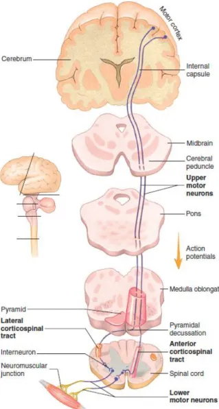
13 The descending tracts control different types of movement (Table 2.1). Tracts in the lateral columns (Figure 2.10 and Figure 2.11) are most important in controlling goal-directed limb movements, such as reaching and manipulating. The lateral corticospinal tract performs an especially important function related to the speed and precision of skilled movements of the hands. Tracts in the ventral columns, such as the reticulospinal tract, have the important role of maintaining posture, balance, and limb position through the control of neck, trunk, and proximal limb muscles.
The lateral corticospinal tract is a good example of how descending pathways are organized. It begins in the cerebral cortex and descends into the brainstem (Figure 2.11). At the inferior end of the pyramids of the medulla oblongata the axons crossover to the opposite side of the body and continue through the spinal cord. Crossover of axons in the brainstem or spinal cord to the opposite side of the body is typical of descending pathways. Thus, the left side of the brain controls skeletal muscles on the right side of the body, and vice versa. The upper motor neurons synapse with interneurons and these, in turn, synapse with lower motor neurons in the brainstem or spinal cord. Lastly, axons of the lower motor neurons extend to the skeletal muscle fibers.
Pathway Function
Direct
Lateral corticospinal Muscle tone and skilled movements, especially of hands.
Anterior corticospinal Muscle tone and movement of trunk muscles. Indirect
Rubrospinal Movement coordination.
Reticulospinal Posture adjustment, especially during
movement.
Vestibulospinal Posture and balance
Tectospinal Movement in response to visual reflexes.
Figure 2.10 – Descending tracts of the spinal cord. Figure adapted from [4].
Anatomical Background António Mendonça 2018
14
2.3 – Spinal Cord Injury
Since the focus of this dissertation consists of the analysis of the gait in subjects with neurological impairment, specifically spinal cord injuries, it is relevant to describe in this subchapter some aspects related with this kind of injuries.
A spinal cord injury (SCI) occurs when the bony protection structure surrounding the cord is damaged by way of fractures, dislocation, burst, compression, hyperextension or hyperflexion. In simple words, a SCI (Figure 2.12) is a global definition for the damage of a spinal cord section that causes changes in its function, either temporary or permanent. Spinal cord injuries can interrupt ascending and/or descending tracts. Reflexes can still function below the level of the injury, but sensations and/or motor functions and reflex modulation may be disrupted. Therefore, it is easy to understand that a SCI can affect severely the performance of daily normal activities, such as walking. Actually, the walking capability of a person after the occurrence of a SCI will strongly depend on the severity (complete or incomplete) and the injury site (which section of the vertebral column was damaged) [1,9].
According to J.E. Lasfargues et al. [8], just in United States, the annual number of traumatic spinal cord injury cases admitted to hospitals was projected to increase from approximately 11 500 in
Figure 2.11 – Direct pathways. Upper motor neuron axons descend to the medulla oblongata. Most axons decussate in the medulla oblongata and descend in the lateral corticospinal tracts in the spinal cord. Some axons continue as the
Anatomical Background António Mendonça 2018
15 1994 to almost 13 400 in 2010. Age adjusted post-hospitalization incidence rate in 1994, in United States, was estimated at approximately 38 per million [8]. Still considering the same article [8], in 2010, there were approximately 247 000 Americans victims of a SCI living in United States. On the other hand, according to A. Singh et al. [42], there are in the central region of Portugal, each year, 58 new cases of SCI considering one million of Portuguese individuals. According to S. Ferro et al. [43], a study conducted at acute-care SCI hospitals and SCI institutes from 11 Italian regions, realized between the 1st of October of 2013 and the 30th of September of 2014, estimated that the crude incidence rate of
traumatic SCI was 14.7 cases per million per year.
The most common cause of spinal cord dysfunction is trauma caused by motor vehicle accidents, falls, shallow diving, acts of violence, and sports injuries. Damage can also occur from various diseases acquired at birth or later in life, from tumors, electric shock, and loss of oxygen related to surgical or underwater mishaps. Most spinal cord injuries that occur can be classified as acute contusions of the cervical portion of the cord and do not completely sever the spinal cord (incomplete injuries) [1].
A SCI is classified as complete or incomplete. An incomplete injury means that the ability of the spinal cord to convey messages to or from the brain is not completely lost. Additionally, this means that, some sensation (even if it’s faint) and movement is possible below the level of injury. A complete injury is characterized by a total lack of sensory and motor function below the level of injury. The absence of motor and sensory function below the level of injury does not necessarily mean that don’t exist remaining intact axons or nerves crossing the injury site, just that they are not functioning conveniently due to the lesion [9].
At the time of SCI, two types of tissue damage occur: (1) primary mechanical damage and (2) secondary tissue damage extending into a much larger region of the cord than the primary damage. The only treatment for the primary damage is prevention itself, such as wearing seatbelts when driving (mainly in automobiles) and not diving into shallow water. Once the accident occurs, there is practically nothing that can be done to repair the primary damage. Secondary spinal cord damage, which begins within minutes of the primary damage, is caused by ischemia (lack of blood supply), edema (fluid accumulation), ion imbalances, the release of “excitotoxins” (such as glutamate), and inflammatory cell invasion. Unlike primary damage, much of the secondary damage can be prevented or reversed if the treatment is prompt. Giving the patient large doses of methylprednisolone, a synthetic anti-inflammatory steroid, within 8 hours of the injury can dramatically reduce the effects of the secondary damage to the cord. Other alternatives for the treatment of the secondary spinal cord damage include the anatomical realignment and stabilization of the vertebral column and still the decompression of the spinal cord. All the rehabilitation procedures applied in medicine intend to retrain patients to use whatever residual connections that still exist across the site of damage [1].
Although the spinal cord was formerly considered incapable of regeneration following severe damage, researchers have learned that most of the neurons of the adult spinal cord survive an injury and begin to regenerate, growing about 1 mm into the site of damage, before regressing to an inactive, atrophic state. The major block to adult spinal cord regeneration is the formation of a scar, consisting mainly of astrocytes, at the site of injury. Myelin in the scar apparently inhibits regeneration. However, and despite the arduous work, researchers are still trying to optimize mechanical tools and chemical factors that might “stimulate” the regeneration of the spinal cord following an injury [1].
Doctors usually assess their patients for spinal cord injuries based on two factors: the location and type of injury a patient has sustained, and the consequent symptoms. Anyone who has fallen, suffered a blow, or lost consciousness may have suffered a SCI. If a person also experiences headaches, loss of movement, tingling, difficulty moving, or difficulty breathing, the conjugation of all these symptoms may be an indicative of the occurrence of a SCI. Yet, no single tests can assess directly all spinal cord injuries. Instead, doctors rely on a variety of protocols that include a clinical evaluation of

![Figure 2.3 – Spinal cord and spinal nerves roots. Figure adapted from [1].](https://thumb-eu.123doks.com/thumbv2/123dok_br/18273296.880822/22.892.234.615.337.990/figure-spinal-cord-spinal-nerves-roots-figure-adapted.webp)
![Figure 2.5 – Spinal cord and spinal nerves with their plexuses and branches of these. Figure adapted from [3]](https://thumb-eu.123doks.com/thumbv2/123dok_br/18273296.880822/24.892.129.768.219.720/figure-spinal-spinal-nerves-plexuses-branches-figure-adapted.webp)
![Figure 2.6 – Dermatome map. Figure adapted from [1].](https://thumb-eu.123doks.com/thumbv2/123dok_br/18273296.880822/25.892.212.651.109.633/figure-dermatome-map-figure-adapted-from.webp)
![Figure 2.7 – Ascending tracts of the spinal cord. Figure adapted from [4].](https://thumb-eu.123doks.com/thumbv2/123dok_br/18273296.880822/27.892.259.642.549.986/figure-ascending-tracts-spinal-cord-figure-adapted.webp)
![Figure 2.9 – Sensory and motor areas of the lateral side of the left cerebral cortex. Figure adapted from [6]](https://thumb-eu.123doks.com/thumbv2/123dok_br/18273296.880822/28.892.104.810.358.808/figure-sensory-motor-lateral-cerebral-cortex-figure-adapted.webp)
![Figure 2.10 – Descending tracts of the spinal cord. Figure adapted from [4].](https://thumb-eu.123doks.com/thumbv2/123dok_br/18273296.880822/30.892.211.683.104.422/figure-descending-tracts-spinal-cord-figure-adapted.webp)
![Figure 2.12 – Illustration of a spinal cord injury. Figure adapted from [15].](https://thumb-eu.123doks.com/thumbv2/123dok_br/18273296.880822/33.892.223.711.169.498/figure-illustration-spinal-cord-injury-figure-adapted.webp)
![Figure 3.2 – Representation of a segmented human gait cycle. Figure adapted from [17]](https://thumb-eu.123doks.com/thumbv2/123dok_br/18273296.880822/39.892.245.646.135.480/figure-representation-segmented-human-gait-cycle-figure-adapted.webp)