UNIVERSIDADE DE LISBOA
FACULDADE DE MEDICINA DE LISBOA
AMYLOID PEPTIDE AND p63:
TWO PRIME TRIGGERS OF NEURAL APOPTOSIS
AND DIFFERENTIATION
Maria Benedita Pereira de Vasconcelos Fonseca
Doutoramento em Ciências Biomédicas (Neurociências), apresentada à Universidade de Lisboa através da Faculdade de Medicina
Supervisor: Cecília M. P. Rodrigues, Ph.D. Co-supervisor: Ana Sebastião, Ph.D.
O presente documento é da exclusiva responsabilidade do seu autor, não cabendo qualquer responsabilidade à Faculdade de Medicina de Lisboa pelos conteúdos nele apresentados
The studies presented in this thesis were performed at the Research Institute for Medicines and Pharmaceutical Sciences (iMed.UL) and Department of Biochemistry and Human Biology, Faculty of Pharmacy, University of Lisbon, under the supervision of Professor Cecília M. P. Rodrigues.
Maria Benedita Pereira de Vasconcelos Fonseca was the recipient of a Ph.D. fellowship (SFRH/BD/43959/2008) from Fundação para a Ciência e Tecnologia (FCT), Lisbon, Portugal. This work was supported by grants PTDC/SAU-FCF/67912/2006, PTDC/SAU-NMC/117877/2010 and Pest-OE/SAU/UI4013/2011 from FCT and FEDER.
De acordo com o disposto no ponto 1 do artigo nº 41 do Regulamento de Estudos Pós-Graduados da Universidade de Lisboa, deliberação nº 93/2006, publicada em Diário da República – II Série nº 153 – 5 de Julho de 2003, o Autor desta dissertação declara que participou na conceção e execução do trabalho experimental, interpretação dos resultados obtidos e redação dos manuscritos.
A impressão desta dissertação foi aprovada pela Comissão
Coordenadora do Conselho Científico da Faculdade de Medicina de
Lisboa em reunião de 18 de junho de 2013.
Agradecimentos
Foi uma longa viagem... Dedico este espaço àqueles que me acompanharam ao longo desta jornada, que me ajudaram e apoiaram a tornar um sonho realidade. Naturalmente, não conseguirei nomear aqui todas as pessoas, mas há no entanto alguns a quem não posso deixar de manifestar o meu apreço e agradecimento sinceros.
Muito especialmente, agradeço à minha orientadora Professora Cecília Rodrigues, um exemplo que para sempre levarei comigo, como pessoa e como profissional. Muito obrigada por me ter orientado, por ter acreditado em mim, por me incentivar e apoiar sempre que precisei, pela sua total disponibilidade, paciência e compreensão.
Agradeço à Professora Ana Sebastião, minha coorientadora, pelo tempo dispendido na análise da tese.
Agradeço à Filipa Nunes, pela sua excelente orientação, por todo o saber que me transmitiu, sempre com muita, muita paciência, dedicação e um sorriso. Obrigada amiga, do fundo do coração. Tive também a oportunidade de ser orientada pela Susana Solá, a quem também quero agradecer a sua exaustiva dedicação e atenção prestadas ao meu projeto, mesmo quando a sua agenda parecia não ter espaço para mais nada. Obrigada Susana, por todo o teu apoio.
Aos meus colegas e ex-colegas do laboratório e sempre amigos, o meu muitíssimo obrigada por me terem acompanhado e ajudado ao longo desta “caminhada”. Agradeço a força e o incentivo constantes, o espírito de grupo e a amizade de todos os colegas. Ao Duarte, um beijinho especial pela nossa amizade, por ter sempre tempo para me escutar, para esclarecer uma dúvida, para ensinar, para as nossas conversas sobre ginásio, para dar um abracinho reconfortante, sempre de uma forma tão sincera. À Joana Xavier, obrigada pelos nossos momentos de desabafo, pela sua ajuda incondicional no laboratório e não só, e pela sua pro-atividade, um exemplo a seguir. Ao André também um agradecimento especial, por ser uma pessoa com um coração tão bom, tão espontâneo e ser tão sorridente. Aqui fica a promessa de enviar-lhe de vez em quando uma caixinha de quindins. À Ana Luísa, o meu reconhecimento por ser sempre tão prestável para todos os colegas do grupo. Obrigada Ana também pela tua ajuda preciosa em momentos de maior aflição no laboratório. Aos meus colegas mais novos,
Pedro Rodrigues, Pedro Dionísio, Diane, Sofia e Marta, um beijinho grande, vocês trouxeram energia nova para o laboratório. Tenho um grande carinho por vocês e desejo-vos grandes sucessos. À Rita, Rui, Joana Amaral, Pedro Borralho, Daniela, Ricardo e Márcia, os meus colegas que me receberam tão bem quando entrei no grupo e me viram “crescer” ao longo destes anos, um agradecimento sincero pelo vosso apoio incondicional, pela atenção dispensada, pelas críticas e opiniões e pelo convívio diário, sempre muito bem dispostos. Agradeço igualmente às Professoras Elsa Rodrigues, Maria João Gama, Margarida Castro Caldas, bem como à Andreia, Inês e ao Miguel, pela sempre boa disposiçao que nos contagia. Manifesto aqui também a minha gratidão à Professora Isabel Bettencourt e a todas as pessoas do iMed.UL que tive oportunidade de conhecer e com quem partilhei os meus momentos, bons e menos bons. À Deolinda, um beijinho com muito carinho, pela seu sorriso diário, pela sua simplicidade e vontade de ajudar.
Aos meus queridos amigos Heloísa, Miguel, Margarida e Vítor, não há palavras para descrever o quão gosto de vocês e o quão importantes são para mim. Eles são como família, os meus queridos companheiros de treino, de sofrimento, de alegrias, de “beca-beca”. Estou eternamente grata a Deus por eles fazerem parte da minha vida.
Aos meus pais e ao meu irmão João, de quem tenho tantas saudades, um beijinho com muito amor e um muito obrigada pelo incentivo permanente e pela compreensão da distância que o trabalho me impediu algumas vezes de minimizar. Um obrigada também aos meus avós, primos e tios, sempre tão presentes. À Adelina, a minha segunda mãe, e ao Rui, Sofia, Leonor e Guilherme, agradeço do fundo do coração todo o carinho e amor que sempre me deram de uma forma tão simples e sincera.
Por fim, um agradecimento muito especial para um ser muito especial. Diogo, a ti dedico esta tese, por todo o amor, pelo suporte emocional, por seres tantas vezes um pai ou uma mãe para mim, para além de namorado, por seres a pessoa que mais admiro neste mundo. O teu olhar e o teu sorriso rasgado e tão teu foram muitas vezes o meu alento em dias menos claros. Eyes do not lie...!!!
Table of Contents
Table of contents...………...…………...………vii Resumo………...…………..xiii Abstract………..………...….xix Abbreviations……….…….….xxiii Publications………...……….…...xxv 1. General Introduction……….….1 1.1. Neural Apoptosis……….………...31.1.1. Apoptosis signalling pathways.……….…....…....4
1.1.1.1. Extrinsic pathway………...…………..…...4
1.1.1.2. Intrinsic pathway……….………...…....5
1.1.2. p53 family of proteins………..…………...……6
1.1.2.1. p53………..………..………....…….8
1.1.2.2. p63 and p73………..……...10
1.1.3. JNK/c-Jun signaling pathway………….………..….…...11
1.1.4. Role of apoptosis in Alzheimer’s disease……….……….………13
1.1.4.1. Aβ production and accumulation……….……….…….…..14
1.1.4.2. Cytotoxicity of Aβ……….…………...………15
1.1.5. Regulation of apoptosis by autophagy……….…..….…..18
1.1.6. Modulation of neural apoptosis by bile acids………....……...21
1.2. Neural Differentiation………..24
1.2.1. Adult NSCs……….………...………...26
1.2.2.1. Proteases………..…………29
1.2.2.2. Bcl-2 family………..…….……..30
1.2.2.3. p53 family………..……….….30
1.2.4. Role of Aβ peptides in neural differentiation………..…………..37
1.2.5. Autophagy during neural differentiation……….………..40
Objectives………43
2. c-Jun Regulates the Stability of Anti-apoptotic ΔNp63 in Amyloid-β-induced apoptosis………..45
2.1. Abstract……….47
2.2. Introduction………..48
2.3. Material and methods………...50
2.3.1. Cell culture and treatments………50
2.3.2. Primary culture of cortical neurons………...51
2.3.3. Measurement of cell death……….51
2.3.4. Plasmids, siRNAs and transfections………..51
2.3.5. Western blot………...52
2.3.6. Statistical analysis………..53
2.4. Results………...53
2.4.1. Aβ-induced cell stress is associated with decreased ΔNp63 and increased c-Jun protein levels………53
2.4.2. TUDCA modulates Aβ-induced changes in ΔNp63 and c-Jun protein levels ……….……….56
2.4.3. c-Jun is required for ΔNp63 degradation after Aβ treatment…………56
2.5. Discussion……….61
3. TAp63γ Demethylation Regulates Protein Stability and Cellular Distribution during Neural Stem Cell Differentiation……….………65
3.1. Abstract……….67
3.3. Material and methods………...69
3.3.1. Ethics statement……….69
3.3.2. Cell culture and differentiation………..70
3.3.3. siRNAs and plasmid transfections……….70
3.3.4. Total, cytosolic and nuclear protein extraction……….71
3.3.5. Immunoblotting……….72
3.3.6. Immunocytochemistry………...72
4.3.7. Immnunoprecipitation assay………..73
3.3.8. Evaluation of apoptosis……….74
3.3.9. Densitometry and statistical analysis……….74
3.4. Results………...74
3.4.1. TAp63γ and JMJD3 levels increase throughout mouse NSC differentiation ………..74
3.4.2. JMJD3 regulates TAp63γ protein in a demethylase activity-dependent manner………...76
3.4.3. TAp63γ manipulation influences neuronal differentiation………..…..77
3.4.4. TAp63γ is directly demethylated by JMJD3 during neural differentiation……….………...79
3.4.5. TAp63γ is stabilized by JMJD3-demethylase activity and accumulates in the nucleus………..………..……81
3.5. Discussion……….83
4. Amyloid β Peptides Promote Autophagy-dependent Differentiation of Mouse Neural Stem Cells………...87
4.1. Abstract……….………89
4.2. Introduction………..90
4.3. Material and methods………...91
4.3.1. Cell culture……….………...91
4.3.2. Cell treatments………..……….92
4.3.4. Dual luciferase assay……….………93
4.3.5. Immunoblotting……….………93
4.3.6. Immunocytochemistry…………..……….94
4.3.7. Flow cytometry analysis………..………..94
4.3.8. Evaluation of cell death……….………95
4.3.9. Densitometry and statistical analysis………..………...96
4.4. Results………...96
4.4.1. Characterization of NSCs isolated from mouse fetal forebrain under differentiating conditions………..……….96
4.4.2. Distinct roles of Aβ1-40 and Aβ1-42 in modulating lineage-specific markers of NSCs………..………..98
4.4.3. Aβ1-40 increases proliferation and neuronal-promoter activity of NSCs………..………..100
4.4.4. Aβ1-40 and Aβ1-42 increase autophagy during differentiation of NSCs………..………..102
4.4.5. Inhibition of autophagy reverts the effects of Aβ1-40 and Aβ1-42 in enhancing lineage-specific markers in NSCs……….………….104
4.5. Discussion………...104
Figures
Figure 1.1. p53 family gene and protein structure ………....……..…...7 Figure 1.2. Autophagy is crucial for intracellular clearance of neurons……….20 Figure 1.3. Tauroursodeoxycholic acid modulates amyloid β-induced toxicity by
inhibiting organelle-drive apoptosis ... 22 Figure 1.4. Monolayer culture system of neural stem cells ………...26 Figure 1.5. p53 in neurogenesis ……….33 Figure 1.6. Pathological versus proneurogenic effects of amyloid β peptides………...39 Figure 1.7. Autophagy is involved in neural differentiation………...42 Figure 2.1. Expression of endogenous p63 isoforms and genotoxic stress-induced
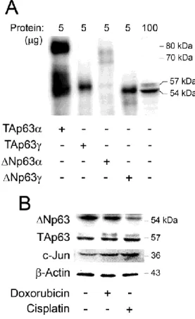
regulation of p63 and c-Jun protein levels in PC12 neuronal-like cells .... 54 Figure 2.2. Aβ-induced cell stress is associated with decreased ΔNp63 and increased
TAp63 and c-Jun protein levels in PC12 neuronal-like cells ... 55 Figure 2.3. TUDCA modulates Aβ-induced apoptotic changes in ΔNp63 and c-Jun
protein levels in PC12 neuronal-like cells and cortical neurons... 57 Figure 2.4. Overexpression of c-Jun downregulates both endogenous and exogenous
levels of ∆Np63 in PC12 neuronal-like cells ... 58 Figure 2.5. Silencing of endogenous c-Jun abrogates Aβ-induced ∆Np63 degradation
and increases exogenous ∆Np63 levels in PC12 neuronal-like cells ... 59 Figure 2.6. c-Jun decreases ∆Np63 half-life in PC12 neuronal-like cells ... 60 Figure 3.1. Endogenous TAp63γ protein levels increase at early stages of neural
differentiation ... 75 Figure 3.2. JMJD3 modulates TAp63γ levels during neural differentiation ... 77 Figure 3.3. Modulation of TAp63γ levels in mouse NSCs affects neuronal
differentiation ... 78 Figure 3.4. TAp63γ and JMJD3 directly associate during neural differentiation ... 80
Figure 3.5. TAp63γ is stabilized by JMJD3 during mouse NSC differentiation... 81 Figure 3.6. TAp63γ is translocated to the nucleus in a demethylase activity-dependent
manner ... 82 Figure 4.1. Monolayer culture system of NSCs and differentiation into major CNS cell
types ... 97 Figure 4.2. Aβ1-40 preferentially promotes neuronal differentiation of NSCs ... 99 Figure 4.3. Aβ1-40 increases the S-phase of the cell cycle and neuronal-promoter
activity in NSCs ... 101 Figure 4.4. Autophagy is increased in Aβ1-40- and Aβ1-42-treated NSCs ... 103 Figure 4.5. The autophagy inhibitor 3-MA abrogated the effects of Aβ1-40 and
Aβ1-42 in neuronal and astrocytic differentiation of NSCs ... 105 Figure 5.1. Graphical abstract ... 112
A doença de Alzheimer (AD) é uma doença neurodegenerativa caracterizada pela ocorrência de perturbações na função sináptica e perda massiva de neurónios nas regiões cerebrais relacionadas com as funções cognitivas, como o hipocampo e o córtex cerebral. A deposição do péptido β amilóide (Aβ) na AD foi desde cedo reportada como sendo um aspeto crucial nos fenómenos patológicos da doença, nomeadamente na formação de placas senis. Vários estudos mostram que o stress oxidativo, a inflamação, a perturbação dos fluxos de cálcio e a morte celular programada estão envolvidos na toxicidade induzida pelo Aβ. Contudo, os mecanismos pelos quais o péptido Aβ desencadeia a morte neuronal não são, ainda, completamente conhecidos. Um melhor conhecimento desses mecanismos básicos, desencadeados pelo péptido Aβ, poderá constituir um importante contributo para retardar, ou até prevenir, a neurodegenerescência associada à AD. Por outro lado, alguns dos fatores associados ao processo apoptótico podem também modular a proliferação e a diferenciação. Os mecanismos regulatórios desta tomada de decisão, quanto ao destino das células, dependem do balanço e da conjugação de sinais de sobrevivência e de morte celular. De facto, o nosso grupo demonstrou já que caspases, calpaínas, p53 e microRNAs associados à apoptose aceleram o processo de diferenciação neural. Além disso, foi recentemente sugerido que o péptido Aβ pode também regular a proliferação e diferenciação de células estaminais neurais (NSCs). Estas células são capazes de originar os principais tipos de células neurais, incluindo neurónios e células da glia, como os astrócitos e os oligodendrócitos. Em cada divisão celular, as NSCs podem seguir destinos celulares diferentes, nomeadamente continuar o processo proliferativo, ou iniciar a diferenciação. Assim, propusémo-nos identificar e caracterizar novos fatores e vias moleculares envolvidas na regulação da apoptose e da diferenciação neural, que sejam relevantes no contexto da AD.
Explorámos, inicialmente, mecanismos moleculares subjacentes à apoptose induzida pelo Aβ em células neuronais PC12 e em neurónios corticais de cultura primária. A incubação com o péptido Aβ resultou na estabilização da isoforma pro- -apoptótica da proteína p63, pertencente à família p53, TAp63, bem como na degradação da isoforma anti-apoptótica, ∆Np63, através de um mecanismo dependente do proteossoma e de cinases de proteínas ativadas pelo mitogénio. Este efeito encontrou-se associado a um aumento dos níveis proteicos de c-Jun, um fator de transcrição envolvido na regulação do destino das células, já anteriormente reportado como sendo modulador da abundância de TAp63. Curiosamente, a pré-incubação das
células com um agente anti-apoptótico, o ácido tauro-ursodesoxicólico (TUDCA), reverteu o efeito do Aβ nas proteínas TAp63 e c-Jun. De igual forma, em células tratadas com compostos genotóxicos, como a cisplatina e a doxorrubina, verificámos um aumento da expressão de c-Jun, associado a uma redução dos níveis proteícos da isoforma ΔNp63. A expressão endógena e ectópica de ΔNp63 foi também dramaticamente reduzida aquando da sobrexpressão de c-Jun. Recorrendo à tecnologia de silencimento com siRNAs, silenciámos c-Jun em células tratadas com Aβ, o que preveniu a degradação da isoforma ΔNp63. Por outro lado, a sobreexpressão de c-Jun originou a redução dos níveis de ΔNp63. Por fim, verificámos que o tempo de semi-vida de ΔNp63 foi significantemente reduzido na presença de c-Jun, confirmando assim o papel de c-Jun na regulação da estabilidade de ΔNp63. Estes resultados indicaram que a abundância de ΔNp63, em resposta à morte apoptótica induzida pelo Aβ, é regulada por um mecanismo dependente de c-Jun.
De seguida, investigámos o envolvimento da proteína p63 na regulação do potencial de proliferação e diferenciação de NSCs. De facto, a proteína p53, homóloga de p63, foi recentemente descrita como fator limitante do potencial proliferativo das células estaminais, desempenhando mesmo um papel crucial durante a neurogénese. Por outro lado, foi também demonstrado que os efeitos pró-neurogénicos da proteína p53 resultam da sua interação com reguladores importantes da diferenciação neural, como a desmetilase JMJD3, específica para o resíduo de lisina-27 da histona 3. Considerando as semelhanças funcionais e estruturais entre as proteínas p53 e p63, investigámos uma possível interação molecular entre a desmetilase JMJD3 e a proteína p63 durante o processo de neurogénese. A proteína p63 já foi descrita como estando envolvida na diferenciação da epiderme e de outros tecidos epiteliais. Contudo, o seu envolvimento durante a diferenciação neural não foi inteiramente esclarecido. Os resultados obtidos demonstraram que a isoforma TAp63γ interage diretamente com a desmetilase JMJD3 e que esta regula os níveis de p63, estabelecendo-se um programa de expressão de marcadores neurais. De facto, a sobrexpressão de JMJD3 promoveu um aumento dos níveis de TAp63γ, de forma dependente da sua atividade de desmetilase. A sobrexpressão de TAp63γ aumentou os níveis do marcador de neurónios β-III tubulina, enquanto que o silenciamento de TAp63γ resultou em efeitos significativamente diferentes. Ensaios de imunoprecipitação permitiram-nos confirmar a interação direta entre TAp63γ e JMJD3, durante a diferenciação de NSCs, assim como a modulação dos níveis de metilação de TAp63γ pela JMJD3. Curiosamente, a JMJD3
regulou também a estabilidade e a distribuição celular da isoforma TAp63γ, bem como o processo de neurogénese regulado pela TAp63γ. Estes resultados clarificam o papel da proteína p63 na diferenciação das células precursoras neurais e demonstram que a isoforma TAp63γ é um alvo direto da JMJD3, sendo esta interação importante para a neurogénese.
Por fim, avaliámos a capacidade de diferentes péptidos Aβ modularem os processos de proliferação e diferenciação de NSCs, clarificando o papel da autofagia nos efeitos mediados pelos péptidos Aβ. De facto, embora considerados neurotóxicos, os péptidos Aβ estão também envolvidos em diversos eventos celulares não patogénicos. Por outro lado, a autofagia tem sido implicada na sobrevivência celular em resposta à toxicidade do Aβ, mas também em processos de diferenciação. Contudo, o seu papel concreto na diferenciação neural continua por esclarecer. Os resultados obtidos demonstraram que os péptidos Aβ1-40 e Aβ1-42 promovem preferencialmente a
neurogénese e a gliogénese, respetivamente, enquanto que o péptido Aβ25-35 não parece
ter qualquer efeito nestes processos. Verificámos também que o papel do Aβ1-40 na
neurogénese é parcialmente dependente da sua função na proliferação de NSCs. Tal foi comprovado pelo aumento da fase S do ciclo celular e pelo aumento da marcação com bromodeoxiuridina em células expostas a Aβ1-40. Além disso, o péptido Aβ1-40
aumentou a atividade do promotor de Tuj1, validando assim os resultados anteriores que mostram o papel importante do péptido Aβ1-40 na diferenciação neuronal. Observámos
também que o processo autofágico parece estar envolvido nos efeitos mediados pelos diferentes péptidos Aβ nas NSCs, independentemente da produção de espécies reativas de oxigénio e da ocorrência de fenómenos apoptóticos. Inibindo a autogafia, através da adição do inibidor autofágico 3-metiladenina, verificou-se que os níveis proteicos de marcadores neuronais e gliais não aumentaram, mesmo na presença dos diferentes péptidos Aβ. Assim, estes resultados suportam e clarificam o papel de diferentes péptidos Aβ na regulação do destino celular das NSCs e sublinham a importância da autofagia no controlo deste processo.
Em conclusão, estes estudos contribuem com dados novos relativamente ao papel do péptido Aβ e da proteína p63 no controlo da decisão entre diferenciação e morte celular, os quais se poderão revelar úteis na modulação do destino celular na AD.
Palavras-chave: Ácido tauro-ursodesoxicólico; Apoptose; Autofagia; Células
Amyloid β (Aβ) peptide accumulation and apoptosis play an important role in the pathogenesis of Alzheimer’s disease. However, the mechanisms by which Aβ mediates neuronal apoptosis are not completely elucidated. Mounting evidence also supports the involvement of specific apoptosis factors in neural stem cell (NSC) differentiation. Therefore, we set to identify and characterize new molecular pathways involved in Aβ-induced regulation of neural apoptosis and differentiation.
First, we further explored the molecular mechanisms of Aβ-induced neuronal death. We found that Aβ elicited stabilization of the pro-apoptotic isoform of p63, TAp63, which in turn was partially inhibited by tauroursodeoxycholic acid. In addition, in response to Aβ-induced apoptosis, the abundance of the anti-apoptotic isoform of p63, ΔNp63, was clearly reduced and tightly regulated in a c-Jun-dependent mechanism.
Next, we investigated whether apoptosis-associated molecule p63, member of the p53 family, might also be involved in differentiation of NSCs. We showed that TAp63γ interacted with the histone H3 lysine 27-specific demethylase JMJD3, a key regulator of neurogenesis, to redirect NSCs to differentiation, as an alternative to cell death. In addition, both TAp63γ and JMJD3 were coordinately regulated to establish a neural-specific gene expression pattern during NSC differentiation. We also found that JMJD3-demethylase activity was crucial in regulating TAp63γ half-life and nuclear accumulation.
Finally, we evaluated the ability of Aβ peptides to modulate NSC proliferation and differentiation, and investigated whether autophagy was involved in Aβ-induced alterations of NSC fate. We showed that Aβ1-40 and Aβ1-42 strongly enhanced
neurogenesis and gliogenesis, respectively, while Aβ25-35 did not influence NSC fate.
Notably, autophagy was implicated in Aβ-mediated effects in NSCs, independently of reactive oxygen species production and apoptosis induction.
In conclusion, the work presented here provides additional insights into the molecular mechanisms implicated in Aβ- and p63-induced cell death signaling pathways, and extends our knowledge in considering these prime triggers of apoptosis as integral components of neural proliferation and differentiation.
Keywords: Amyloid β peptide; Apoptosis; Autophagy; c-Jun; Neural stem cells;
Abbreviations
3-MA 3-methyladenine
AD Alzheimer’s disease Aβ amyloid β
AIF apoptosis-inducing factor
AP autophagosome
AP-1 activator protein-1
Apaf-1 apoptosis protease-activating factor 1
APP amyloid precursor protein
ATG autophagy-related
Bcl-2 B-cell lymphoma-2
Bcl-xL B-cell leukemia/lymphoma extra long
BH Bcl-2 homology
BrdU bromodeoxyuridine
C99 99-residue membrane-bound fragment
CHX cycloheximide
CNS central nervous system
Cyt c cytochrome c
DG dentate gyrus
EGF epidermal growth factor
ESC embryonic stem cells
FBS fetal bovine serum
FGF fibroblast growth factor
KO knockout
NGF nerve growth factor
GFAP glial fibrillary acidic protein
GFP green fluorescent protein
IAP inhibitor of apoptosis protein
Id1 inhibitor of differentiation 1
JNK c-Jun N-terminal kinase
LC3 microtubule-associated protein light chain 3
LDH lactate dehydrogenase
MAP2 microtubule-associated protein 2
MAPK mitogen-activated protein kinase
MeK methylated lysine
miRNAs microRNAs
MOMP mitochondrial outer membrane permeabilisation
NeuN neuronal nuclei
NG2 neuronal/glial 2
NPCs Neuronal precursor cells
NSCs neural stem cells
NTR neurotrophin receptor
PBS phosphate buffer saline
PC12 rat pheochromocytoma
PI3K phosphatidylinositide 3 kinase
PS1 presenilin-1
PS2 presenilin-2
PTM post-translational modifications
RG radial glia
ROS reactive oxygen species
SGZ subgranular zone
siRNA short-interference RNA
SVZ subventricular zone
TNF tumor necrosis factor
TrkA tyrosine kinase receptor type 1
TUDCA tauroursodeoxycholic acid
Publications
The present thesis was mostly based on work that has been published in international peer-reviewed journals:
Fonseca MB, Nunes AF, Rodrigues CMP. c-Jun regulates the stability of
anti-apoptotic ∆Np63 in amyloid β-induced apoptosis. Journal of Alzheimer's Disease 2012; 28 (3): 685-94.
Fonseca MB, Nunes AF, Solá S, Rodrigues CMP. p63 demethylation by JMJD3
modulates p63 stability and cellular distribution during neural stem cell differentiation.
Plos One 2012; 7 (12): e52417.
Fonseca MB, Solá S, Xavier JM, Dionísio P, Rodrigues CMP. Amyloid β peptides
promote autophagy-dependent differentiation of mouse neural stem cells. Molecular
Neurobiology 2013; June [Epub ahead of print].
The following manuscripts have also been published during the course of the Ph.D. studies:
Viana RJ, Fonseca MB, Ramalho RM, Nunes AF, Rodrigues CM. Organelle stress sensors and cell death mechanisms in neurodegenerative diseases. CNS Neurological
Disorders - Drug Targets 2010; 9 (6): 679-92.
Nunes AF, Amaral JD, Lo AC, Fonseca MB, Viana RJS, D'Hooge R, Rodrigues CMP.. TUDCA, a bile acid, attenuates amyloid precursor protein processing and amyloid β deposition in APP/PS1 Mice. Molecular Neurobiology 2012; 45 (3): 440-54.
1. General Introduction
1.1 Neural Apoptosis
The term apoptosis was first introduced in 1972 by Kerr et al. to describe the morphological processes leading to controlled cellular destruction (Kerr et al., 1972). The apoptosis term has a Greek origin, meaning "falling off” or “dropping off", in analogy to leaves falling off trees or petals dropping off flowers. This analogy emphasizes that the death of living matter is a necessary part of the life cycle of organisms, being apoptosis an extremely coordinated and highly efficient form of cell death that plays a considerable role during physiological conditions, including development, differentiation, proliferation and function of the immune system; and in the removal of harmful cells.
During development of the central (CNS) and peripheral nervous systems, naturally occurring neuronal death is required during a specific time window to ensure the establishment of a correct match between the neuronal numbers and the size of the target tissue and for morphogenetic processes, such as neural tube closure (Oppenheim, 1991). The ultimate survival of any given neuron is determined by its capacity to sequester sufficient amounts of target-derived trophic factors, such as the nerve growth factor (NGF) (Kaplan and Miller, 2000). NGF binds to the neurotrophic tyrosine kinase receptor type 1 (TrkA) on the terminal axonal arbor and mediates cellular survival signaling. A second neurotrophin receptor (NTR), the p75NTR, plays an antagonistic role in this system, promoting the elimination of the neurons that do not compete for target-derived NGF. The initial overproduction of neurons, followed by death of some, represents an adaptive process that provides enough neurons to form nerve cell circuits that are precisely matched to their functional specifications. Neuronal loss subsequent to this development window is still physiologically appropriate but may also contribute to neurological deficits. Notably, the cell death machinery has vital components of the maintenance and differentiation programs of adult stem cells (Aranha et al., 2009; Solá et al., 2012), which must be restrictively activated to assure differentiation efficiency, and carefully regulated to avoid cell loss. Therefore, it is not surprising that during differentiation, specific cellular changes occur in a similar manner to those observed during apoptosis (Chasis and Schrier, 1989). In addition, Biebl et al. (Biebl et al., 2000) have also reported that apoptosis may have an important regulatory function by eliminating
supernumerous cells from neurogenic regions, and may thus contribute to a self-renewal mechanism in the adult mammalian brain. Furthermore, several studies have demonstrated that neuronal apoptosis also occurs as a pathological event following excitotoxic, ischemic or traumatic nervous system injury. Finally, there is increasing evidence that apoptotic cell death is one of the mechanisms leading to neuronal loss in neurodegenerative diseases (Yuan and Yankner, 2000), including Huntington’s disease, Parkinson’s disease, and Alzheimer’s disease (AD) (Lassmann et al., 1995; Ohyagi et al., 2005; Smale et al., 1995; Tang et al., 2005; Yao and Wood, 2009).
Deposition of amyloid β (Aβ) peptide in AD was earlier thought to initiate the pathological cascade of this disease, including the formation of senile plaques and neurofibrillary tangles (NFTs), neuronal loss, and dementia. Previous studies have demonstrated that Aβ causes neurotoxicity and neuronal cell apoptosis in vivo and in
vitro (Alvarez et al., 2004; Cancino et al., 2008; Laurén et al., 2009). However, the
mechanisms of Aβ-induced apoptosis remain to be clarified.
1.1.1 Apoptosis signaling pathways
Classic apoptosis consists in at least two phases, initiation and execution, which in turn results in the activation of cysteine-dependent aspartate directed proteases, termed caspases. Most morphological changes of apoptotic cells are caused by caspases, in a sequence of biochemical events. The death receptor (extrinsic) and mitochondrial (intrinsic) apoptosis pathways represent the canonical routes of caspase activation during the initiation phase (Kroemer et al., 2007).
1.1.1.1. Extrinsic pathway
Death receptors belong to the tumor necrosis factor (TNF) superfamily of cell surface receptors, and contain a cytoplasmic protein motif termed death domain that enables receptors to engage the cell apoptotic machinery. Caspase activation by the death receptor involves the binding of extracellular death ligands, such as Fas ligand (FasL) or TNFα to transmembrane death receptors, causing the recruitment and oligomerization of the adapter molecule Fas-associating death domain-containing protein (FADD) within the death-inducing signaling complex. Oligomerized FADD, in turn, binds the procaspase-8 and -10, causing their dimerization and activation
(Debatin and Krammer, 2004). Cytosolic and active caspase-8 and -10 then mediate the activation of the effector caspases-3, -6 and -7, causing further caspase activation events, and finally substrate proteolysis and cell death (Debatin and Krammer, 2004; Mattson, 2006).
1.1.1.2. Intrinsic pathway
Mitochondria play a key role in the regulation of apoptosis, collecting and integrating both pro- and anti-apoptotic signals, such as pro- and anti-apoptotic B-cell lymphoma-2 (Bcl-2) family proteins, p53, kinases, phosphatases, reactive oxygen species (ROS), calcium overload, viral proteins or toxins. Unlike in extrinsic apoptosis, caspase activation is not always a requirement for mitochondrial-induced apoptosis, although caspases accelerate the process. The mitochondrial pathway of apoptosis begins with mitochondrial outer membrane permeabilization (MOMP), and subsequent release of apoptotic factors from the mitochondria, including cytochrome c (cyt c), second mitochondria-derived activator of caspase/direct inhibitor of apoptosis protein (IAP) binding protein with a low pI (Smac/DIABLO) and Omi stress-regulated endoprotease/high temperature requirement protein A2 (Omi/HtrA2), which act through caspase activation; and the caspase independent apoptosis-inducing factor (AIF) and endonuclease G (EndoG) (reviewed in (Kroemer et al., 2007)).
This process is strongly regulated by Bcl-2 family proteins. The anti-apoptotic members of this family have four Bcl-2 homology (Oda et al.) regions that mediate protein-protein interactions (Youle and Strasser, 2008) and contain transmembrane domains that mediate their insertion into the outer membranes of mitochondria as well as into the endoplasmic reticulum (Kelly and Strasser, 2011). This group includes Bcl-2, Bcl-w, Mcl-1, A1, BOO/DIVA, NR-13 and Bcl-xL, which are responsible for inhibition of both MOMP and release of
apoptotic factors from the mitochondria. The pro-apoptotic Bcl-2 proteins Bax, Bak, Bok, Bcl-xS, Bcl-gL and Bfk are multi-domain proteins with up to four BH
domains, while Bid, Bad, Bik, Hrk, Puma, Noxa, Bmf and Bim have only the BH3 domain, hence called BH3-only proteins. The pro-apoptotic Bcl-2 family members reside in the cytosol but following death signaling, they are translocated to mitochondria to promote MOMP. Bad translocates to mitochondria and forms a pro-apoptotic complex with Bcl-xL. This translocation may also be inhibited by
survival factors that induce the phosphorylation of Bad, leading to its cytosolic sequestration (Pradelli et al., 2010). Bax and Bim, in turn, translocate to mitochondria in response to death stimuli or survival factor withdrawal.
Upon release from mitochondria, cyt c, together with dATP and the apoptotic protease-activating factor-1 (Apaf-1), form an activation complex with caspase-9, the apoptosome. Another level of regulation is provided by the IAP family members that promote survival signaling pathways and interfere with the activation of caspases (Fulda and Vucic, 2012). Smac/DIABLO and Omi/HtrA2, in turn, can sequester and/or degrade IAPs, thereby facilitating caspase activation. AIF and EndoG do not interact with caspases and instead translocate to the nucleus to promote DNA fragmentation and chromatin condensation.
Although apparently independent, in some instances, extrinsic death signals can crosstalk with the intrinsic pathway through caspase-8-mediated proteolysis of the BH3-only protein Bid. Truncated Bid (tBid) can promote mitochondrial cyt c release and assembly of the apoptosome, comprising ATP, seven molecules of Apaf-1 and the same number of caspase-9 homodimers (reviewed in (Kroemer et al., 2007)).
1.1.2. p53 family of proteins
The p53 family includes the products encoded by the p53 gene and its homologues, p63 and p73. The three members of the family share a high level of similarity, which allows p63 and p73 to transactivate p53-responsive genes causing cell cycle arrest and apoptosis (Dötsch et al., 2010; Murray-Zmijewski et al., 2006). The complexity of the family has been enriched by the use of alternative promoters, splicing and translational sites (Moll and Slade, 2004; Murray-Zmijewski et al., 2006). Consequently, several protein isoforms with distinct N- and C- termini are encoded. Transcribing from P1 promoter principally gives rise to full-length isoforms with the transactivation domain (TAD), including p53, TAp63 and TAp73, whereas using the alternative P2 promoter produces N-terminal truncated isoforms without TAD, such as ΔNp53, ΔNp63 and ΔNp73 (Bourdon, 2007) (Fig. 1.1).
Combining the alternative splicing with different promoter usage, additional protein isoforms of p53, p63 and p73 arise. The p73 gene expresses at least 7 alternatively spliced C-terminal isoforms (α, β, γ, δ, ε, ζ, η) and at least 4
alternatively spliced N-terminal isoforms, which contain different parts of the transactivation domain.
Figure 1.1. p53 family gene and protein structure. The main p53 family protein domains include the transactivation domain (TAD), DNA binding domain (DBD), oligomerization domain (OD), and sterile alpha motif (SAM). All p53 family proteins produce two groups of mRNAs controlled by different promoters. The isoforms generated may or may not contain the transactivation domain, depending on whether they are generated by the activity of the promoter P1 (TA forms) or from the promoter P2 (∆N forms). Adapted from Allocati et al., 2012.
Similarly to p73, the human and mouse p63 gives rise to three different splice variants that differ in their C-termini: a full-length α form; a β form that is truncated after exon 12; and a γ form that lacks exons 12-14 and uses an additional exon 15. Each of these isoforms may or may not contain the TAD, depending on whether they are generated by the activity of the promoter upstream of exon 1 (P1) (TA forms) or from the alternative promoter in intron 3 (∆N forms). Although ∆N isoforms of p63 do not activate transcription, they can act dominant negatively and inhibit transactivation by TAp63, TAp73 and p53 proteins, through a different transactivation domain present in their distinct N-terminal end (Helton et al., 2006).
1.1.2.1. p53
p53 functions mainly as a DNA-binding, sequence-specific transcription factor, which has well-established roles in promoting neuronal cell death during development (Tedeschi and Di Giovanni, 2009), and in adult individuals after exposure to stress and/or DNA damage. Indeed, the p53 protein is a downstream target of the DNA damage signaling network that is expressed at low levels under physiological conditions and whose activity is promoted by a wide range of stress signals; once activated, and depending on cell type and cell environment, p53 stimulates the expression of multiple pro-apoptotic genes involved in either reversible cell cycle arrest or apoptosis, including p21, GADD45, Bax, and many other, as well as targets of p63 and p73 (Irwin and Kaelin, 2001; Melino et al., 2002; Mills, 2006; Vousden and Lu, 2002).
The pro-apoptotic activity of p53 is often evident after irreversible damage in circumstances of exposure to genotoxic stress, oxidative stress or NGF withdrawal in developing or mature cortical and sympathetic neurons (Aloyz et al., 1998; Anderson and Tolkovsky, 1999; Bonaguidi et al., 2011; Cregan et al., 2004; Song and Xu, 2007). In addition, p53 regulates the expression of proteins that modulate its own activation and stability, such as the E3 ligase Mdm-2, forming multiple positive and negative feedback loops (Harris and Levine, 2005). In fact, in response to DNA damage, the p53 protein accumulates upon a number of post-translational modifications (Sims and Reinberg), which reduce its affinity for Mdm-2 and hence favor its stabilization (Kruse and Gu, 2009). Alternatively, Mdm-2-repressing mechanisms such as those mediated by the onco-suppressor
protein ADP ribosylation factor (ARF), which acts by sequestering Mdm-2 and preventing the Mdm-2-p53 interaction in response to oncogenic stress, can stabilize p53 (Iwakuma and Lozano, 2003).
Lethal proteins whose promoter contains a functional p53 responsive element of the activator type include pro-apoptotic members of the Bcl-2 protein family such as Bad, Bak, Bax, Bid, Bok, Noxa, and PUMA, as well as other components of the mitochondrial apoptotic pathway like the cytosolic adaptor Apaf-1, AIF, AIF-homologous mitochondria-associated inducer of death, p53-regulated apoptosis-inducing protein 1, oxidative stress-induced growth inhibitor 1, which has recently been discovered to contribute to p53-dependent MOMP in response to DNA damage, and caspase-6 (Galluzzi et al., 2011). Moreover, a number of genes are transcriptionally repressed by p53. These genes code for negative regulators of MOMP, such as Bcl-2, Bcl-xL, and Mcl-1, as well as for
anti-apoptotic proteins that operate downstream of mitochondria (Galluzzi et al., 2011).
Curiously, until recently, p53 was believed to promote MOMP only by transactivating pro-apoptotic genes and/or by repressing genes that exert anti-apoptotic effects. Recently, it has become clear that the cytoplasmic pool of p53 mediates a direct apoptogenic effect at mitochondria by physically interacting and inhibiting anti-apoptotic Bcl-2 family members (Chipuk et al., 2005; Chipuk et al., 2004; Moll et al., 2005; Morselli et al., 2008; Vaseva et al., 2009), and directly stimulating pro-apoptotic proteins, including mitochondria Bak (Pietsch et al., 2008), cytosolic Bax (Chipuk et al., 2004), Bad (Jiang et al., 2006), and Bid (Song et al., 2009). Finally, caspase-dependent proteolysis of p53 may also generate fragments that translocate to mitochondria and induce MOMP, thereby activating a feedforward loop for the amplification of the apoptotic signal (Robles et al., 2001).
1.1.2.2. p63 and p73
Much like p53, both p63 and p73 are clearly implicated in apoptosis regulation (Rocco and Ellisen, 2006). The TAp63 isoforms are pro-apoptotic, while the ΔNp63 isoforms are anti-apoptotic, similarly to the TA and ΔN isoforms of p73 (Flores et al., 2002; Wu et al., 2003).
Several studies have highlighted an essential role of endogenous TAp73 in regulating apoptosis in response to DNA damage (Jost et al., 1997; Lin et al., 2004). Upon this stress, not only ∆Np73 but also ∆Np63α (Chatterjee et al., 2008; Müller et al., 2006; Westfall et al., 2005) are rapidly degraded. Thus, they do not exert their dominant negative effect on p53, TAp63 and TAp73, allowing cell cycle arrest and apoptosis to proceed. Moreover, previous studies have already shown that treatment of cells with various DNA-damaging agents resulted in increased expression of TAp63 and TAp73 isoforms (Hershkovitz Rokah et al., 2010; Sayan et al., 2007b; Yao et al., 2010c).
p73 is a target of the tyrosine kinase c-Abl in response to DNA damage induced by cisplatin or ionizing radiation, thereby resulting in apoptosis (Agami and Bernards, 2000; Gong et al., 1999; Yuan et al., 1999). In turn, the role of p63 in apoptosis pathways is still under debated. Gressner et al. (Gressner et al., 2005) described the role of TAp63α in activating apoptosis through death receptor signaling and mitochondria by activating transcription of p53 targets (Testoni et al., 2006). In fact, it was shown that TAp63 can induce the expression of death receptors including Fas, TNF-R, and TRAIL-R, as well as pro-apoptotic Bcl-2 family members, such as Bax (Gressner et al., 2005). Furthermore, inhibition of TAp63 function results in chemoresistance. Sayan et al. (Sayan et al., 2007b) have also demonstrated that after induction of apoptosis, the transactivation-inhibitory domain of the ∆Np63α isoforms is cleaved by activated caspases. Cleavage of ∆Np63α relieves its inhibitory effect on transcriptionally active p63 proteins, and the cleavage of TAp63α results in production of a TAp63 protein with enhanced transcriptional activity, promoting apoptosis.
Nevertheless, the involvement of p63 and p73 in neural p53-mediated apoptosis is still controversial (Benchimol, 2004). In fact, there is intense debate on whether and how p63 and p73 interact with p53 in apoptosis context. During development, the p53 family has been considered pivotal in determining the life
versus death of developing peripheral sympathetic neurons (Jacobs et al., 2005;
Pozniak et al., 2000; Walsh et al., 2004). p53 has a partial but essential role for efficient neuronal apoptosis in vivo (Aloyz et al., 1998; Jacobs et al., 2005; Slack et al., 1996). TAp63 can induce neuronal apoptosis on its own, but p53 requires TAp63, indicating that p63 is dominant to p53 during developmental sympathetic neuronal death (Jacobs et al., 2006). In addition, analysis of the embryonic
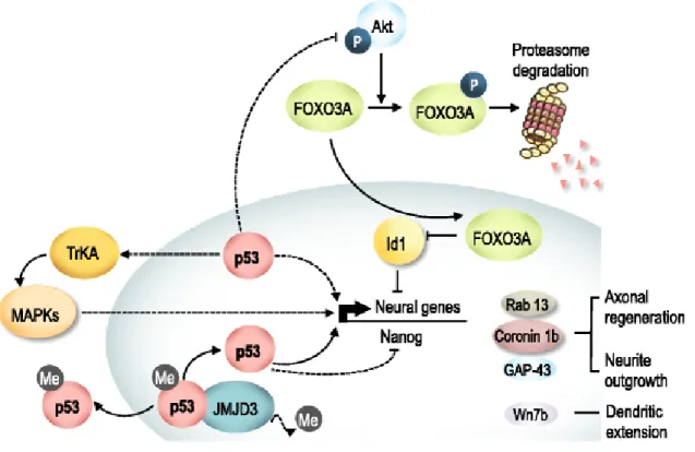
superior cervical ganglion shows that TAp63 is also essential for naturally occurring sympathetic neuronal death in vivo. Sympathetic neuron survival is largely regulated by pro-apoptotic signals deriving from p75NTR and pro-survival signals deriving from the NGF/TrkA receptor (Kaplan and Miller, 2000). In addition, NGF withdrawal and p75NTR activation converge on TAp63 and p53, which then activate Bax gene and protein expression to induce mitochondrial apoptosis (Jacobs et al., 2005). Interestingly, Dugani et al. (Dugani et al., 2009) have also demonstrated that the ∆Np63 isoform is crucial for the survival of neural precursors, namely embryonic cortical precursors, and newly born neurons, antagonizing the pro-apoptotic actions of p53. NGF induced anti-apoptotic ∆Np73 protein functions, at least in part, by antagonizing the pro-apoptotic actions of p53 (Pozniak et al., 2000), and potentially TAp63. Thus, p73 acts as an essential pro-survival protein (Lee et al., 2004; Pozniak et al., 2000). Thus, coordinated regulation of the abundance of p53 family proteins appears to be critical in influencing the outcome of neural apoptosis.
1.1.3. JNK/c-Jun signaling pathway
Several stimuli including growth factors, proinflammatory cytokines, neurotransmitters, cell-matrix interactions, physical and chemical stresses activate MAPK signaling pathways (Chang and Karin, 2001; Ip and Davis, 1998; Karin et al., 1997; Whitmarsh and Davis, 1996). In mammalian cells, the MAPK family is comprised of 3 groups: c-Jun N-terminal kinase (JNK), extracellular signal-regulated kinase, and p38. The JNK pathway is a central stress signaling pathway implicated in neuronal plasticity, regeneration and death (Herdegen et al., 1997). Signaling cascades activate the activator protein-1 (AP-1) through the phosphorylation of distinct substrates. AP-1 is linked to various cellular responses, including proliferation, differentiation, oncogenic transformation and apoptosis (Angel and Karin, 1991; Shaulian and Karin, 2002). The AP-1 capacity to control several biological processes is primarily due to its structural and regulatory complexity. In fact, AP-1 is a collective term referring to dimeric basic/leucine zipper transcription factors that belong to several subfamilies, including the Jun (Estus et al., 1997) subfamily. These DNA binding proteins may form homodimers or heterodimers with each other (Chinenov and Kerppola, 2001). c-Jun is an essential regulator of both cell survival and death, with its levels being increased upon exposure to a variety of stress
signals and chemotherapeutic agents as well as growth factors (Eferl and Wagner, 2003). Its activity is regulated by phosphorylation at serines 63 and/or 73 through JNKs (Dérijard et al., 1994; Ham et al., 1995; Kyriakis et al., 1994).
The detailed mechanisms by which c-Jun regulates apoptosis is not fully understood. The role of c-jun in apoptosis may be stimulus- and cell type-specific. JNK increases the ability of c-Jun to activate the transcription of target genes (Estus et al., 1994; Ham et al., 1995) that are more proximal to promote caspase activation and neuron apoptosis (Edwards and Tolkovsky, 1994; Putcha et al., 2000). Indeed, induction of pro-apoptotic Bcl-2 family members Bim (Whitfield et al., 2001) and death protein 5/harakiri were shown to be JNK/c-Jun-dependent in neurons (Harris and Johnson, 2001). In addition, some studies have reported that after NGF withdrawal in sympathetic neurons, c-Jun protein levels increased and the N-terminal c-Jun transcriptional activation domain became more phosphorylated (Eilers et al., 1998; Eilers et al., 2001), in correlation with an increase in JNK activity (Bruckner et al., 2001; Eilers et al., 1998). Moreover, microinjection of an expression vector for a c-Jun dominant negative mutant, which inhibited AP-1 activity, resulted in protection of neurons against NGF withdrawal-induced death, whereas overexpression of wild-type c-Jun was sufficient to induce apoptosis in the presence of NGF (Estus et al., 1994; Ham et al., 1995). The role for c-Jun in CNS neurons has also been reported, namely in cerebellar granule neurons (Watson et al., 1998). In addition, the c-Jun pathway has also been implicated in the induction of apoptosis in differentiated PC12 cells deprived of NGF (Xia et al., 1995), in striatal neurons treated with neurotoxic concentrations of dopamine (Luo et al., 1998), or in a hippocampal neuron cell line transfected with an expression vector for polyglutamine-expanded Huntingtin (Liu, 1998).
Curiously, JNK may also phosphorylate p53 (Fuchs et al., 1998), and subsequently up-regulate Bax (Miyashita and Reed, 1995). However, the interplay between p53 and c-Jun in regulating neuronal survival and death genes remains to be determined. Regarding p73, it was already demonstrated that JNK and c-Jun is required for the stabilization of TAp73 (Jones et al., 2007; Toh et al., 2004) and degradation of ∆Np73 under stress stimulation (Dulloo et al., 2010). Importantly, c-Jun has also been shown to regulate the abundance of p63. Yao et al. (Yao et al., 2010b) showed that the transcriptional activity of the TAp63 promoter and TAp63
protein levels were both up-regulated by an increased c-Jun activity in the Hep3B human hepatocellular carcinoma cell line.
However, since c-Jun null mice die during embryonic development at embryonic day 12.5 (E12.5) due to hepatic failure (Hilberg et al., 1993), in vivo studies focused on the role of c-Jun in the nervous system were only possible due to knockout (KO) mice models for the three mammalian JNK genes, Jnk1, Jnk2, and
Jnk3. JNK3 is mainly expressed in the CNS (Junyent et al., 2011), whereas JNK1 and
JNK2 are expressed in many other tissues (Ip and Davis, 1998). In fact, c-Jun phosphorylation and neuronal cell death was inhibited in neurons from JNK3 null mice (Bruckner et al., 2001). Finally, JNK1/JNK2 double KO mice, but not single, showed that both JNKs are required for developmental cell death in the neural tube, whereas in the developing cortex they promote neuronal survival (Kuan et al., 1999; Sabapathy et al., 1999). Thus, although JNKs and c-Jun may be pro-apoptotic in many neuronal cell types, they can have other cellular roles.
1.1.4. Role of apoptosis in Alzheimer’s disease
AD is the most common human age-related, sporadic neurodegenerative disorder, characterized by a global, progressive and irreversible cognitive decline that is strongly correlated with synaptic dysfunction and death of neurons from hippocampus and associated regions of the cerebral cortex. The affected brain shows extracellular deposits of Aβ peptides, known as amyloid or senile plaques (Mattson, 2004), and intracellular accumulation of paired helical filaments that form NFTs (McKee et al., 1990), consisting of polymerized hyperphosphorylated tau protein (Spillantini and Goedert, 1998). Although activation of apoptotic signaling has been reported in AD brains (Raynaud and Marcilhac, 2006), the precise intracellular signaling pathways by which Aβ peptides trigger cell death are not fully understood.
The study of apoptosis in neurodegenerative diseases has become a challenging task and a controversial area of research, especially because synaptic loss and electrophysiological abnormalities typically precede cell loss. In addition, given the slow progression of most neurodegenerative diseases, in contrast with the rapid progression of a cell through apoptosis, the detection of representative apoptotic neurons in AD patients could be difficult. Further, apoptotic processes may be terminated or not yet initiated by the time of tissue examination. In fact, the collected
tissue is usually representative of the end-stage disease, being removed much later than it would be ideal to treat patients and prevent neuronal cell death. Finally, the criteria used to classify the type of cell death as apoptosis has often been based on morphological assessment and biochemical assays, which may also account for other forms of programmed cell death.
Although many AD mouse models have been reported, there is no single model that exactly mimics human AD (Elder et al., 2010). Thus, it is important to keep in mind the limitations when research data generated from using these AD animal models are interpreted.
Knowledge of the production and degradation pathways acting on the toxic proteins that cause AD, and various other proteinopathies, could help in the design and improvement of therapeutic strategies. Specifically, the reduction of Aβ load has given many insights into drug development strategies for potential therapies in AD aimed at preventing Aβ formation or accelerating its degradation. β- and γ-secretases have been considered as possible drug targets. Targeting secretases may, however, have unforeseen effects because a physiological role for Aβ has also been proposed (Atwood et al., 2003; Chen and Dong, 2009; Pearson and Peers, 2006).
1.1.4.1. Aβ production and accumulation
The amyloid precursor protein (APP) in mammals belongs to a family of conserved type I membrane proteins that include APP, APP-like protein 1 (Aydin et al.) and 2 (APLP2) (Aydin et al., 2012). Although the APP family is abundantly expressed in the brain and APLP1 expression is restricted to neurons (Lorent et al., 1995), APP and APLP2 can also be found in the majority of other tissues. The human
APP gene encodes a single transmembrane polypeptide, whose primary structure
contains a signal peptide for secretion, a large extracellular N-terminal domain, a transmembrane domain, and a cytoplasmic C-terminal tail.
During normal development, APP can undergo amyloidogenic or non-amyloidogenic processing via cleavage by three different secretases, α-, β-, and y-secretases (Ling et al., 2003). In the non-amyloidogenic pathway, cleavage of APP occurs via presenilin-containing α-secretase complex, which cleaves within Aβ sequence and consequently abrogates Aβ formation, leading to the release of a large soluble extracellular fragment (Mattson), which can be neuroprotective, and retention of an 83-residue membrane-bound fragment (C83). Alternatively, APP can be
cleaved by β-secretase, which cuts at the N-terminus of Aβ, generating a smaller extracellular fragment (Mattson) and a 99-residue membrane-bound fragment (C99). Peptide fragments C83 and C99 may then undergo conformational alterations and become targets for the presenilin (PS1/PS2)-dependent γ-secretase, which cleaves within the plasma membrane domain. C99 proteolysis generates Aβ1-40 and Aβ1-42
(Selkoe and Wolfe, 2007). The cleavage of either C83 or C99 by γ-secretase may also generate an amyloid intracellular domain (AICD), which moves to the nucleus where it may modulate transcription of target genes.
Aβ is produced as a monomer, but readily aggregates to form multimeric complexes. Therefore, the term “soluble Aβ” is generally applied either to newly generated, cell-secreted Aβ or to that fraction of tissue or synthetic Aβ that is taken into the aqueous phase of a non-detergent containing extraction buffer. In contrast, “misfolded” and “aggregated” Aβ are terms used to describe very early, nonspecific changes in Aβ folding states or solubility states, respectively. In addition, “oligomeric” Aβ refers to peptide assemblies with limited stoichiometry, such as dimmers and trimers, while protofibrils are structures of intermediate order between aggregates and fibrils (Gandy, 2005).
Aβ peptide is detected in human cerebrospinal fluid (CSF) as a range of isoforms between 38 and 43 amino acids in length. The predominant isoforms secreted are Aβ1-40 (90%) whereas Aβ1-42 isoform represents less than 10%
(Thinakaran and Koo, 2008). In patients with AD, the relative proportions of Aβ1-40
and Aβ1-42 change to approximately 50% each (Mehta et al., 2001). The accumulation
of Aβ is progressive and strongly accepted as an important contributor to the neuronal dysfunction and loss (Hardy and Selkoe, 2002). The pattern and distribution of the different Aβ species varies among the different topographical lesions.
1.1.4.2. Cytotoxicity of Aβ
Several studies suggest that Aβ-induced oxidative stress leads to apoptotic neuronal cell death that can be inhibited by antioxidants (Behl et al., 1994; Mattson and Goodman, 1995; Pillot et al., 1999). In fact, many lines of evidence suggest that mitochondria dysfunction and oxidative damage have a central role in aging-related neurodegenerative diseases, being key regulators of cell survival and death (Danial and Korsmeyer, 2004).
The involvement of apoptosis in AD has been corroborated by studies showing that Aβ alters the expression of the Bcl-2 family of apoptosis-related genes (Yao et al., 2005) and that survival pathways play a pivotal role in preventing Aβ-mediated apoptosis (Watson and Fan, 2005). On the other hand, studies in an anti-NGF transgenic mouse model and PC12 cells suggest that discontinued or limited NGF supply induces an overproduction of Aβ peptides, triggering downstream apoptotic cell death (Capsoni et al., 2000; Matrone et al., 2008). Furthermore, p75NTR has been consistently linked to changes occurring in AD (Schliebs, 2005; Sotthibundhu et al., 2009), including cell death of basal forebrain cholinergic neurons, where this receptor is highly expressed (Dechant and Barde, 2002). Both in vitro and
in vivo evidence suggests that the JNK/c-Jun cascade is a critical event during
neuronal death in AD (Anderson et al., 1994; Kihiko et al., 1999; Viana et al., 2010), where JNK mediates the activation of several molecules, including caspase-2 (Viana et al., 2010), p53 (She et al., 2002), and Bcl-2 family members (Sorenson, 2004) and potentiate inflammatory responses via AP-1 activation (Manning and Davis, 2003). Viana et al. (Viana et al., 2010) showed that Aβ exposure in PC12 cells resulted in activation and nuclear translocation of JNK, and caspase-2 activation. Caspase-2 triggers apoptosis through activation of the mitochondrial apoptosis pathway. The induction of c-Jun was shown to occur in vitro after Aβ treatment (Estus et al., 1997; Iwasaki et al., 1996; Kihiko et al., 1999) and in AD brains (Anderson et al., 1994; Anderson et al., 1996), suggesting a possible requirement for this transcription factor in Aβ toxicity.
Curiously, p53 family members have also been associated with Aβ-induced neuronal apoptosis. The first indication that the p53 family may play a role in AD came in 1996 with the demonstration that intracellular Aβ up-regulates p53 in the brains of transgenic mice overexpressing the Aβ fragment of APP (LaFerla et al., 1996). The use of pifithrin-α, an inhibitor of p53-dependent gene transcription, was shown to protect neurons against Aβ-induced apoptosis (Culmsee et al., 2001; Hooper et al., 2007). p53 was also found to be up-regulated in the brain of AD patients [43, 44]. In 2004, Caricasole et al. (Caricasole et al., 2004) showed that Aβ activates the expression of the soluble Wnt antagonist Dickkopf-1 (Caricasole et al.), a p53 target gene. Knockdown of Dkk1 in primary neurons, in turn, almost completely blocked Aβ-induced tau phosphorylation, implicating the p53 family in the “amyloid cascade” pathway.
More recently, it has been demonstrated that p53 participates in Aβ-induced apoptosis of PC12 neuronal cells through modulation of Bax expression (Ramalho et al., 2004). Furthermore, Aβ-induced apoptosis in rat primary cortical neurons was associated with translocation of Bax to the mitochondria, followed by cyt c release, caspase activation, and DNA and nuclear fragmentation (Solá et al., 2003). Interestingly, a novel biological function of intracellular Aβ 1-42 has been suggested, where it acts as a transcription factor for the p53 promoter,
enhancing p53-dependent neuronal apoptosis in AD (Ohyagi et al., 2005).
Several reports have also demonstrated p73 involvement in apoptosis triggered by Aβ25-35, being probably vital for mediating the process of AD (Zhang
et al., 2011). It has been shown that in primary cortical neurons generated from p73 KO mice, the activity of JNK increased, while JNK inhibition decreased tau phosphorylation in these neurons (Wetzel et al., 2008). Since ΔNp73, and not TAp73, can bind and inhibit JNK, the loss of ΔNp73 forms may result in tau phosphorylation and neurodegeneration. As mentioned before, Aβ peptide activates c-Abl and increases p73 levels (Alvarez et al., 2004; Vázquez et al., 2009), while the Swedish mutant form of APP increases expression from both the TA and ΔN promoters of the p73 gene, only inducing an overall increase in the TA forms (Vázquez et al., 2009). Surprisingly, the mechanisms regulating p63 levels under Aβ-mediated apoptosis have yet to be elucidated.
Aβ peptides accumulate within mitochondria, strongly affecting their function and morphology, particularly in the synaptic compartment (Balietti et al., 2012). Increasing evidence indicates that the mitochondrial dysfunction is an important early factor in the development of AD-like pathology (Blass and Gibson, 2006; Yao et al., 2009). Accumulation of Aβ in mitochondria appears to be also associated with diminished enzymatic activity of respiratory chain complexes III and IV, and reduction of oxygen consumption (Caspersen et al., 2005).
Finally, several studies have demonstrated that Aβ triggers the formation of tau pathology. Inflammatory processes are strongly correlated with AD onset and progression in humans, and could have a pivotal role in disease etiology (Kitazawa et al., 2005; Krstic and Knuesel, 2013). Interestingly, it has been suggested that different forms of Aβ may be responsible for inducing tau hyperphosphorylation in different settings (Blurton-Jones and Laferla, 2006; LaFerla, 2010).
1.1.5. Regulation of apoptosis by autophagy
Autophagy is a cellular process in which isolated membrane sequesters part of the cytoplasm to form a double-membrane vesicle, called autophagosome (AP) that fuses with lysosomes for the degradation of its contents, long-lived cytoplasmic proteins or damaged organelles, by acidic lysosomal hydrolases, maintaining normal cell homeostasis (Nixon and Yang, 2011). There are at least three types of autophagy; macroautophagy (delivery of cytosolic contents to the lysosome by autophagosomes), microautophagy (inward invagination of the lysosomal membrane) and chaperone-mediated autophagy (direct translocation across the lysosomal membrane). Among these, macroautophagy referred to as autophagy has been the most studied. Autophagy-related (Atg) proteins are a set of evolutionarily conserved gene products, originally identified in yeast and followed by the identification of homologs in higher eukaryotes (Huang and Klionsky, 2002), which are required for autophagy.
Among Atg proteins, one subset is essential for AP formation, and is referred to as the “core” molecular machinery (Xie and Klionsky, 2007). In mammalian cells, most of the Atg proteins are observed in isolated membranes but not in complete APs (Longatti and Tooze, 2009; Tooze and Razi, 2009). Only microtubule-associated protein light chain 3 (LC3), a mammalian homolog of yeast Atg8, is known to exist in APs, and therefore, this protein serves as marker for APs and is widely used to monitor autophagy (Kabeya et al., 2000).
Since autophagy can block apoptosis, one might expect that their regulation would be tightly coordinated. Curiously, the same proteins can regulate both processes. Some molecular connections occur upstream of the autophagic and apoptotic machinery itself, where signaling pathways regulate both processes (Thorburn, 2008). p53, which is a potent inducer of apoptosis, can also induce autophagy through increased expression of a direct p53 target gene called DRAM (Crighton et al., 2006). Similarly, activation of the phosphatidylinositide 3 kinase (PI3K)/Akt pathway, which is well-known to inhibit apoptosis, also inhibits autophagy (Arico et al., 2001). Thus, important signaling pathways simultaneously increase or decrease both autophagy and apoptosis. In addition, proteins that are themselves central components of the apoptosis machinery, including Bcl-2 family proteins and FADD regulate both processes directly. In addition, in normal cytoprotective autophagy, activation of JNK can up-regulate Beclin-1 (Li et al.,