Internet Journal of Medical Update
Journal home page: http://www.akspublication.com/ijmu
Original Work
Implications of zinc on fetal neural tube defects
Rajeev Vats*
ᴪ, R K Sharma** and A Sharma***
*School of Biological Sciences, University of Dodoma, Dodoma, Tanzania
**Department of Zoology, Kurukshetra University, Kurukshetra, India
***Department of Neurosurgery, G B Pant Hospital, New Delhi, India
(Received 01 September 2011 and accepted 31 March 2012)
ABSTRACT: Zinc is essential for normal growth and differentiation in all mammalian species and it is reported that folic acid supplementation has reduced the incidence of neural tube defects (NTD). It is still considered one of the important congenital malformations having wide implications. Zinc deficiency has been reported to produce NTD in animals. The present study was undertaken to evaluate zinc status of newborn babies with NTD and their mothers. Blood samples were taken from 287 mothers and their babies having NTD and from 110 controls visiting hospitals and health clinics. Zinc level as μg/ml for blood and serum and μg/g for cell mass were determined on GBC 932 atomic absorption spectrophotometer (Australia) by fluorometery. The mean maternal blood, serum and cell mass concentration in NTD group (14.56 ± 1.34 μg/ml, 0.6 ± 0.01 μg/ml, 5.64 ± 0.35 μg/gm respectively) were significantly lower than those of the control mothers (24.15 ± 2.95 μg/ml, 0.72 ± 0.03 μg/ml, 7.37 ± 0.44 μg/gm respectively). There is a significant decrease in the concentration of Zinc in newborns having NTD (15.65 ± 3.18
μg/ml, 0.56 ± 0.08 μg/ml, 5.11 ± 0.18 μg/gm respectively) as compared with normal newborns (28.04 ± 1.1 μg/ml, 0.59 ± 0.08 μg/ml and 6.08 ± 0.29 μg/gm respectively). Maternal nutritional zinc deficiency in newborns and their mothers is thought to be one of the factors responsible for NTD. However, the lowered zinc concentration may be influencing the causation of NTD. More investigations on zinc status in mothers during antenatal period, especially in the prenatal development and antenatal zinc status including normal babies and NTD babies are required at population level.
KEY WORDS: Neural Tube Defect; Trace Element; Zinc INTRODUCTIONᴪ
Neural tube defects (NTD) constitute a significant proportion of all congenital malformations in human beings. Extensive work has been done so far but little progress has been made in finding the etiology of some of them (anencephaly & spina bifida) and on zinc deficiency as a basis for generating etiological hypotheses by various epidemiological studies.1 Association between vitamins especially folic acid, Vitamin B122-4 and
other trace elements like copper and selenium is also reported to reduce the incidence of NTD, but generally they are not prescribed by the
ᴪCorrespondence at: #1382 Sector-13, Urban
Estate, Kurukshetra-136118, India; Phone: 00 91 174 425486; Email: vatsr71@gmail.com
clinicians/practitioners during pregnancy.5 A number of prospective & retrospective human studies have provided evidence that folic acid supplementation to pregnant women lowers the incidence of NTD in the offspring6-8. Folic acid is widely used by pregnant women but NTD is still an important congenital malformation having wide implications. This may be due to the fact that deficiency of folic acid alone is not responsible for all kind of NTD and many other factors (nutritional & genetic) are responsible in the etiology of various kinds of NTDs9-12.
20
showed that chicks hatched from hens fed a zinc deficient diet were weak, and died within four days. Blamberg et al16 also found grossly deformed embryos. A similar range of skeletal defects was observed by Keinholz et al17. Zinc transfer to fetus is reduced resulting in higher percentage of stillbirths and lower viability of piglets. Reproductive aspects of zinc deficiency in pigs are well documented 18.
Zinc is essential for the growth and development of the fetus and plays a critical role in many cellular reactions, including gene transcription and cell division and differentiation. The inadequate intake of zinc is associated with NTDs in both animals and humans19. The essentiality of zinc in the formation of the neural tube is further supported by the observation that women with acrodermatitis enteropathica, a disorder of impaired zinc absorption from the intestine, are at high risk for babies with NTDs20.
Zinc deficiency in humans and animals causes teratological, genetic and medical abnormalities. Human requirement of zinc seems to be high during periods of rapid growth, such as embryonic life, infancy, puberty, pregnancy and tissue repair. During gestation, deficiency of this trace element even if transitory, is teratogenic and associated with chromosomal abnormalities and alters cognitive functions21. Newborns with NTDs have significantly low level of serum zinc, supporting zinc deficiency as an association of NTDs22-24. Zeyrek et al25 reported that low maternal zinc and high copper during pregnancy may be responsible for NTDs. Over the last three decades, zinc has been recognized to play a vital role in almost all aspects of living systems either directly or indirectly26.
METHODOLOGY
In the present study 287 newborns with NTD (Meningomyelocele-132, Meningocele-47, Lipomeningocele-15, Spina bifida occulta-41 and
Encephalocele-52) have been studied and their mothers served as our cases and 110 apparently normal full term babies and their mothers served as controls selected from the same population from the year 1997-2000. A detailed maternal obstetric history, age and socio-economic status were noted, and newborns with NTD examined carefully with the help of a pediatrician. Maternal socio-economic status was classified according to Kuppuswamy socio-economic status score27.
Venous blood (5.0 ml each) from all newborns and their mothers was drawn from the antecubital vein, using stainless steel needles (disposable syringes) and collected in trace element free plastic vials. All necessary precautions were taken to avoid contamination. One hour after collection, the blood was centrifuged and the clear serum was transferred to plastic vials. The sera were stored frozen at -20°C until analyzed.
The blood from each case was divided into three groups. In group (I) whole blood, in group (II) blood serum and in group (III) packed cell mass was subjected to extraction of zinc. All the samples were digested in long necked round bottom flasks with triple acid (conc. HNO3, 70% perchloric acid
and conc. H2SO4 - 10:3:1). This process was
executed by heating the contents till most of the triple acid mixture evaporated from the flask. The contents of each flask were then washed with triple distilled water and were stored in plastic vials at 4oC for further analysis. The zinc levels as ng/ml were determined by GBC 932 spectrophotometer28. All the data were given as arithmetic means, SD, SEM and ranges. The 95% confidence intervals (CI) for the population means were also shown. T-test was used to compare the Zinc levels of the subjects29.
RESULT
The result of the Zinc concentration in blood, cell mass and serum in the mothers having NTD newborns and the mothers having normal newborns are shown in Table 1. The mean value of Zinc concentration of control group is 24.15 ± 2.95
μg/ml for blood, 7.37 ± 0.44 μg/gm for cell mass and 0.719 ± 0.035 μg/ml for serum as compared to the mothers of NTD newborns who showed a zinc level of 14.56 ± 1.34 μg/ml, 5.64 ± 0.35 μg/gm and 0.606 ± 0.0163 μg/ml respectively. All the values were significantly lower (p<0.05) in the mothers of newborns having NTD.
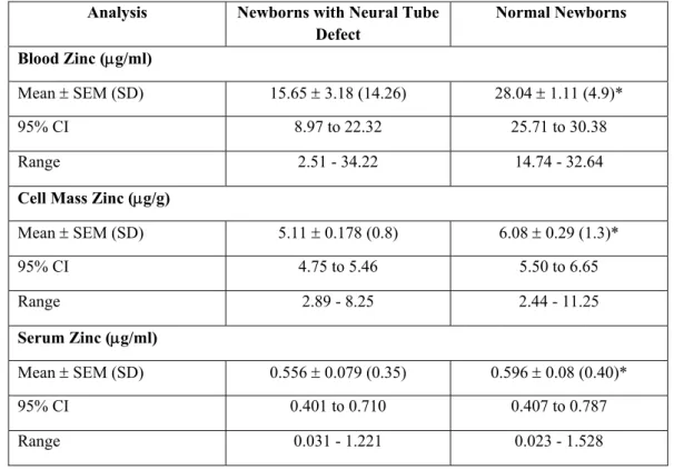
The concentration of zinc was also significantly lower in newborns with NTD and is summarized in table 2. The zinc concentrations in blood of normal newborns, in cell mass and serum was 28.04 ± 1.11
μg/ml, 6.08 ± .288 μg/gm and 0.596 ± 0.08 μg/ml, in comparison to NTD newborns having zinc concentration 15.65 ± 3.18; 5.11 ± 0.178 and 0.556
Table 1: Maternal Zinc Status of Healthy and Newborns with NTD Analysis Mothers of Newborns with
NTD
Control Mothers with Normal Newborns Blood Zinc (μg/ml)
Mean ± SEM (SD) 14.56 ± 1.34 (5.99) 24.15 ± 2.95 (13.2)*
95% CI 11.93 to 17.18 18.46 to 29.83
Range 6.15 - 32.73 3.65 - 46.48
Cell Mass Zinc (μg/g)
Mean ± SEM (SD) 5.64 ± 0.357 (1.6) 7.37 ± 0.44 (2.0)*
95% CI 4.93 to 6.34 6.44 to 8.29
Range 0.256 - 3.57 2.14 - 11.87
Serum Zinc (μg/ml)
Mean ± SEM (SD) 0.606 ± 0.016 (0.02) 0.719 ± 0.035 (0.16)*
95% CI 0.575 to 0.638 0.648 to 0.789
Range 0.17 – 1.96 0.268 - 1.30
*p<0.05
Table 2: Zinc Status in Normal Newborns and those with NTD Analysis Newborns with Neural Tube
Defect
Normal Newborns Blood Zinc (μg/ml)
Mean ± SEM (SD) 15.65 ± 3.18 (14.26) 28.04 ± 1.11 (4.9)*
95% CI 8.97 to 22.32 25.71 to 30.38
Range 2.51 - 34.22 14.74 - 32.64
Cell Mass Zinc (μg/g)
Mean ± SEM (SD) 5.11 ± 0.178 (0.8) 6.08 ± 0.29 (1.3)*
95% CI 4.75 to 5.46 5.50 to 6.65
Range 2.89 - 8.25 2.44 - 11.25
Serum Zinc (μg/ml)
Mean ± SEM (SD) 0.556 ± 0.079 (0.35) 0.596 ± 0.08 (0.40)*
95% CI 0.401 to 0.710 0.407 to 0.787
Range 0.031 - 1.221 0.023 - 1.528
*p<0.05
DISCUSSION
The present study has demonstrated that the concentration of zinc is significantly lower in
22
parts in nucleic acid metabolism are zinc dependent. In humans zinc deficiency has been reported to be associated with growth retardation30,31,10. Physiological changes during pregnancy increase nutritional requirements. During pregnancy iron is commonly supplemented and it seems that iron does interfere with zinc absorption in the intestine32. It is also reported that zinc absorption tends to increase during pregnancy33 and it is possible that some other trace elements interfere. Folic acid supplementation is generally recommended decreasing the incidence of NTD2. Zinc deficiency in animals has been shown to produce CNS malformations34.
Zinc deficiency during early pregnancy in rats is also reported to produce abnormal blastocyst35, increase the rate of reabsorption and a high incidence of congenital malformation and teratogenecity, particularly fetal neural tube defects such as anencephaly36. Thus it is reasonable to assume that zinc deficiency produces a similar set of changes in humans as observed in rats in most of the earlier studies and suggests a basic common metabolic mechanism of trace elements in mammals in general. As such it is difficult to say anything pertaining to the mechanism whether it is terata or necrosis or both responsible for such congenital malformation in humans. Pregnant women suffering from acrodermatitis enteropathica (AE), an inborn error of zinc metabolism, exhibit high frequency of fetal deaths and malformed infants particularly with neural tube defects36-39. The pathogenesis of AE is the result of impaired intestinal zinc absorption and such patients exhibit low serum lipid and arachidonic acid increased IgA and defective prostaglandin synthesis38,40. The malformation observed in the present study can be attributed to the metabolic alterations that are the result of deficient zinc supply to the developing fetus.
The development of the mammalian fetus depends on the constant supply of nutrients from the mother41. Thus the maintenance of pregnancy implies an increased ingestion of zinc to meet increasing demands of the trace elements of fetus42. As the gross need of fetal growth especially in the 3rd trimester of pregnancy are often in excess of the assimilative capacities of the mother and thus imply a net maternal loss of these nutrients43, if the nutrient deficient diets with low level of trace elements are continued during pregnancy. The meagre stored zinc level in maternal tissue is possibly responsible for the low supply across the placenta to the developing fetus possibly lead to congenital anomalies. The low level of zinc during pregnancy also leads to the manifestation of Cd toxicity44 or toxicity of Pb, Hg and certain drugs and alcohol45. It is, therefore, suggested that a
constant supply and monitoring of zinc is a must during pregnancy.
The mechanisms responsible for these defects or abnormal development are thought to be based on depression of nucleic acid synthesis13. Prolongation of the mitotic interval and reduction in the number of neural cells early in development could combine to produce a variety of malformations. The specific nature of the defect would then depend on the state of presumptive area of the primitive tube when any given developmental process began. Defects in the closure of neural tube and other problems in differentiation of CNS were observed in early chick embryo explants cultured in zinc deficient media46. In addition, mesodermal differentiation and growth are also altered.
In the present study, both the newborn babies with NTDs and their mothers had significantly low levels of Zn when compared to the control group. The results showed that in comparison with the control group, the mothers who had given birth to babies with NTD had low levels of zinc. This is in line with the findings of Cengiz et al 47. This difference between the two groups may be related to the levels of zinc in the environment and their nutrition. There are important interactions between trace elements and vitamins at the level of intestinal absorption. Although in normal pregnancies, possibly because of the placental transfer to the baby, serum zinc level decreases48, in the present study the serum level of Zn in the mothers who had infants with NTD was significantly lower in comparison to the control group.
In conclusion, as the etiology of NTD is thought to be multifactorial, a lack or an excess of trace elements and the interactions between vitamins and trace elements may play a significant role in its development. The results of the present study indicate that Zn supplements may be important in the prevention of NTD. Large scale prenatal zinc supplementation trials are therefore recommended for further confirmation as the association between zinc deficiency and NTD needs further investigation.
ACKNOWLEDGEMENTS
The authors are thankful to the Senior Residents, Department of Pediatric Neurosurgery, G.B Pant Hospital, New Delhi for the collection of blood samples of the patients and Director, DWR, Karnal for the use of Atomic Absorption spectrophotometer. The assistance given by the University Grants Commission, New Delhi to Dr. Rajeev Vats is acknowledged.
REFERENCES
occurrence of neural tube defects in California. Am J Epidemiol. 1999 Sep;150(6):605-16. 2. Wald NJ. Folic acid and neural tube defects.
Bibl Nutr Dieta. 2001;55:22-33.
3. Steen MT, Boddie AM, Fisher AJ, et al. Neural-tube defects are associated with low concentrations of cobalamin (vitamin B12) in amniotic fluid. Prenat Diagn. 1998 Jan;18(6):545-55.
4. Borman B, Cryer C. Fallacies of international and national comparisons of diseases occurrence in the epidemiology of neural tube defects. Teratology. 1990 Oct;42(4):405-12. 5. Guvenc H, Karatas F, Guvenc M, et al. Low
level of selenium in mothers and their newborns in pregnancies with neural tube defects. Pediatrics. 1995 Jan;95(6):879-82. 6. Campbell NR. How safe are folic acid
supplements? Arch Intern Med. 1996 Aug;156(15):1638-44.
7. Gonzalez MJ, Schmitz KJ, Matos MI, et al. Folate supplementation and neural tube defects: a review of a public health issue. P R Health Sci J. 1997 Dec;16(4):387-93.
8. Locksmith GJ, Duff P. Preventing neural tube defects: the importance of periconceptional folic acid supplements. Obstet Gynecol. 1998 Jan;91(6):1027-34.
9. Scott JM, Kirke PN, Weir DG. The role of nutrition in neural tube defects. Annu Rev Nutr. 1990;10:277-95.
10. Rathi SS, Srinivas M, Grover JK, et al. Zinc levels in women and newborns. Indian J Pediatr. 1999 Sep-Oct;66(5):681-4
11. Melvin EC, George TM, Worley G, et al. Genetic studies in neural tube defects. NTD collaborative group. Pediatr Neurosurg. 2000 Jan;32(1):1-9.
12. Srinivas M, Gupta DK, Rathi SS, et al. Association between lower hair zinc levels and neural tube defects. Indian J Pediatr. 2001 Jan;68(6):519-22.
13. Hurley LS. Developmental Nutrition. Englewood Cliffs, NJ: Prentice-Hall 1980. 14. Hurley LS. Teratogenic aspects of manganese
zinc, and copper nutrition. Physiol Rev. 1981 Apr;61(2):249-95.
15. Turk DE, Sunde ML, Hoekstra WG. Zinc deficiency experiments with poultry. Poul Sci. 1959;38:1256.
16. Blamberg, DL, Blackwood WB, Supplee WC, et al. Effect of zinc deficiency in hens on hatchability and embryonic development. Proc Soc Exp Biol Med. 1960 Jan;104:217-20. 17. Keinholz EW, Turk DE, Sunde ML, et al.
Effects of zinc deficiency on the diets of hens’. J Nutr. 1961 Oct;75:211-21.
18. Wegger I, Palludan B. Zinc metabolism in swine with special emphasis on reproduction.
In: Trace element Metabolism in Man and Animals (Kirchgessner M, Ed) 1978;3:428-35. 19. Tamura T, Goldenberg RL. Zinc nutriture and
pregnancy outcome. Nutr Res. 1996;16:139-81.
20. Mambidge KM, Neldner KH, Walravens PA. Letter: Zinc, acrodermatitis enteropathica and congenital malformations. Lancet. 1975 Mar;1(7906):577-8.
21. Nair N, Bedwal RS, Mathur RS. Biological trace elements: Their prophylactic, prognostic an etiological values. Cell Biology (News Letter). 1989;2 (1):1-13.
22. Carrillo-Ponce Mde L, Martinez-Ordaz VA, Velasco-Rodriquez VM, et al. Serum lead, cadmium, and zinc levels in newborns with neural tube defects from a polluted zone in Mexico. Reprod Toxicol. 2004 Dec;19(2):149-54.
23. Golalipour MJ, Mansourian AR, Keshtkar A. Serum zinc levels in newborns with neural tube defects. Indian Pediatr. 2006 Sep;43(9):809-12.
24. Dey AC, Shahidullah M, Mannan MA, et al. Maternal and neonatal serum zinc level and its relationship with neural tube defects. J Health Popul Nutr. 2010 Aug:28(4):343-50.
25. Zeyrek D, Soran M, Cakmak A, et al. Serum copper and zinc levels in mothers and cord blood of their newborn infants with neural tube defects: a case-control study. Indian Pediatr. 2009 Aug;46(8):675-80.
26. Hurley LS, Swenerton H. Lack of mobilization of bone and liver zinc under teratogenic conditions of zinc deficiency in rats. J Nutr. 1971 May;101(5):597-603.
27. Park K. Hospital sociology. Socioeconomic status scale. In: Prark K, ed. Park’s Textbook of Preventive and Social Medicine. 15th ed. Jabalpur; Banarsi Dass Bhanot Press, 1997;458-60.
28. Ludmilla D. Chemical analysis by Atomic Absorption Spectroscopy. Varian Techtron Pvt. Ltd. Melbourne, Australia, 1976.
29. Guilford JP, Fruchter B. Fundamental statistics in psychology and education. McGraw-Hill International Book Co. London, 1981.
30. Meadows NJ, Ruse W, Smith MF, et al. Zinc and small babies. Lancet. 1981 Nov;2(8256):1135-7.
31. Simmer K, Thompson RPH. Maternal zinc and intrauterine growth retardation. Clin Sci. 1985;68:478-82.
24
33. King JC. Effect of reproduction on the bioavailability of calcium, zinc and selenium. J Nutr. 2001 Apr;131(4suppl):1355S-8S.
34. Warkany J, Petering HG. Congenital malformations of the central nervous system in rats produced by maternal zinc deficiency. Teratology. 1972 Jan;5(3):319.
35. Hurley LS, Shrader RE. Abnormal development of preimplantation rat eggs after three days of maternal zinc deficiency. Nature. 1975 Apr;254(5499):427-9.
36. Bedwal RS, Bahuguna A. Zinc, copper and selenium in reproduction. Experientia. 1994 Jul;50(7):626-40.
37. Mambidge KM, Neldner KH, Walravens PA. Letter: Zinc acrodermititis enteropathica, and congenital malformations. Lancet. 1975 Mar;1(7906):577-8.
38. Salton MH, Jenkins DM. Maternal and fetal plasma zinc concentration and fetal abnormalities. Br J Obstet Gynecol. 1982 Jan;89(1):56-8.
39. Hurley LS, Keen CL, Lonnerdal B. Aspects of trace element interactions during development. Fed Proc. 1983 Apr;42(6):1735-9.
40. Lonnerdal B, Keen CL, Hurley LS. Zinc binding ligands and complexes in zinc metabolism. In: Advances in Nutrition Research, Ed. HH, Draper, Plenum Press, New York. 1984;139-68.
41. Bedwal RS, Nair N, Mathur RS. Effects of zinc deficiency and toxicity on reproductive
organs, pregnancy and lactation - a review. Trace Elem Med.1991;8(2):89-100.
42. Colombo R. Metabolic adaptations in white rat foetuses. Acta Embryol Exp (Palermo). 1979;3:349-66.
43. Williams RB, Davis NT, McDonald I. The effects of pregnancy and lactation in copper and zinc retention rat. Br J Nutr.1977 nov;38(3):407-16.
44. Chisolm JC, Handorf CR. Zinc, cadmium, metallothionein and progesterone: do they participate in the etiology of pregnancy induced hypertension? Med Hypotheses. 1985 Jul;17(3):231-42.
45. Jamesons S. Zinc nutrition and human pregnancy. In: Zinc deficiency in human subjects. Eds. Jameson. Alan R. Liss, Inc. New York. 1983;53-69.
46. Iniquez C, Casas J, Carreres J. Effects of Zinc deficiency on the chick embryo blastoderm. Acta Anat (Basel). 1978;101(2):120-9.
47. Cengiz B, Soylemez F, Ozturk E, et al. Serum zinc, selenium, copper, and lead levels in women with second-trimester induced abortion resulting from neural tube defects: a preliminary study. Biol Trace Elem Res. 2004 Mar;97(3):225-35.