Frequency of Chromosomal Abnormalities in
Products of Conception
Frequência de anomalias cromossômicas em
material de aborto
Thaís Mesquita Alves Teles
1Carolina Maria Marques de Paula
1Mariana Gontijo Ramos
1Helena B. B. L. Martins da Costa
2,3Cyntia Roberta Almeida Andrade
2Sarah Abreu Coxir
3Maria Lectícia Firpe Penna
11Department of Biomedical Sciences, College of Human, Social and Health Sciences, Universidade FUMEC, Belo Horizonte, MG, Brazil 2Department of Molecular Biology, Códon Biotechnology Laboratory,
Belo Horizonte, MG, Brazil
3Department of Cytogenetics, Códon Biotechnology Laboratory, Belo Horizonte, MG, Brazil
Rev Bras Ginecol Obstet 2017;39:110–114.
Address for correspondence Maria Lectícia Firpe Penna, PhD, Rua Cobre, 200, Belo Horizonte, MG, Brazil (e-mail: [email protected]).
Keywords
►
spontaneous
abortion
►
aneuploidies
►
QF-PCR
►
cytogenetics
Abstract
Purpose
To describe the frequencies of chromosomal abnormalities found in
abor-tion material, and to observe its correlaabor-tion to maternal age.
Methods
A retrospective study was conducted based on data obtained from the
data-bank of a medical genetics laboratory in Belo Horizonte, MG, Brazil. A total of 884 results
from products of conception analysis were included, 204 of which were analyzed by
cytogenetics, and 680 by molecular biology based on quantitative
fl
uorescence polymerase
chain reaction (QF-PCR). The frequency of individual chromosomal aberrations and the
relationship between the presence of anomalies and maternal age were also evaluated.
Results
The conventional cytogenetics technique was able to detect 52% of normal
and 48% of abnormal results in the analyzed material. Quantitative
fl
uorescence
polymerase chain reaction revealed 60% of normal and 40% of abnormal results
from the samples evaluated by this method. The presence of trisomy 15 was detected
only by cytogenetics, as it was not included in the QF-PCR routine investigation in the
laboratory. A signi
fi
cant increase in abnormal results was observed among women
aged 35 years or older compared with younger women (
p
¼
0.02).
Conclusion
Chromosomal aberrations are still a major cause of spontaneous
abor-tion, and the conventional cytogenetics technique is ef
fi
cient for miscarriage material
analysis, but molecular methods such as QF-PCR are adequate complementary
strategies to detect the major chromosomal anomalies, leading to technical reports
with reliable results.
received
June 5, 2016
accepted
January 9, 2017
published online
March 15, 2017
DOI http://dx.doi.org/ 10.1055/s-0037-1600521.
ISSN 0100-7203.
Copyright © 2017 by Thieme-Revinter Publicações Ltda, Rio de Janeiro, Brazil
Introduction
Miscarriage is defined by the World Health Organization (WHO) as the premature loss of a fetus before the 20th week of pregnancy, or, if the gestational age is unknown, theloss ofan embryo or fetus weighing less than 400 g.1 Spontaneous pregnancyloss is the most common complication of pregnancy, and occurs in12–15% of clinically recognized pregnancies. The chance ofa couple experiencing two consecutivelosses is of 2 to 4%, but most women who have miscarriages can give birth to a healthy child later in life.2
The etiology of abortion is multifactorial, and may involve endocrine, anatomic, immunological, infectious, environ-mental and genetic factors.3 Chromosomal abnormalities have been reported in 50% of spontaneously aborted fetuses of clinically recognized pregnancies, and can be divided in two basic groups: numerical and structural anomalies. These can involve one or more autosomal, sex or both chromo-somes simultaneously.4,5
The most frequent autosomal anomaly observed in speci-mens from spontaneous losses is trisomy 16 (thought to be lethal and incompatible with full fetal development), followed by other autosomal aneuploidies and X monosomy.6,7As fetal chromosomal abnormalities are largely responsible for the inefficiency of human reproduction and its associated burdens, it is necessary to perform laboratory investigations of the products of conception (POC) using different diagnostic tech-niques to help to understand the possible causes of miscarriage and to provide adequate assistance for future pregnancies.8–10 The analysis of POC has been traditionally performed by cytogenetic karyotyping through the microscope examina-tion of banded chromosomal preparaexamina-tions, detecting
numer-ical and structural alterations. Molecular cytogenetic diagnostic tests are based on studying the fetal karyotype directly at the DNA level, and use DNA extracted from fetal cells, not requiring tissue culture and allowing the analysis of specimens fixed in ethanol, formaldehyde or included in paraffin.11,12Some of these techniques arefluorescence in situ hybridization (FISH), multiplex ligation-dependent probe amplification (MLPA) and quantitative fluorescent polymerase chain reaction (QF-PCR).
All reproductive losses should be investigated by cytoge-netics, considering that conventional karyotyping has been the gold standard for the chromosomal investigations of POC. This method allows the detection of structural (transloca-tions, deletions and inversions) as well as numerical chro-mosomal aberrations. However, it is a laborious, time consuming procedure that can lead to a significant amount of cases with no results, as it depends on human cells in active process of replication. The rate of culture failure is of 10–40%, as POC tissues are frequently macerated, contami-nated orfixated in alcohol or formaldehyde.12,13Molecular cytogenetic techniques can be used to study POC abnormali-ties, as they do not require cell culture; however, the results could be limited because only numerical chromosomal alter-ations can be identified on the analyzed chromosomes.14
The genetic studies of POC provide important information for the genetic counseling of couples who experience preg-nancy failure, as they help to elucidate the possible causes of fetal losses, indicating if any chromosomal abnormality was responsible for the miscarriage. They can also indirectly suggest if one of the parents could be the carrier of any structural disorder.12,14
Resumo
Objetivos
Descrever a frequência de anomalias cromossômicas encontradas em
material de aborto, e observar se estas estão relacionadas com a idade materna.
Métodos
Foi realizado um estudo retrospectivo no banco de dados de um laboratório
de genética médica em Belo Horizonte, MG. O estudo incluiu 204 resultados avaliados
por citogenética, e 680 resultados por biologia molecular baseada em reação em
ensaio
fl
uorescente da reação em cadeia da polimerase (QF-PCR), totalizando um
número de 884 análises. A frequência de diferentes anomalias cromossômicas e a
relação entre a presença de anomalias e a idade materna também foi avaliada.
Resultados
A citogenética convencional foi capaz de detectar 52% de resultados
normais e 48% de resultados anormais no material analisado. A QF-PCR revelou 60% de
resultados normais e 40% de anormais nas amostras avaliadas por esta técnica. A
presença da trissomia 15 foi detectada por citogenética, mas até então não era incluída
na investigação por QF-PCR no laboratório. Um aumento signi
fi
cativo na quantidade de
resultados anormais foi observado em mulheres com idade de 35 anos ou mais, quando
comparado a mulheres mais jovens (
p
¼
0,02).
Conclusão
As aberrações cromossômicas são causas importantes de abortos
espontâ-neos, e o estudo citogenético é e
fi
caz para a análise das amostras de material de
aborto, mas as técnicas moleculares, como a QF-PCR, representam métodos
com-plementares adequados para detectar as principais anomalias cromossômicas,
possi-bilitando a liberação de laudos com resultados con
fi
áveis.
Palavras-chave
The aim of this study was to describe the frequencies of chromosomal abnormalities found in abortion material, and to determine if there is a correlation between the presence of aberrations and maternal age.
Methods
Study Type and Samples
An observational, retrospective study was conducted to describe the results obtained from miscarriage material analysis performed by a private medical genetics laboratory in Belo Horizonte, MG, Brazil. The laboratory performs tests in miscarriage material received from different regions of Brazil, covering the whole country. The results were obtained from the laboratory databank, and, besides maternal age, no personal information from the patients was included. As the laboratory receives material from different medical facilities with limited information, clinical data such as gestational age at abortion and clinical history of the parents was not available. A total of 884 results from miscarriage material analysis performed between January 2011 and Decem-ber 2015 was included.
Sample Analysis
Miscarriage material analysis techniques were performed by professional staff, according to the laboratory routine, using conventional cytogenetics, or QF-PCR.
Conventional cytogenetics was performed in samples containing tissue that had fetal origin, using standard culture, harvesting and staining conditions, by an experienced cytoge-netics technician. A total of 204 cytogecytoge-netics results were obtained and included in the study. The aberrations and karyotypes were classified according to the International Sys-tem for Human Cytogenetic Nomenclature 2013 (ISCN 2013).15 In case of cell growth failure by the cytogenetics tech-nique, and according to the laboratory demand, the QF-PCR molecular technique was performed, as previously de-scribed.16For detection of chromosome aberrations, markers for sexual X and Y, and for autosomal 13, 16, 18, 21 and 22 chromosomes were performed by experienced professionals, according to the laboratory routine. A total of 680 QF-PCR results were available. These included the samples with cell growth failure in cytogenetics that were investigated by molecular QF-PCR, and the miscarriage material samples unsuitable for, or in which cytogenetics was not demanded.
Statistical Analysis
The results were presented as frequency values according to the different technique performed (cytogenetics or molecu-lar biology), and distributed according to the chromosomal alterations found by each method and total sample results. The relation between the frequency of chromosomal anom-alies and maternal age was analyzed by logistic regression using the statistical software Minitab 17.3.1 (State College, PA, USA). Significance was set atp<0.05.
The study was submitted and approved by the Ethics Committee of the institution under protocol CEP nº 1.346.235, on December 1, 2015.
Results
A total of 884 results from miscarriage material samples was included in the study. ►Table 1 shows the frequency of normal and abnormal results obtained by conventional cytogenetics and molecular biology QF-PCR technique. From the total sample of 884, 368 (42%) cases of chromosome abnormalities were detected, while 516 (58%) cases had no detected alterations. Cytogenetics was able to identify 52% of normal results (106 out of 204 tested), and QF-PCR, 60% (410 out of 680 tested). Cytogenetics showed 48% (98 out of 204 tested) of abnormal results, and molecular biology detected 40% (270 out of 680 tested) of abnormal cases, considering the chromosomes analyzed by this technique.
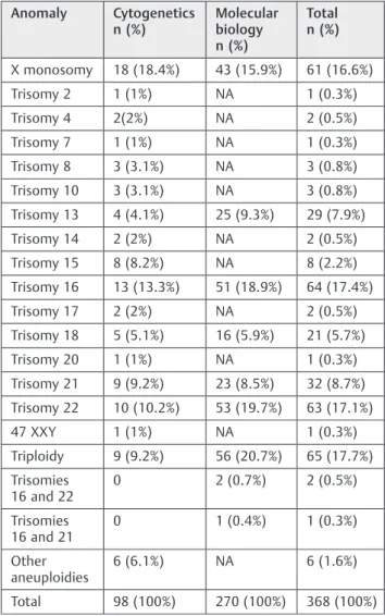
The chromosomal abnormalities identified by each meth-od are demonstrated in►Table 2.
Cytogenetics results showed 18.4% X monosomy, 13.3% trisomy 16, and 10.2% trisomy 22. It also revealed that 8.2% of the abnormalities were trisomy15. Molecular biology results were 15.9% for X monosomy, 18.9% for trisomy 16, and 19.7% for trisomy 22. Chromosome 15 was not evaluated by QF-PCR in this study (►Table 2).
Considering all the samples, trisomy was the most com-mon chromosome aberration, accounting for 63% (232 out of 368) of the abnormalities. The most frequent was trisomy 16 (17.4%), followed by trisomy 22 (17.1%). Monosomy accounted for 16.6% (61 out of 368) of the anomalies, and polyploidies, for 18.8% (69 out of 368) (►Table 2 and ►Table 3).
The relationship between aneuploidy and maternal age is shown in►Fig. 1. In the young maternal age group (n¼452), 62.2% (281) had normal results, while abnormalities were found in 171 cases (37.8%). For the advanced maternal age group (n¼432), 235 (54.4%) normal results were observed, compared with 197 (45.6%) abnormalfindings. This difference was considered statistically significant (p¼0.02) (►Fig. 1).
Discussion
The analysis of chromosomal abnormalities in POC is useful to determine the possible causes of miscarriage, and to provide information and counseling for couples regarding future pregnancies.
This retrospective study showed the presence of aneuploi-dies in 42% of all the analyzed samples. When the different techniques were considered, the results were similar (40% by QF-PCR and 48% by cytogenetics). A recent study by Coelho et al17 demonstrated the presence of aneuploidies in 54.6% of miscarriage material from Brazilian patients, analyzed by Table 1 Normal and abnormal results observed by cytogenetics and molecular biology techniques
Cytogenetics (n¼204)
Molecular biology (n¼680)
Total (n¼884)
Normal 106 (52%) 410 (60%) 516 (58%)
QF-PCR. Jenderny15 reported a frequency of 61% abnormal results in POC analyzed by cytogenetics and QF-PCR in the German population. Other studies found similar (48%) or lower frequencies (36%) of total chromosomal aberrations.18,19
The present study revealed that the main chromosomal abnormality detected in abortion material was trisomy, followed by triploidy and monosomy X. These results corroborate with other studies that demonstrated similar results, showing that trisomies, especially those involving chromosomes 16 and 22, are implicated in spontaneous abortion.17,20–25
An important point noted in the present study was that trisomy 15 was found in 8.2% of the samples analyzed by conventional cytogenetics. Similar values were observed by Coelho et al17(14.1%), Moraes et al20 (9%), Subramaniyam et al21(13.5%), and Romero et al25 (7,7%), suggesting that trisomy 15 is recurrent in POC samples. Until the present moment, chromosome 15 markers have not been included in routine QF-PCR analysis of abortion material at the labora-tory. However, the results showed that a marker for chromo-some 15 should be included in the molecular analysis, improving the quality of the released technical reports.
Conventional cytogenetics technique remains highly recommended for spontaneous miscarriage analysis. How-ever, this method has certain disadvantages, such as a long laboratory cycle, labor intensity and culture failure, espe-cially when the tissues obtained from patients are not well preserved.17,26
The QF-PCR technique is regarded as a highly accurate, low cost and rapid diagnostic method to facilitate the detec-tion of clinically relevant chromosome aberradetec-tions. However, some limitations of the technique are the difficulty to offer a correct diagnosis for mosaicism, small deletions, transloca-tions and duplicatransloca-tions.15,17
Since our laboratory miscarriage material is received from different medical facilities, the QF-PCR method plays an important role as a reliable method for the detection of aneuploidies, when culture fails or when the karyotype is not possible due to inadequate material preservation.
Many reports have suggested that advanced maternal age is an important factor related to chromosomal aneuploi-dies.22Our study showed a significant increase in the rate of aneuploidy in the advanced maternal age group when com-pared with the young maternal age group (p¼0.02). Bastos et al6 and Jia et al22 also suggest a maternal age-related increase in chromosomal anomalies. Hormonal changes during the aging process, as decreased production of pro-gesterone can lead to increased rates of miscarriage in women older than 35 years.27
In conclusion, chromosomal aberrations are still a major cause of miscarriage, and the conventional cytogenetics study Table 2 Frequency of chromosomal anomalies
Anomaly Cytogenetics n (%)
Molecular biology n (%)
Total n (%)
X monosomy 18 (18.4%) 43 (15.9%) 61 (16.6%)
Trisomy 2 1 (1%) NA 1 (0.3%)
Trisomy 4 2(2%) NA 2 (0.5%)
Trisomy 7 1 (1%) NA 1 (0.3%)
Trisomy 8 3 (3.1%) NA 3 (0.8%)
Trisomy 10 3 (3.1%) NA 3 (0.8%)
Trisomy 13 4 (4.1%) 25 (9.3%) 29 (7.9%)
Trisomy 14 2 (2%) NA 2 (0.5%)
Trisomy 15 8 (8.2%) NA 8 (2.2%)
Trisomy 16 13 (13.3%) 51 (18.9%) 64 (17.4%)
Trisomy 17 2 (2%) NA 2 (0.5%)
Trisomy 18 5 (5.1%) 16 (5.9%) 21 (5.7%)
Trisomy 20 1 (1%) NA 1 (0.3%)
Trisomy 21 9 (9.2%) 23 (8.5%) 32 (8.7%)
Trisomy 22 10 (10.2%) 53 (19.7%) 63 (17.1%)
47 XXY 1 (1%) NA 1 (0.3%)
Triploidy 9 (9.2%) 56 (20.7%) 65 (17.7%)
Trisomies 16 and 22
0 2 (0.7%) 2 (0.5%)
Trisomies 16 and 21
0 1 (0.4%) 1 (0.3%)
Other aneuploidies
6 (6.1%) NA 6 (1.6%)
Total 98 (100%) 270 (100%) 368 (100%)
Abbreviation: NA, Not analyzed by QF-PCR.
Table 3 Groups of chromosomal anomalies
Anomaly Total n (%)
Trisomy 232 (63%)
Polyploidy 69 (18.8%)
X monosomy 61 (16.6%)
Others 6 (1.6%)
Total 368 (all)
is highly recommended, as it can detect different types of chromosomal abnormalities. Molecular biology techniques, such as QF-PCR, are important complementary methods that can be effective to detect the main chromosomal anomalies, and may be used in combination with cytogenetics to allow the release of technical reports with reliable results.
References
1 Zegers-Hochschild F, Adamson GD, Mouzon J, et al. Glossário revisado da terminologia das Técnicas de Reprodução Assistida (TRA. 2009Comitê Internacional para Monitorização da Tecnolo-gia Reprodutiva Assistida (ICMART) e Organização Mundial da Saúde (OMS). Caracas: Red Latinoamericana de Reproducción Asistida; 2009
2 Rolnik DL, Carvalho MHB, Catelani ALPM, et al. Cytogenetic analysis of material from spontaneous abortion. Rev Assoc Med Bras (1992) 2010;56(06):681–683
3 Hogge WA, Byrnes AL, Lanasa MC, Surti U. The clinical use of karyotyping spontaneous abortions. Am J Obstet Gynecol 2003; 189(02):397–400, discussion 400–402
4 Gonçalves RO, Santos WVB, Sarno M, Cerqueira BAV, Gonçalves MS, Costa OLN. Chromosomal abnormalities in couples with recurrent first trimester abortions. Rev Bras Ginecol Obstet 2014;36(03):113–117
5 Hyde KJ, Schust DJ. Genetic considerations in recurrent pregnancy loss. Cold Spring Harb Perspect Med 2015;5(03):a023119 6 Bastos R, Ramalho C, Dória S. Prevalence of chromosomal
ab-normalities in spontaneous abortions or fetal deaths. Acta Med Port 2014;27(01):42–48
7 Isfer EV, Sanchez RC, Saito M. Medicina fetal: diagnóstico pré-natal e conduta. Rio de Janeiro: Revinter; 1996
8 López AGA, Huerta SB, Galván RH, Posadas RA, del Ángel AG, González PG. Diagnóstico citogenético en aborto espontáneo del primer trimestre. Ginecol Obstet Méx 2003;79(12):779–784 9 Liu S, Song L, Cram DS, et al. Traditional karyotyping vs copy
number variation sequencing for detection of chromosomal abnormalities associated with spontaneous miscarriage. Ultra-sound Obstet Gynecol 2015;46(04):472–477
10 Lathi RB, Gray Hazard FK, Heerema-McKenney A, Taylor J, Chueh JT. First trimester miscarriage evaluation. Semin Reprod Med 2011;29(06):463–469
11 Trask BJ. Human cytogenetics: 46 chromosomes, 46 years and counting. Nat Rev Genet 2002;3(10):769–778
12 Pena SDJ, Costa HBBLM, Carvalho ERF, Sturzeneker R. Investigação genética dos abortamentos espontâneos pelo DNA. Rev Méd Minas Gerais 2003;13(03):164–173
13 Pinto Junior W. Diagnóstico pré-natal. Cien Saude Colet 2002; 7(01):139–157
14 Vieira SR, Ferrari LP. Investigação de alterações citogenéticas em abortos espontâneos: um retrospecto de 2006 a 2011. Cad Esc Saúde 2013;2(10):1–20
15 Jenderny J. Chromosome aberrations in a large series of sponta-neous miscarriages in the German population and review of the literature. Mol Cytogenet 2014;7:38
16 Diego-Alvarez D, Garcia-Hoyos M, Trujillo MJ, et al. Application of quantitativefluorescent PCR with short tandem repeat markers to the study of aneuploidies in spontaneous miscarriages. Hum Reprod 2005;20(05):1235–1243
17 Coelho FF, Marques FK, Gonçalves MS, Almeida VC, Mateo EC, Ferreira AC. Detection of aneuploidies in spontaneous abortions by quantitativefluorescent PCR with short tandem repeat mar-kers: a retrospective study. Genet Mol Res 2016;15(03): Doi: 10.4238/gmr.15038617
18 Shearer BM, Thorland EC, Carlson AW, Jalal SM, Ketterling RP. Reflexfluorescent in situ hybridization testing for unsuccessful product of conception cultures: a retrospective analysis of 5555 samples attempted by conventional cytogenetics and fluorescent in situ hybridization. Genet Med 2011;13(06): 545–552
19 Zou G, Zhang J, Li XW, He L, He G, Duan T. Quantitativefluorescent polymerase chain reaction to detect chromosomal anomalies in spontaneous abortion. Int J Gynaecol Obstet 2008;103(03):237–240 20 Moraes AC, Moron AF, Hashimoto EM, et al. Cytogenetic and molecular evaluation of spontaneous abortion samples. Rev Bras Ginecol Obstet 2005;27(09):554–560
21 Subramaniyam S, Pulijaal VR, Mathew S. Double and multiple chromosomal aneuploidies in spontaneous abortions: A single institutional experience. J Hum Reprod Sci 2014;7(04): 262–268
22 Jia CW, Wang L, Lan YL, et al. Aneuploidy in early miscarriage and its related factors. Chin Med J (Engl) 2015;128(20):2772–2776 23 Shen J, Wu W, Gao C, et al. Chromosomal copy number analysis on
chorionic villus samples from early spontaneous miscarriages by high throughput genetic technology. Mol Cytogenet 2016;9:7 24 Nicolaides KH. First-trimester screening for chromosomal
abnormalities. Semin Perinatol 2005;29(04):190–194
25 Romero ST, Geiersbach KB, Paxton CN, et al. Differentiation of genetic abnormalities in early pregnancy loss. Ultrasound Obstet Gynecol 2015;45(01):89–94
26 Tekcan A, Tural S, Elbistan M, Kara N, Guven D, Kocak I. The combined QF-PCR and cytogenetic approach in prenatal diagno-sis. Mol Biol Rep 2014;41(11):7431–7436