Major Article
Corresponding author: Dr. Shahla Mansouri.
e-mail: smansouri@kmu.ac.ir
Received 29 June 2017
Accepted 20 December 2017
Clonal relationships, antimicrobial susceptibilities,
and molecular characterization of extended-spectrum
beta-lactamase-producing
Escherichia coli
isolates from
urinary tract infections and fecal samples in Southeast Iran
Zahra Hashemizadeh
[1],
Davood Kalantar-Neyestanaki
[1],[2]and
Shahla Mansouri
[1],[3][1]. Department of Microbiology and Virology, School of Medicine, Kerman University of Medical Sciences. Kerman, Iran. [2]. Student Research Committee, School of Medicine, Kerman University of Medical Sciences, Kerman, Iran.
[3]. Research Center for Infectious Diseases and Tropical Medicine, Kerman, Iran.
Abstract
Introduction: Multidrug-resistant (MDR) Escherichia coli, a species that is a leading cause of urinary tract infections (UTIs) and is a major global public health concern. This study was designed to detect the differences in antibiotic resistance patterns, the
production and type of extended spectrum β-lactamases (ESBLs), and the clonal relationships among E. coli isolates from UTIs and fecal samples. Methods: Antibacterial resistance was determined by the disk diffusion method. ESBL, carbapenemase, and
AmpC-producing isolates were detected phenotypically. Then, the ESBL genes were sequenced to detect the type. Enterobacterial repetitive intergenic consensus-polymerase chain reaction (ERIC-PCR) was performed on the ESBL-positive isolates. Results: The most common effective antibacterial agents were colistin, imipenem, and amikacin. Among the isolates, 204 (56.6%)
were MDR. Of the 163 ESBL-positive isolates, 11 (6.7%) produced AmpC, and the frequencies of beta-lactamase-positive
genes were as follows: blaCTX-Mgroup1, 76%; blaTEM1, 74.8%; blaSHV12, 1.2%; and blaOXA1, 12.88%. ERIC PCR showed a diverse
pattern, suggesting that clonal spread of E. coli in this area is uncommon, and that most of the infecting strains are endogenous. Conclusions: The high rates of antibacterial-resistant and MDR isolates are quite important since these strains can act as source of resistant bacteria that can be spread in the community. Controlling antibiotic use, against inappropriate use and abuse, in the community and continuous surveillance of emerging resistance traits are critical to controlling the spread of resistance.
Keywords: Escherichia col.Antimicrobial resistance. ESBL. AmpC beta-lactamas.
INTRODUCTION
Escherichia coli is found in the normal microbiota of the human intestinal tract, and it has an important role in protecting against pathogenic bacteria1. An estimated 70-90%
of community-acquired urinary tract infections (UTIs) and 50% of hospital-acquired UTIs are caused by E. coli2. Most nosocomial UTIs are caused by endogenous strains, although exogenous infections from other patients or other sources have also been documented3.
Many antibacterial agents, such as beta-lactams and
fluoroquinolones, are recommended for the treatment of UTIs4. However, inappropriate use and overuse of antibacterial agents have resulted in the emergence of multidrug-resistant (MDR) isolates; therefore, the drugs previously used to treat patients,
such as fluoroquinolones, trimethoprim/sulfamethoxazole,
and cephalosporin, have become ineffective4. Beta-lactams are the most commonly used antimicrobial agents for the treatment of UTIs5. The main mechanism of resistance to
beta-lactams is the production of beta-lactamase enzymes5.
Beta-lactamase enzymes, such as extended spectrum beta-lactamases (ESBLs), metallo-beta-beta-lactamases (MBLs), and
AmpC beta-lactamase, have been reported in gram-negative bacteria6. When these resistant bacterial isolates cause hospital-
and community-acquired infections, treatment is difficult since
the therapeutic options are limited5. Beta-lactamase genes are usually encoded on mobile genetic elements that can be easily transmitted between different bacterial species or strains7. Therefore, epidemiological studies are crucial for monitoring drug resistance among bacterial isolates and determining the distribution of drug resistance in the community. In this study,
the antibacterial resistance profiles, frequency of ESBL genes,
and clonal relationships among isolates from fecal samples and the urinary tracts of inpatients and outpatients with UTIs
was determined to find better strategies for the control and
METHODS
Study population and bacterial isolates
From October 2014 to June 2015, 351 E. coli strains (100 from inpatients with UTIs and 151 from outpatients with UTIs) were isolated in Kerman, Iran. The patients were considered inpatients when they stayed in a given hospital ward for
treatment for ≥48h and/or within 30 days of hospital discharge,
otherwise they were considered outpatients. Outpatients were those who visited the emergency ward with acute symptoms or were referred to local private laboratories for urine culture. All
outpatients were not previously hospitalized due to any disease8. Fecal samples (n=100) were collected by individual rectal swabs from volunteers without any digestive problems and were
transported to the laboratory in Cary-Blair transport medium and
then placed overnight in buffered peptone water at 37°C. The samples were then cultivated on MacConkey agar (CONDA,
Co., SPAIN). Lactose-positive isolates on MacConkey agar were selected for biochemical confirmatory tests for E. coli.
The criteria for selection were no history of hospitalization and
signs or symptoms of UTI or gastrointestinal symptoms, such as nausea, vomiting, abdominal cramps, diarrhea, fever, and
headache. All isolates were confirmed to be E. coli by the results of standard biochemical tests, including gram-negative,oxidase
negative, catalase positive, lactose positive, MR positive, VP negative, Indole positive, and acid/acid and gas positive on
TSI medium9.
Determination of antibacterial susceptibility and ESBL, AmpC, and carbapenemase production
The standard disk diffusion method was used to determine
the antibacterial susceptibility profiles of the isolates to 11
antibacterial agents10. The following disks (obtained from
MAST Chemical Company, England) were used in this study: amoxicillin/clavulanic acid (AUG, 30/10µg), cefotaxime (CTX, 30µg), ceftazidime (CAZ, 30µg), cefepime (CPM, 30µg), imipenem (IMP, 10µg), ciprofloxacin (CIP, 10µg), nalidixic acid (NA, 30µg), trimethoprim (TM, 5µg), piperacillin/tazobactam (PIP, 100/10µg), amikacin (AK, 30µg), and gentamicin (GM, 10µg). MDR isolates were defined as described by
Magiorakos et al.11. The European Committee on Antimicrobial
Susceptibility Testing (EUCAST) guidelines were used to test
for susceptibility to colistin (CO, 10µg)12. However, the MIC
by E-test (HiMedia, Co., India) was determined for isolates that were not susceptible to colistin in the disk diffusion for
confirmation12,13. ESBL-producing isolates were identified by the combined disk method with clavulanic acid and
extended-spectrum cephalosporins (CPD, CTX, and CAZ; 30µg alone and in combination with 10µg of clavulanic acid)14. The AmpC disk test was used to detect AmpC beta-lactamases as described
by Black et al.15. The modified Hodge test (MHT) was used to detect carbapenemase-producing isolates10. E. coli ATCC 25922 and Pseudomonas aeruginosa ATCC 27853 were used as standard strains in the antibacterial susceptibility testing, and
Klebsiella pneumoniae ATCC 700603 was used as the positive
control for the ESBL test.
Polymerase chain reaction to detect beta-lactamase genes
The boiling method was used to prepare template DNA for
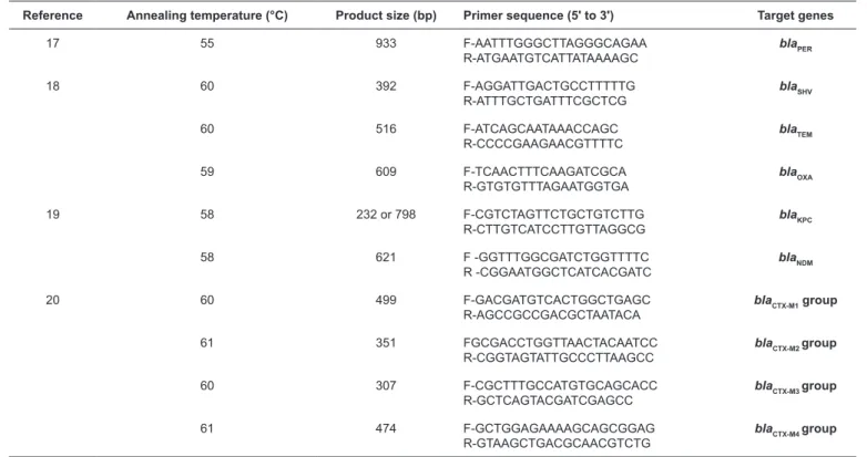
PCR as described by Qi et al.16. The oligonucleotide primers used to detect blaCTX-M group 1-4, blaTEM, blaSHV, blaOXA, blaPER,
blaKPC, and blaNDM are listed in Table 117-20. PCR amplification
was performed in a total volume of 50µL containing 1µL of each primer (10pM), 25µL of DNA Polymerase Master Mix RED (Ampliqon, Co., Denmark), 2µL of DNA, and 21µL of DNase and RNase free water in a FlexCycler PCR Thermal Cycler (Analytik Jena, Germany) under the following cycling
conditions: an initial denaturation step at 95°C for 5min followed by 30 cycles of denaturation at 95 °C for 1min, annealing at 55-61°C for 1min (Table 1), extension at 72°C for 1min, and a
final extension step at 72°C for 5min. The PCR products were
separated by electrophoresis in a 1.5% agarose gel in 0.5 Tris,
EDTA, Boric acid (TBE) buffer. For each ESBL group, amplicons from randomized selected strains were sequenced (Macrogen, South Korea). The obtained nucleotide sequences were compared with those in the GenBank database for homologous nucleotide sequences by BLAST (www.ncbi.nih.gov/BLAST program).
Molecular typing of ESBL isolates by ERIC-PCR
Enterobacterial repetitive intergenic consensus-polymerase chain reaction (ERIC-PCR) was used to determine the clonal relationships among the isolates using the ERIC2 primer (5ʹ-AAGTAAGTGACTGGGGTGAGC-3ʹ)21. The ERIC-PCR
amplifications were carried out in a total volume of 25µL containing 1µL of ERIC2 primer (10pM), 12.5µL of DNA Polymerase Master Mix RED, 1µL of DNA template, and 10µL of DNase and RNase free water (SinaClon BioScience, Co., Iran) in a FlexCycler PCR Thermal Cycler under the following
cycling conditions: an initial denaturation step at 95°C for 5min followed by 30 cycles of denaturation at 95°C for 1min, annealing at 59°C for 1min, and extension at 72°C for 2min,
and a final extension step at 72°C for 10min. The obtained PCR
fragments were separated by electrophoresis in a 1.5% agarose
gel, and the gel was analyzed by using Gel Compare software (BioMérieux, Applied Maths, Belgium) using a cut off of 70%
to discriminate isolates22.
Statistical analysis
For data analysis, SPSS version 20.0 (IBM, Armonk, USA)
was used. Fisher’s exact test was used for data analysis and to
evaluate the significance of differences. P value less than 0.05 was considered statistically significant.
RESULTS
Antibacterial susceptibility
All the tested isolates were susceptible to colistin. Although 10 isolates showed intermediate susceptibility to colistin by the
disk diffusion method, the E test results showed that all isolates were sensitive, with an MIC ≤2µg/ml. The other most effective
drugs were imipenem and amikacin (Table 2). There were
Reference Annealing temperature (°C) Product size (bp) Primer sequence (5ʹ to 3ʹ) Target genes
17 55 933 F-AATTTGGGCTTAGGGCAGAA
R-ATGAATGTCATTATAAAAGC
bla
PER
18 60 392 F-AGGATTGACTGCCTTTTTG
R-ATTTGCTGATTTCGCTCG
bla
SHV
60 516 F-ATCAGCAATAAACCAGC
R-CCCCGAAGAACGTTTTC
bla
TEM
59 609 F-TCAACTTTCAAGATCGCA
R-GTGTGTTTAGAATGGTGA
bla
OXA
19 58 232 or 798 F-CGTCTAGTTCTGCTGTCTTG
R-CTTGTCATCCTTGTTAGGCG
bla
KPC
58 621 F -GGTTTGGCGATCTGGTTTTC
R -CGGAATGGCTCATCACGATC
bla
NDM
20 60 499 F-GACGATGTCACTGGCTGAGC
R-AGCCGCCGACGCTAATACA
bla
CTX-M1 group
61 351 FGCGACCTGGTTAACTACAATCC
R-CGGTAGTATTGCCCTTAAGCC
bla
CTX-M2 group
60 307 F-CGCTTTGCCATGTGCAGCACC
R-GCTCAGTACGATCGAGCC
bla
CTX-M3 group
61 474 F-GCTGGAGAAAAGCAGCGGAG
R-GTAAGCTGACGCAACGTCTG
bla
CTX-M4 group
TABLE 1: PCR Primers used to amplify ESBL genes in Escherichia coli strains isolated from urinary tract infections and fecal samples.
PCR: polymerase chain reaction; ESBL: extended spectrum β-lactamases.
Resistant strains (n %) Antimicrobial agent (µg/disk)
P value fecal flora
(n=100)
outpatients (n=151)
inpatients (n=100)
0.009 49 49.0 85 56.2 70 70.0 Amoxicillin-clavulanic acid (30/10)
0.05 81 81.0 102 67.5 75 75.0 Trimethoprim (5)
0.03 80 80.0 98 64.9 73 73.0 Nalidixic acid (30)
0.08 49 49.0 63 41.7 56 56.0 Cefotaxime (30)
0.3 47 47.0 65 43.0 53 53.0 Cefepime (30)
0.8 0 0.0 0 0.0 0 0.0 Colistin (10)
0.05 42 42.0 61 40.3 55 55.0 Ceftazidime (30)
0.07 9 9.0 8 5.2 4 4.0 Imipenem (10)
0.2 2 2.0 10 6.6 6 6.0 Amikacin (30)
0.04 13 13.0 33 21.8 27 27.0 Piperacillin/tazobactam (100/10)
0.1 48 48.0 63 41.7 55 55.0 Ciprofloxacin (10)
0.7 16 16.0 24 16.0 19 19.0 Gentamicin (10)
TABLE 2: Antibacterial resistant patterns of 351 Escherichia coli isolates from in patients and outpatients with urinary tract infections and fecal samples.
and fecal samples, and the resistance rates were significantly
higher in inpatient samples than in outpatient and fecal samples
(p ≤ 0.05). For TM and NA, a significantly higher rate of
resistance was observed in fecal samples than in the inpatient
and outpatient isolates (p ≤ 0.05). There was no difference with
respect to gender or age.
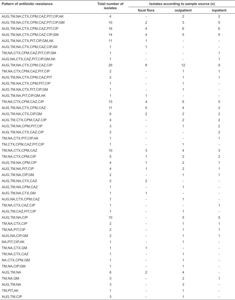
The rate of MDR, defined as resistance to at least three
classes of antimicrobial agents, was high, and the MDR phenotype was detected in 61%, 55.6%, and 57% of the inpatient, outpatient, and fecal isolates, respectively (Table 3).
Pattern of antibiotic resistance Total number of isolates
Isolates according to sample source (n)
fecal flora outpatient inpatient
AUG,TM,NA,CTX,CPM,CAZ,PIT,CIP,AK 4 - 2 2
AUG,TM,NA,CTX,CPM,CAZ,PIT,CIP,GM 10 2 3 5
AUG,TM,NA,CTX,CPM,CAZ,PIT,CIP 16 4 6 6
AUG,TM,NA,CTX,CPM,CAZ,CIP,GM 14 4 5 5
AUG,TM,NA,CTX,PIT,CIP,GM,AK 11 1 -
-AUG,TM,NA,CTX,CPM,CAZ,CIP,AK 1 1 -
-TM,NA,CTX,CPM,CAZ,PIT,CIP,GM 1 - - 1
AUG,NA,CTX,CAZ,PIT,CIP,GM,AK 1 - - 1
AUG,TM,NA,CTX,CPM,CAZ,CIP 29 8 12 9
TM,NA,CTX,CPM,CAZ,PIT,CIP 2 - 1 1
AUG,TM,NA,CTX,CPM,CAZ,PIT 2 - 1 1
AUG,TM,NA,CTX,CPM,PIT,CIP 1 - 1
-AUG,TM,NA,CTX,PIT,CIP,GM 1 - 1
-AUG,TM,NA,PIT,CIP,GM,AK 1 1 -
-TM,NA,CTX,CPM,CAZ,CIP 15 4 6 5
AUG,TM,NA,CTX,CPM,CAZ 11 5 4 2
AUG,TM,NA,CTX,CIP,GM 6 2 2 2
AUG,TM,CTX,CPM,CAZ,CIP 4 - 2 2
AUG,TM,NA,CPM,PIT,CIP 3 - 1 2
AUG,TM,NA,CTX,CAZ,CIP 2 - - 2
TM,NA,CTX,PIT,CIP,AK 1 - - 1
TM,CTX,CPM,CAZ,PIT,CIP 1 - 1
-TM,NA,CTX,CPM,CAZ 10 3 4 3
TM,NA,CTX,CPM,CIP 5 1 2 2
AUG,TM,NA,CPM,CIP 4 1 2 1
AUG,TM,NA,PIT,CIP 4 1 2 1
AUG,TM,NA,CIP,GM 2 - 1 1
AUG,TM,NA,CTX,CAZ 2 2 -
-AUG,TM,NA,CPM,CAZ 1 - 1
-AUG,TM,NA,CTX,GM 1 1 -
-AUG,NA,CTX,CPM,CAZ 1 - 1
-TM,NA,CTX,CAZ,CIP 1 - - 1
AUG,TM,CAZ,PIT,CIP 1 - 1
-AUG,TM,NA,CIP 10 - 5 5
TM,NA,CTX,CIP 2 - 1 1
TM,NA,PIT,CIP 2 - 1 1
AUG,NA,CIP,GM 2 - 1 1
NA,PIT,CIP,AK 1 - 1
-TM,NA,CTX,GM 1 1 -
-TM,NA,CTX,CAZ 1 - 1
-NA,CTX,CPM,GM 1 - 1
-TM,NA,CIP,GM 1 - 1
-AUG,TM,NA 6 2 4
-TM,NA,GM 3 - 2 1
AUG,TM,NA 3 - 3
-TM,PIT,AK 1 - 1
-AUG,TM,CIP 3 - 1
-UTIs: urinary tract infections; AUG: amoxicillin-clavulanic acid; TM: trimethoprim; NA: nalidixic acid; CTX: cefotaxime; CPM: cefepime; CO: colistin; CAZ: ceftazidime; IMP: imipenem; AM: amikacin; PIT: piperacillin/tazobactam; CIP: ciprofloxacin; GM: gentamicin.
followed by AUG, TM, NA, CTX, CPM, CAZ, and CIP (20
isolates, 9.8%; Table 3). The antimicrobial resistance patterns
of 24 isolates were unique.
Detection of ESBLs, AmpC, carbapenemase, and blagenes
The frequency of ESBL-positive isolates, as determined by the
combined disk test, was 46.4% (163) for all isolates, and was 53% (53), 41% (62), and 48% (48) for inpatient, outpatient, and fecal
isolates, respectively. Isolates from inpatients had a significantly higher rate of ESBLs compared to outpatient and fecal samples (P=0.000). Among the 163 ESBL-positive isolates, 11 (6.74%)
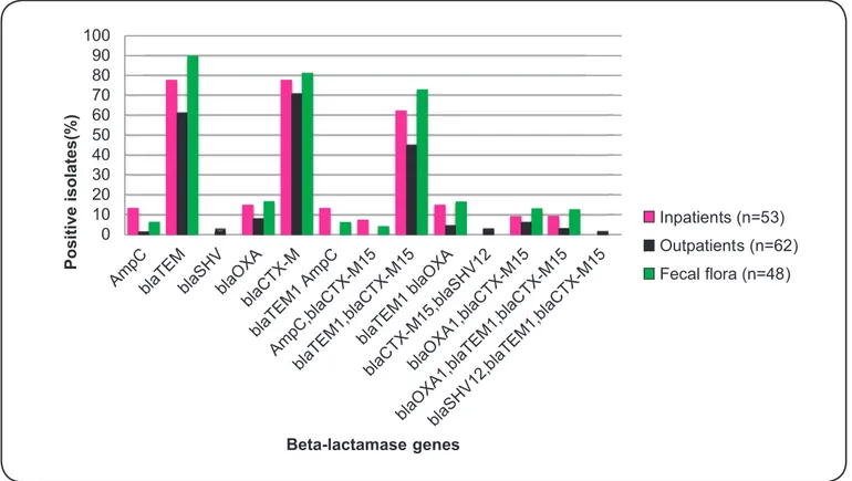
isolates were AmpC producers. The MHT was negative for all imipenem-resistant isolates. The rates of detection for the
following ESBL genes by PCR: blaTEM, blaSHV, blaOXA, and bla
CTX-Mgroup1 were 74.8% (122), 1.2% (2), 12.88% (21), and 76% (124), respectively (Figure 1). The other tested beta-lactamase genes,
blaPER, blaCTX-Mgroup2,3,4, blaKPC, and blaNDM, were not detected in any of the tested isolates. The rates of AmpC and blaTEM were
significantly higher in isolates from inpatients than in isolates from outpatients (P=0.003; Figure 1). Sequencing of the bla
CTX-Mgroup1, blaTEM, blaOXA, and blaSHV ESBL genes confirmed that they were blaCTX-Mgroup1, blaTEM1, blaOXA1, and blaSHV12, respectively.
Genetic comparison of the
ESBL-producing Escherichia coli isolates by ERIC-PCR
The ERIC-PCR amplified sequences produced a
well-separated, sharp band in agarose electrophoresis in the range
0 10 20 30 40 50 60 70 80 90 100
Positive
isolates(%
)
Beta-lactamase genes
Inpatients (n=53)
Outpatients (n=62)
Fecal flora (n=48)
FIGURE 1: Distribution of ESBL genes and AmpC beta-lactamases among 163 ESBL-producing Escherichia coli isolates from urinary tract infections and
fecal flora. ESBL: extended spectrum β-lactamases.
of 100-3000bp as estimated with the Gene Ruler 100bp Plus DNA Ladder (Thermo Scientific, Co. Canada). The amplified sequences were analyzed by the UPGMA program (UPGMA
clustering UN weighted pair-group method with arithmetic
averages) using Gel Compare software (version 4.0). In the data
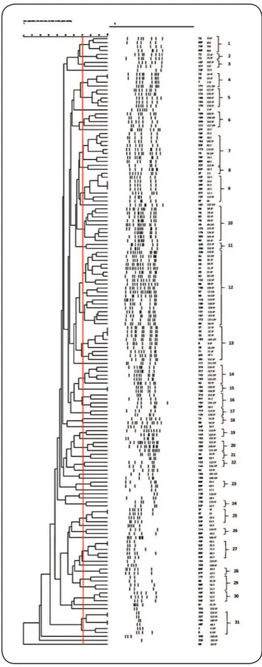
analysis, a cutoff of 70% was used due to the high diversity of the isolates. With this cutoff, 31 clusters and 17 singletons were detected. The dendrogram showed one major cluster comprising 19 isolates, with 14 from inpatients and 5 from outpatients. The second major cluster included 8 UTI isolates, which were mostly from inpatients. The other clusters contained a combination of isolates from inpatient and outpatient UTIs and fecal samples (Figure 2).
DISCUSSION
UTIs are a major public health problem, both in hospitals and in the community. Drug resistance in E. coli, due to
horizontal gene transfer, has resulted in the emergence of MDR isolates, which are difficult to treat. The bacteria that cause most UTIs are of an endogenous origin; therefore, the fecal flora is
important, and is likely a source of resistance genes. In this study, the antimicrobial resistance of fecal and UTI isolates were compared. E. coli is an important cause of uncomplicated UTIs in women, and it is also one of the most common causes of septicemia and meningitis in newborn infants, diarrhea
in immunocompromised and debilitated patients8. In recent years, the emergence of antimicrobial resistance has become an increasingly common factor in the management of patients with UTIs. Antibiotic resistance in E. coli isolates from women
with uncomplicated UTIs has been reported in Germany, Spain,
Sweden, and the UK23, and higher rates of resistance have been
reported from non-European countries, such as India, China,
and South Korea24-26. Our results also showed a high rate of resistance to various antimicrobial agents.
In a study performed in this area in 2002, the rates of resistance to various antimicrobial agents was much lower than those observed in the present study, and the sensitivity rates to
GM, CTX, ZOX (ceftizoxime), and FM (nitrofurantoin) for
fecal and UTI isolates were greater than 90%27. This marked increase in resistance rates in a short time should be taken very seriously, and steps should be taken to reduce the rate of this increase. According to Walker et al.28 and based on other available literature, the rate of trimethoprim sensitivity remains high for most MDR E. coli isolates from UTIs. Commensal
E. coli in the gastrointestinal tract are regarded as an important source of antibiotic-resistance genes, and these organisms can transfer resistance genes to other bacteria when exposed to antimicrobial agents29. In this study, the rates of resistance
to amoxicillin-clavulanic acid and piperacillin/tazobactam were significantly higher in UTI isolates than in fecal isolates (P ≥ 0.05). This demonstrates the important role of the fecal flora as a source of drug resistance in the community. The
presence of antimicrobials in animal food for growth promotion and prophylaxis may be responsible for the emergence of antibacterial resistance strains in humans30,31. Simultaneous resistance to 5-9 antimicrobial agents was detected in 79.4% of the isolates, while resistance to 3-4 antimicrobial agents was detected in 17.1% of the isolates. These results indicate that there is a shortage of available drugs for the treatment of UTIs.
ESBL-producing bacterial strains have spread worldwide, and nosocomial infections with ESBL-producing E. coli
isolates are responsible for high rates of morbidity and mortality in infected patients32. Therefore, early detection and
identification of ESBL-producing isolates are necessary to
control the infection and prevent the spread of these organisms
in hospitals and in the community. Significant associations have been reported for UTIs caused by ESBL-producing E. coli and male sex, residence in a nursing home, diabetes mellitus, and recurrent UTIs33. Other risk factors have also been reported, such as indwelling urinary catheters, old age, ventilation, and organ transplants or other surgery, as well as high-dose or long-time treatment with antimicrobials, especially beta-lactams and
fluoroquinolones33. In the present study, 46.4% of the isolates
were ESBL producers. However, isolates from outpatients had a significantly lower rate of ESBL production compared to isolates from inpatients and fecal samples. We did not find any difference in ESBL production according to age or gender. A high rate of ESBL-producing E. coli isolates from fecal samples has been
reported by other researchers, and Leflon et al.30 reported an
increase in the rate, from 0.6% to 6%, over a five-year period in Paris, France. In the present study, we observed relatively
FIGURE 2: Dendrogram generated with ERIC-PCR data for ESBL-positive
Escherichia coli isolates from UTIs and fecal flora. ERIC-PCR:
high numbers of isolates producing blaCTX-Mgroup1 (76%) and
blaTEM (74.8%). The presence of blaCTX-Mgroup1 and blaTEM were
significantly higher in isolates from fecal samples and UTIs of inpatients (P=0.0004). Recently, a higher rate of blaCTX-M has been reported in enteric bacteria, especially E. coli and Klebsiella pneumoniae, likely since the genes encoding these enzymes are
located on plasmids or other mobile genetic elements. Because
CTX-M family genes are present on plasmids or other mobile
genetic elements, these enzymes have become predominant
worldwide and are substituting for the more common SHV and
TEM7,32. The most common type of CTX-M reported in various geographic regions is CTX-M15; these strains are usually MDR and are detected not only in nosocomial infections but also in the community4,24,32, and this result is in accordance with our finding
of a high frequency of CTX-M15-producing E. coli isolates. The blaCTX-M-positive isolates in this study were MDR, showing
resistance to fluoroquinolones, trimethoprim, tetracycline, and
aminoglycosides, as well as non-carbapenem beta-lactamases, as was reported by Woodford & Ward in 200434. It has been suggested that CTX-M15 was disseminated by clonal expansion of the pandemic E. coli clone ST13124, which could also be the source of the genes observed in this study.
The genetic diversity of ESBL-producing E. coli isolates,
as determined by ERIC-PCR, revealed the presence of diverse
clonal groups (Figure 2). Similar results were observed according to the antimicrobial resistance patterns, with 42 different combinations of antimicrobial resistance. These results suggested that different strains of E. coli are widely distributed in this area, and empiric therapy is not recommended for the treatment of UTIs in this region.
Finally, we suggest that antimicrobial susceptibility testing should be performed to select the best available drug for the treatment of patients with UTIs. Due to high impact of CTX-M
on public health, such as the difficultly in treating community-acquired UTIs, the spread of isolates producing ESBLs, especially
CTX-M15 beta-lactamase, needs to be closely monitored. It could be concluded that in this area, inappropriate use or misuse of antimicrobial agents may be a cause of the high resistance rates instead of clonal spread of resistant strains.
Financial support
This research was supported by Kerman University of Medical
Sciences & Health Services grant no 94/497.
Conflict of interest
The authors declare that there is no conflict of interest.
REFERENCES
1. Riley L. Pandemic lineages of extraintestinal pathogenic Escherichia coli. Clin Microbiol Infect. 2014;20(5):380-90.
2. Pitout J. Extraintestinal pathogenic Escherichia coli: a combination of virulence with antibiotic resistance. Front Microbiol. 2012;3:(9):1-7.
3. Ezejiofor T. Environmental factors in renal disease: the contribution of urinary tract infections in Nigerian renal disease burden-research and minireview. Clin Microbiol. 2016;5(2):1-15.
4. Van der Bij AK, Peirano G, Pitondo-Silva A, Pitout JD. The presence of genes encoding for different virulence factors in clonally related
Escherichia coli that produce CTX-Ms. Diagn Microbiol Infect Dis. 2012;72(4):297-302.
5. Paterson DL, Bonomo RA. Extended-spectrum β-lactamases:
a clinical update. Clin Microbiol Rev. 2005;18(4):657-86.
6. Bush K. Overcoming β-lactam resistance in Gram-negative pathogens. Future Med Chem. 2016; 8(9):921-4.
7. Chong Y, Ito Y, Kamimura T. Genetic evolution and clinical impact
in extended-spectrum β-lactamase-producing Escherichia coli and
Klebsiella pneumoniae. Infect Genet Evol. 2011;11(7):1499-504.
8. Toval F, Köhler C-D, Vogel U, Wagenlehner F, Mellmann A,
Fruth A, et al. Characterization of Escherichia coli isolates from hospital inpatients or outpatients with urinary tract infection. J Clin Microbiol. 2014;52(2):407-18.
9. Mahon CR, Lehman DC, Manuselis G.Textbook of diagnostic microbiology. 3rd edition. Saint Louis, MO: Saunders Elsevier; 2007.
10. Clinical and Laboratory Standards Institute (CLSI). Performance Standards for Antimicrobial Susceptibility Testing; Twenty-Fourth
Informational Supplement. CLSI M100-S24. 2014;34(1):50-57.
11. Magiorakos AP, Srinivasan A, Carey RB, Carmeli Y, Falagas ME,
Giske CG, et al. Multidrug-resistant, extensively drug-resistant and
pandrug-resistant bacteria: an international expert proposal for
interim standard definitions for acquired resistance. Clin Microbiol
Infect. 2012;18(3):268-81.
12. The European Committee on Antimicrobial Susceptibility Testing
(EUCAST). Clinical breakpoints. Breakpoint tables for bacteria. Breakpoint tables for interpretation of MICs and zone diameters.
Version 7.1, 2017. Available from: http://www.eucast.org/clinical_
breakpoints/
13. Galani I, Kontopidou F, Souli M, Rrkatsina PD, Koratzanis E,
Deliolanis J, et al. Colistin susceptibility testing by E-test and disk
diffusion methods. Int J Antimicrob Agents. 2008;31(5):434-9. 14. Weldhagen GF, Poirel L, Nordmann P. Amber class A
extended-spectrum β-lactamases in Pseudomonas aeruginosa: novel
developments and clinical impact. Antimicrob Agents Chemother. 2003;47(8):2385-92.
15. Black JA, Moland ES, Thomson KS. AmpC disk test for detection of plasmid-mediated ampc-lactamases in enterobacteriaceae
lacking chromosomal AmpC-β-lactamase. J Clin Microbiol.
2005;43(7):3110-13.
16. Qi C, Pilla V, Jessica HY, Reed K. Changing prevalence of Escherichia coli with CTX-M–type extended-spectrum
β-lactamases in outpatient urinary E. coli between 2003 and 2008. Diagn Microbiol Infect Dis. 2010;67(1):87-91.
17. Jiang J, Zhang Z, Li M, Zhou D, Ruan F, Lu Y. Detection of
extended-spectrum β-lactamases in clinical isolates of Pseudomonas aeruginosa. Antimicrob Agents Chemother.2006;50(9):2990-5. 18. Colom K, Pérez J, Alonso R, Fernández-Aranguiz A, Lariño E,
Cisterna R. Simple and reliable multiplex PCR assay for detection
of blaTEM, blaSHV and blaOXA–1 genes in Enterobacteriaceae. FEMS Microbiol Lett. 2003;223(2):147-51.
19. Poirel L, Walsh TR, Cuvillier V, Nordmann P. Multiplex PCR for
detection of acquired carbapenemase genes. Diagn Microbiol Infect
Dis. 2011;70:119-23.
among clinical isolates of Klebsiella pneumoniae at Labbafinejad
Hospital, Tehran, Iran.Microb Drug Resist. 2010;16(1):49-53. 21. Meacham KJ, Zhang L, Foxman B, Bauer RJ, Marrs CF. Evaluation
of genotyping large numbers of Escherichia coli isolates by
enterobacterial repetitive intergenic consensus-PCR. JClin Microbiol. 2003;41(11):5224-6.
22. Ardakani MA, Ranjbar R. Molecular typing of uropathogenic E. coli strains by the ERIC-PCR method. Electron Physician.
2016:8(4):2291-6.
23. Kahlmeter G, Ahman J, Matuschek E. Antimicrobial resistance of
Escherichia coli causing uncomplicated urinary tract infections: a
European update for 2014 and comparison with 2000 and 2008. Infect
Dis Ther. 2015;4(4):417-23.
24. Park SH, Byun JH, Choi SM, Lee DG, Kim SH, Kwon JC, et al.
Molecular epidemiology of extended-spectrum β-lactamase-producing Escherichia coli in the community and hospital in Korea: emergence
of ST131 producing CTX-M-15. BMC Infect Dis. 2012;12(1):1-11.
25. Xia S, Fan X, Huang Z, Xia L, Xiao M, Chen R, et al. Dominance
of CTX-M-type extended-spectrum β- lactamase (ESBL)-producing Escherichia coli isolated from patients with community-onset and
hospital- onset infection in China. PLoS One. 2014;9(7):e100707.
26. Lakshminarayana SA, Chavan SKD, Prakash R, Sangeetha S. Bacterial pathogens in urinary tract infection and antibiotic susceptibility pattern
from a Teaching Hospital, Bengaluru, India.Int J Curr Microbiol Appl Sci.2015;4(11):731-6.
27. Mansouri S, Shareifi S. Antimicrobial resistance pattern of Escherichia coli causing urinary tract infections, and that of human fecal flora, in the Southeast of Iran. Microb Drug Resist. 2002;8(2):123-8.
28. Walker E, Lyman A, Gupta K, Mahoney MV, Snyder GM, Hirsch
EB. Clinical management of an increasing threat: outpatient urinary
tract infections due to multidrug-resistant uropathogens. Clin Infect Dis. 2016;63(7):960-5.
29. Melo DB, Menezes APO, Reis JN, Guimarães AG. Antimicrobial
resistance and genetic diversity of Escherichia coli isolated from
humans and foods. Braz J Microbiol. 2015;46(4):1165-70.
30. Leflon-Guibout V, Blanco J, Amaqdouf K, Mora A, Guize
L, Nicolas-Chanoine MH. Absence of CTX-M enzymes but
high prevalence of clones, including clone ST131, among fecal
Escherichia coli isolates from healthy subjects living in the area of
Paris, France. J Clin Microbiol. 2008;46(12):3900-5.
31. Marshall BM, Levy SB. Food animals and antimicrobials: impacts on human health. Clin Microbiol Rev. 2011;24(4):718-33.
32. Rodríguez-Baño J, Navarro MD, Romero L, Muniain MA, Perea
EJ, Pérez-Cano R, et al. Clinical and molecular epidemiology of extended-spectrum β-lactamase-producing Escherichia coli as
a cause of nosocomial infection or colonization: implications for
control. Clin Infect Dis. 2006;42(1):37-45.
33. Briongos-Figuero LS, Gómez-Traveso T, Bachiller-Luque P,
Domínguez-Gil González M, Gómez-Nieto A, Palacios-Martín T, et al. Epidemiology, risk factors and comorbidity for urinary tract infections caused by extended-spectrum beta-lactamase (ESBL)-producing enterobacteria. Int J Clin Pract. 2012;66(9):891-6.
34. Woodford N, Ward ME, Kaufmann ME, Turton J, Fagan EJ, James D, et al. Community and hospital spread of Escherichia coli producing CTX-M extended-spectrum β-lactamases in the UK.