IDENTIFICATION AND ANTIMICROBIAL RESISTANCE OF MICROFLORA COLONIZING FERAL PIG (SUS
SCROFA) OF BRAZILIAN PANTANAL.
Lessa, SS1; Paes, RCS1; Santoro, PN1, Mauro, RA2; Vieira-da-Motta, O1*
1
Laboratório de Sanidade Animal,Setor de Doenças Infecto-Contagiosas, Universidade Estadual do Norte Fluminense Darcy Ribeiro, Campos dos Goytacazes, RJ, Brasil; 2Centro Nacional de Pesquisa de Gado de Corte/Embrapa, Campo Grande, MS,
Brasil.
Submitted: April 13, 2010; Approved: January 13, 2011.
ABSTRACT
Antimicrobial resistance of bacteria is a worldwide problem affecting wild life by living with resistant
bacteria in the environment. This study presents a discussion of outside factors environment on microflora of
feral pigs (Sus scrofa) from Brazilian Pantanal. Animals had samples collected from six different body sites
coming from two separated geographic areas, Nhecolandia and Rio Negro regions. With routine biochemical
tests and commercial kits 516 bacteria were identified, with 240 Gram-positive, predominantly staphylococci
(36) and enterococci (186) strains. Among Gram-negative (GN) bacteria the predominant specimens of Enterobacteriaceae (247) mainly represented by Serratia spp. (105), Escherichia coli (50), and Enterobacter
spp. (40) and specimens not identified (7). Antimicrobial susceptibility was tested against 17 drugs by agar
diffusion method. Staphylococci were negative to production of enterotoxins and TSST-1, with all strains
sensitive towards four drugs and highest resistance toward ampicillin (17%). Enterococci presented the
highest sensitivity against vancomycin (98%), ampicillin (94%) and tetracycline (90%), and highest
resistance pattern toward oxacillin (99%), clindamycin (83%), and cotrimoxazole (54%). In GN the highest
resistance was observed with Serratia marcescens against CFL (98%), AMC (66%) and AMP (60%) and all
drugs was most effective against E. coli SUT, TET (100%), AMP, TOB (98%), GEN, CLO (95%), CFO, CIP (93%). The results show a new profile of oxacillin-resistant enterococci from Brazilian feral pigs and suggest
a limited residue and spreading of antimicrobials in the environment, possibly because of low anthropogenic
impact reflected by the drug susceptibility profile of bacteria isolated.
Key words: Brazilian Pantanal, feral pig, antibiotic resistance pattern.
INTRODUCTION
Pantanal is one of the most important wetlands ecosystems
in the world comprehending a geographical region in the
central South America continent, which border limit includes
Brazil, Paraguay and Bolivia. Cyclical flooding characterizes
the region and Brazilian Pantanal embraces the biggest part of
the area with 140.000 km2 (15). Water environment has been shown to be the most efficient niche for exchange of genes of antimicrobial resistance among microorganisms and selection
for resistance is proportional to time of exposure of bacteria to
antimicrobial in the environment (2). Antimicrobial resistant
bacteria have emerged around the world, and together with this
phenomena the increasing of human mortality (17). The way
bacteria acquire resistance may vary and for enterococci most of the cases of resistance is acquired throughout chromosomal
mutation or gene acquisition (5). Fecal bacteria may survive in
soil and one can speculate that the contact of feral pigs with
environment could result in the exchange of resistant
microorganisms after contact with other animals, since these
agents may be present in all sort of environment, such as in
contaminated soil (3, 43). In domestic animals, such as in pigs
farms several studies showed the prevalence of resistant bacteria around world (1, 11, 43) and in this context the wild
life may represent a risk for human and domestic animals (33).
It was also showed the association of use of antibiotics as a
group medication in pig farms and colonization of methicillin
resistant Staphylococcus aureus (MRSA) in pig and the transmission between different properties in The Netherlands
(46). Gram-negative bacteria (GN) can also be found in a
diverse myriad of samples, but water, soil and feces represent the main source of contamination, and although fecal coliforms
such as E. coli may not survive for long period in extra-intestinal conditions their presence may indicate recent fecal
contamination generated by warm-blooded animals, including
humans (21). The use of drugs in animal also may influence in
microorganisms antimicrobial resistance profile, including the
environment contamination (38, 40, 49). Although Schierack
and colleagues (41) declared that no data are available from E. coli microflora from wild boars, pathogenic strains of E. coli O 157:H7 and Campylobacter spp. were isolated from fecal samples of feral pigs in the central coast of California – USA,
and contamination of environment was discussed involving
these animals as a potential risk factor for the spread of food
borne pathogens contamination and crop fields damages (23,
24), besides shedding zoonotic pathogens in surface water (6). It is also assumed that feral pigs may play a role in
transmission zoonotic agents in Australia (33). Some other
enterobacteria, such as non-fecal coliforms, and other groups of
GN bacteria, characterized by their psychrotrophic nature and
simple nutritional requirements, such as Pseudomonas,
Acinetobacter, Serratia, Enterobacter, Proteus and Vibrio, in
addition to the enterococci, may be recovered from
environmental samples and enable them to persist for
prolonged periods in environments such as water collections
and soil, representing important contamination pathways (47).
These microbes are common in the intestinal microbiota but in
special conditions they became opportunistic and because of
this characteristic they are known as amphibionts (29). It has
been proposed by several authors that antibiotic resistance patterns (ARPs) of Escherichia coli (27, 32) and fecal streptococci (19, 50, 51) can be used as phenotypic
“fingerprints” to determine the source of fecal pollution in
natural waters or food. This study aimed to identify microflora
colonizing feral pigs (Sus scrofa) of Brazilian Pantanal,
localized in the Nhecolândia and Rio Negro wetlands areas and
to examine their ARPs against drugs tested and staphylococci
pathogenicity.
MATERIAL AND METHODS
Sample collection
The samples were collected in the sub region of
Nhecolandia, Mato Grosso do Sul State (MS), Brazil
(18°59’20”S and 56°37’07”W, see figure bellow), from 34
feral pigs (20 females and 14 males) in January 2006, from 12
animals (9 females and 3 males) in october 2008, and 10 animals (3 females and 7 males) in august 2008 in the sub
region of Rio Negro (19º30’18”S and 55º36’44”W) (Figure 1).
Feral pigs were live-captured in traps and all animals were
humanely contended and then released after sampling.
Commercial swabs (Copan Diagnostics, Italy) were used to
collect samples from oral cavity, nasal cavity, ear canals, anus,
transported to the laboratory.
Figure 1. Brazilian wetlands showing with subregions according. Source: EMBRAPA http://www.cpap.embrapa.br
/agencia/fazendas/fazesub.htm
Strains Isolation and Identification
The material was inoculated on chocolate agar (Acumedia, USA) supplemented by 5% defibrinate sterile horse blood and
suplement VX at 37°C/24hs. Colonies were identified by Gram
staining, cultured in blood agar (Acumedia, USA) and
incubated at 37°C/24hs. Colony morphology, size,
pigmentation and hemolytic pattern were observed, and tested
for catalase (Sigma, USA) and oxidase production.
Enterobacteriaceae strains were inoculated on MacConkey agar (Acumedia, USA) and identified by IMVIC and complementar tests of urease, manitol, DNase, lisina, sacarose,
xilose, H2S, arabinose, maltose, inositol, and EMB agar.
Hemolytic ability of E. coli strains was tested in 5% sheep blood agar.
Differentiation among the species of genera Streptococcus
was conducted by tolerance test to 6,5% NaCl, growth in bile,
esculin hydrolysis, production of pyrrolidonyl arylamidase
(PYR) enzyme (PROBAC, Brazil). As controls strains
Enterococcus faecalis ATCC29212 from Fiocruz-RJ, Brazil, and Streptococcus dysgalactiae, isolated from cow milk in the Laboratory of Animal Sanity/CCTA/UENF. Micrococcaceae genera was differentiated by oxidase test (Difco, USA),
susceptibility to bacitracin and furazolidone, with
Staphylococcus aureus ATCC25923 and Micrococcus luteus ATCC4698 used as controls. Staphylococci pathogecity was
evaluated by testing for DNase production (DNase agar,
Merck, Germany), coagulase production in rabbit plasma coagulase tube test (Difco, USA), and hemolysis in blood agar
(Acumedia, USA) with Staphylococcus aureus ATCC25923 and S. epidermidis ATCC12228, used as positive and negative controls, respectively.
Commercial kits mini Api ID32 Staph, Api ID32E and
rapid ID32 Strep (bioMérieux, France) with support of
automated software (MiniApi, bioMérieux, Italy) were used for
strains identification.
Toxin detection in staphylococci
For enterotoxin production by staphylococci strains
SET-RPLA (Oxoid, Denka Seiken, Japan) was used to detect
SEA-SEE, and immunodifusion test to detect TSST-1 by using
specific rabbit polyclonal anti-TSST-1 affinity purified
antibodies and purified staphylococcal TSST-1 toxin (12) as
antigen and positive control.
Antimicrobial assays
Susceptibility antimicrobial was realized by the disk
diffusion method according to NCCLS (31) in Mueller Hinton
agar-MHA (Acumedia, USA). For enterococci, MHA was
supplemented with 5% defibrinated sheep blood.
(CFO, 30 g), clyndamicin (CLI, 2 g), erytromicin (ERI,
15 g), gentamicin (GEN, 10 g), oxacyllin (OXA, 1 g),
penicillin G (PEN, 10UI), cotrymoxazole (SUT,
25 g),tetracycline (TET, 30 g) and vancomycin (VAN, 30 g).
For GN the antimicrobial tested included
amoxicillin+clavulanic acid (AMC, 20/10µg), ampicillin
(AMP, 10µg), cephalotin (CFL, 30µg), cephoxitin (CFO,
30µg), ciprofloxacin (CIP, 5µg), chloramphenicol (CLO,
30µg), enrofloxacin (ENO, 10µg), gentamicin (GEN, 10µg),
clotrimoxazole (SUT, 25µg), tetracycline (TET, 30µg),
tobramycin (TOB, 10µg). All tests were assayed in triplicate.
RESULTS AND DISCUSSION
The feral pig (Sus scrofa), one of the world's worst
invasive species, was introduced to the Brazilian Pantanal
about 200 years ago and is thought to compete with the native species, such as white-lipped peccary (Tayassu pecari) and
collared peccary (Pecari tajacu). However, the competitiveness
among these three species seemed not to occur, but feral pigs
(Sus scrofa) may, nevertheless, impact the wildlife community
in other ways as predators of eggs, by destruction of vegetation
through rooting, or by functioning as disease reservoirs (15).
Contact, throughout encounters, between these animals was
observed (15), but no information about possible transmission of microorganisms was described so far. Although feral pigs
from this environment have the habit of mud bath and frequent
contact with water collections in natural environment, the
scope of genera of bacteria isolated was restrict in number with
the approach used in this work. Others have investigated the
microbiota of feral pigs from different countries, including
pathogenic bacteria (33, 34, 45). After bacteriological routine
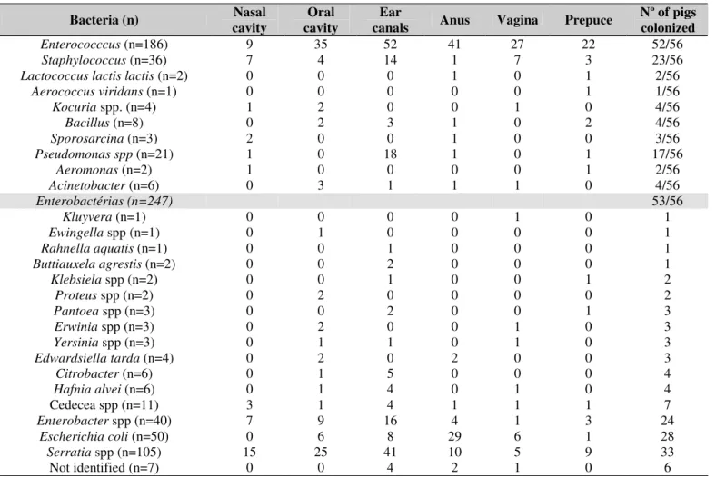
processing of swabs, 516 specimens were isolated, with 240 Gram-positive bacteria, among them 36 Staphylococcus and 186 Enterococcus identified. The methodology used also identified one strain of Aerococcus viridans, two Lactococcus
lactis subsp. Lactis, three Sporosarcina, four Kocuria spp. and eight Bacillus spp.. Gram-negative bacteria classification resulted in 276 strains, with two Aeromonas spp., six Acinetobacter, 21 Pseudomonas spp. and 247 (Table 1). Serratia spp. (n=105) and E. coli (n=50) were the GN species most prevalent in the study which were isolated from all body
sites investigated. Environment may interfere on microbiota
and involves factors such as water content, and the practice of
using poultry litter in agriculture for crops nutrient purposes
may not impact soil community of fecal indicator bacteria of
farms, as observed under drought conditions (25). Neither fecal
or water samples were examined in the present work, but
studies showed that only 10 bacterial isolates are required to determine the most common clones in fecal samples (42), one
can assume that the results showed may reflect the microbiota
of feral pigs studied. E. coli may colonize specific intestinal sections (16). In Germany, the study of with 21 hunted feral
pigs described clones of E. coli isolated from intestinal sections, all with different antimicrobial susceptibility profile
when compared with susceptible strains isolated from domestic
pigs (41). Strains of E. coli isolated in the present study had no hemolytic ability as observed in sheep blood agar, and contrary
to other observations that found only one E. coli from jejunum
portion of wild boar in Germany (41), and in accordance to
others, commensal E. coli strains rarely contain virulence genes
(10).
All Staphylococcus strains were submitted to classification
by Api system, resulting in S. simulans (1), S. saprophyticus (1), S. xylosus (1), S. warneri (1), S. epidermidis (1), S. haemolyticus (3), S. chromogenes (5), S. hyicus (7), S. sciuri (11), and five coagulase-negative Staphylococcus. Studies from
van Dijck and van de Voorde (45) found S. aureus and Poeta et
al. (34) did not isolate staphylococci from wild life boars from forests of Belgium and Portugal, respectivelly. However, the
Table 1. Bacteria isolated from feral pigs (Sus scrofa) from Brazilian Pantanal, frequency of body colonization and number of animals, in the period of 2007 and 2008.
Bacteria (n) Nasal
cavity
Oral cavity
Ear
canals Anus Vagina Prepuce
Nº of pigs colonized
Enterococccus (n=186) 9 35 52 41 27 22 52/56
Staphylococcus (n=36) 7 4 14 1 7 3 23/56
Lactococcus lactis lactis (n=2) 0 0 0 1 0 1 2/56
Aerococcus viridans (n=1) 0 0 0 0 0 1 1/56
Kocuria spp. (n=4) 1 2 0 0 1 0 4/56
Bacillus (n=8) 0 2 3 1 0 2 4/56
Sporosarcina (n=3) 2 0 0 1 0 0 3/56
Pseudomonas spp (n=21) 1 0 18 1 0 1 17/56
Aeromonas (n=2) 1 0 0 0 0 1 2/56
Acinetobacter (n=6) 0 3 1 1 1 0 4/56
Enterobactérias (n=247) 53/56
Kluyvera (n=1) 0 0 0 0 1 0 1
Ewingella spp (n=1) 0 1 0 0 0 0 1
Rahnella aquatis (n=1) 0 0 1 0 0 0 1
Buttiauxela agrestis (n=2) 0 0 2 0 0 0 1
Klebsiela spp (n=2) 0 0 1 0 0 1 2
Proteus spp (n=2) 0 2 0 0 0 0 2
Pantoea spp (n=3) 0 0 2 0 0 1 3
Erwinia spp (n=3) 0 2 0 0 1 0 3
Yersinia spp (n=3) 0 1 1 0 1 0 3
Edwardsiella tarda (n=4) 0 2 0 2 0 0 3
Citrobacter (n=6) 0 1 5 0 0 0 4
Hafnia alvei (n=6) 0 1 4 0 1 0 4
Cedecea spp (n=11) 3 1 4 1 1 1 7
Enterobacter spp (n=40) 7 9 16 4 1 3 24
Escherichia coli (n=50) 0 6 8 29 6 1 28
Serratia spp (n=105) 15 25 41 10 5 9 33
Not identified (n=7) 0 0 4 2 1 0 6
Antimicrobial susceptibility
Thirteen strains (36%) of Staphylococcus spp. were sensitive toward all drugs tested. The S. xylosus strain colonizing the prepuce of one animal showed multiple
resistance toward amoxicillin, penicillin, ampicillin and erythromycin (Table 3). Ampicillin was the most ineffective
drug against staphylococci with resistance observed in 17% of
strains followed of erythromycin (14%). Bagcigil et al. (8) showed that 38% S. aureus isolated from nasal cavity of pigs, dogs, horses and cattle were erythromycin resistant in
Dennmark, mostly animals living in farms and in frequent
contact with macrolid drugs, and all strains belonging to a
clonal group expressing the gene ermC. Armand-Lefevre et al.
(4) studying S. aureus in pig farmers found high resistance to erythromycin among the isolates from farmers (66%),
compared to controls (10% resistant), while 38% of the isolates
from pigs were intermediate resistant toward the drug. The
cause of staphylococci ampicillin and erythromycin resistance found the present study is to be investigated, since domestic
pigs were not investigated yet in the area investigated.
Data from 186 isolates of Enterococcus in the present study showed high sensibility to vancomycin (98%), ampicilin
(94%), tetracyclin (90%), penicillin G (83%), amoxicilin (70%)
and cephalotin (69%), and with high resistance toward
oxacillin (99%), clindamycin (83%) and cotrimoxazole (54%)
Enterococcus strains from feral pigs toward 11 antimicrobial drugs, observed higher resistance against erythromycin
(48,5%), tetracycline (44,8%) and ciprofloxacin (17,9%) and
lower resistance against ampicillin (3,7%), cloranphenicol
(4,5%), estreptomycin (6,7%) and kanamycin (9%). The results
in the present work with enterococci resistance toward
erythromycin was 13%, and lower than that observed against
the same drug in animals from Portugal (48,5%). Poeta et al. (34), observed 44,8% of tetracycline resistance among the
isolates, while in the present work the level of resistance was
practically insignificant (6%), while resistance against
ampicillin presented results compatible, with 6% resistance in
the present work against 3,7% in the Portuguese enterococci
isolates.
Table 3. Antimicrobial susceptibility, in percentage, of Gram-positive bacteria isolated from feral pig (Sus scrofa) of Brazilian pantanal, in the period of 2007 and 2008.
Enterococcus (n=186) Staphylococcus (n=36) Kocuria (n=4)
R I S R I S R I S
AMO 30 0 70 8 0 92 25 0 75
AMP 6 0 94 17 0 83 0 0 100
CFL 12 19 69 0 0 100 0 0 100
CFO 46 22 33 6 0 94 25 0 75
CLI 83 5 11 8 11 81 0 0 100
ERI 13 60 27 14 31 56 25 0 75
GEN 24 16 60 0 0 100 0 0 100
OXA 99 0 1 6 0 94 0 0 100
PEN 17 0 83 8 0 92 25 0 75
SUT 54 5 41 3 3 94 50 0 50
TET 6 4 90 0 0 100 0 0 100
VAN 0 2 98 0 0 100 0 0 100
The species E. faecalis is known as one of the main resistant against drugs from strains isolated from domestic pigs
in different countries (1, 20, 49). Enterococcus faecalis and E.
faecium present natural resistance to several antimicrobial drugs, including aztreonam, cotrimoxazole, clindamicin and
cephalosporins, and habitually, lower sensibility toward
aminoglycosides and penicillin G, moderate sensibility toward
ampicillin and cloranphenicol, but high sensibility toward
glycopeptides (22). Otherwise, when resistant to the last drugs
the Enterococcus represent an epidemiological risk, since the genes may be transferible to other bacteria (5). There is no reference to clindamycin resistance in enterococci isolated
from pigs.
The level of resistance toward cotrimoxazole in
enterococci was also discussed by others studying domestic
pigs. Aubry-Damon et al. (7) associated a predominance of enteric bacteria resistant to drugs, among them cotrimoxazole,
from pig farmer workers in France, and compared with isolates
from pigs. The strains isolated from controls (no pig farmers) were sensitive to cotrimoxazole, suggesting the transmission of
resistant bacteria for pig farmers.
Among 186 isolates from enterococci from feral pig of
Brazilian Pantanal, three strains presented intermediate profile
toward vancomycin. The plasmid gene vanA, responsible for the high resistance to this drug may be transferable to humans
and animals (36, 37). A study with E. faecalis and E. faecium isolated from humans and pigs in Dennmark showed that 17%
of the pigs isolates and only 1,5% from humans isolates were
characteristic of fecal coliforms (9). Enterococci may also change their antimicrobial profile according to environmental water contamination with antibiotic residue detection in surface water and groundwater from swine plant operations (38).
Both Lactococcus lactis lactis strains presented sensitivity to most antibiotics tested, and one strain was resistant to clindamycin and other intermediate toward cephoxitin. Aerococcus viridans strains were sensitive against all drugs, except toward oxacillin, which presented resistance profile.
Natural or intrinsic and acquired antibiotic resistance in enterococci was described as inherent characteristics of species of the genus or a consequence of insusceptibilities to physicochemical and environmental factors, but no mention about resistance to penicillin or their derivative is credited to enterococci unless overproduction of penicillin-binding protein (PBP) occurs (26). According to CASFM (Comité de l’Antibiogramme de la Société Française de Microbiogie) (13), enterococci may be a naturally oxacillin resistant bacteria. This is accordance with the results observed in this work, since virtually all enterococci strains presented resistance toward oxacillin. All together, these data indicate that the enterococci oxacillin resistance phenotype may be considered a stable genetic trait in this species isolated from feral pigs in Brazilian Pantanal, and never observed by others before. This alleged enterococci oxacillin resistance genetic trait deserves more investigations.
According to Table 2, for GN bacteria the susceptibility towards drugs tested showed that the bacteria with highest resistance was Serratia marcescens, with 98% resistance toward Cephalotin, 66% toward amoxicillin+clavulanic acid and 60% toward ampicillin. E. coli was the most sensitive with 10% resistance profile toward AMC and 7% toward CFL. Schierack and colleagues (41) found no resistance among E. coli strains from feral pigs, while strains from domestic pigs were more resistant. GN bacteria in the gut can present different profile toward drugs, resistance against tetracycline was higher than other drugs in E. coli (18). Taking the data from resistance profile of GN bacteria in the study and with other published data in domestic pigs, one can infer that anthropomorphic pressure in Brazilian Pantanal environment is low. Others have observed that cattle-ranching activities may favor feral pigs and the current anthropogenic changes in the landscape could lead to changes in competitive dynamics between these animals and native species (15), but exchange of bacteria and influence of such activity on resistance profile of microorganisms is yet to be studied. Cattle are considered the primary reservoir of E. coli O157 (28), but fecal
shedding by other domestic livestock and wildlife has been
described (35, 39) and cattle-ranching and agriculture practice for food purposes activities in California could be affected by surface water visited by feral pigs and, consequently, containing pathogenic bacteria (23, 24).
Table 2. Antimicrobial susceptibility, in percentage, of 233 Enterobacteriaceae isolated from feral pig (Sus scrofa) of Brazilian Pantanal, in the period of 2007 and 2008.
Serratia marcescens (n=97)
Enterobacter spp.
(n=35) Cedecea (n=11) outras (n=48) E. coli (n=42)
R I S R I S R I S R I S R I S
AMC 66 19 15 20 9 71 27 0 73 10 23 67 10 7 83
AMP 60 15 25 20 20 60 27 0 73 31 13 56 2 0 98
CFL 98 0 2 29 14 57 36 9 55 27 17 56 7 17 76
CFO 8 12 79 17 11 71 9 9 82 29 6 65 2 5 93
CIP 1 4 95 3 11 86 0 9 91 4 13 83 0 7 93
CLO 2 11 87 3 17 80 0 9 91 2 15 83 0 5 95
ENO 2 18 80 3 26 71 0 9 91 2 29 69 5 10 86
GEN 1 0 99 6 0 94 0 0 100 8 10 81 0 5 95
SUT 2 0 98 0 14 86 0 0 100 21 2 77 0 0 100
TET 51 30 20 3 3 94 9 0 91 8 13 79 0 0 100
In the literature no information is available on microbiota
of feral pigs from Brazilian Pantanal. The environmental aspect emphasized in this work is based on the necessity to know the
drug resistance of this microbiota to propose a possible
interference of human activities in that environment. The study
presented may reveal that controversial aspects on bacterial
resistance towards drugs may occur specially in areas with
association of heavy pressure of livestock and agricultural
activities, or natural resistance is inherent to wild microorganisms associated to wild animals. However, most of
the isolates were sensitive to drugs tested in this study and the
results may reflect a regional characteristic of Brazilian
wetlands like Pantanal, with cyclic water seasons reflecting on
drug profile of microorganisms living in that environment,
suggesting dispersion of residues of any kind of contamination,
including antimicrobial drugs.
ACKNOWLEDGEMENTS
To FAPERJ (E-26 103.097/2008-JCNE), UNIDERP and
CNPq for financial support for OVM and FAPERJ for grant to
the first author. To Dr. Luis Simeão do Carmo from Federal
University of Minas Gerais, Brazil, for supplying TSST-1
immunodifusion kits. To Fiocruz-RJ for supplying ATCC strains. To technical support of M.L.B. Amaral and G.N.
Teixeira from LSA/UENF.
REFERENCES
1. Aarestrup, F.M.; Agerso, Y.; Gerner–Smidt, P.; Madsen, M.; Jensen, L. B. (2000). Comparison of antimicrobial resistance phenotypes and resistance genes in Enterococcus faecalis and Enterococcus faecium from humans in the community, broilers, and pigs in Denmark. Diagn. Microbiol. Infect. Dis. 37, 127–137.
2. Ali Abadi, F.S.; Lees, P. (2000). Antibiotic treatment for animals: effect on bacterial population and dosage regimen optimization. Int. J. Antimicrob. Agents 14, 307-313.
3. Andrews, R.E.; Johnson, W.S; Guard, A.R; Marvin, J.D. (2004). Survival
of Enterococci and Tn916-like conjugative transposons in soil. Can. J. Microbiol. 50, 957–966.
4. Armand-Lefevre, A.; Ruimy, R.; Andremont, A. (2005). Clonal comparison of Staphylococcus aureus isolates from healthy pig farmers, human controls, and pigs. Emerg. Infect. Dis. 11 (5), 711-714.
5. Arthur, M.; Courvalin, P. (1993). Genetics and mechanisms of glycopeptide resistance in enterococci. Antimicrob. Agents Chemother. 37, 1536–1571.
6. Atwill, E.R.; Sweitzer, R.A.; Pereira, M.G.; Gardner, I.A.; van Vuren D.; Boyce W.M. (1997). Prevalence of and associated risk factors for shedding Cryptosporidium parvum oocysts and Giardia cysts within feral pig populations in California. Appl. Environ. Microbiol. 63, 3946–3949. 7. Aubry-Damon, H.; Grenet, K.; Sall-Ndiaye, P.; Che, D.; Cordeiro, E.;
Bougnoux, M.-E.; Rigaud, E.; Le Strat, Y.; Lemanissier, V.; Armand-Lefèvre, L.; Delzescaux, D.; Desenclos, J.C.; Liénard, M.; Andremont, A. (2004). Antimicrobial Resistance in Commensal Flora of Pig Farmers.
Emerg. Infect. Dis. 10 (5), 873-879.
8. Bagcigil, F.A.; Moodley, A.; Baptiste, K.E; Jensen, V.F; Guardabassi, L. (2007). Occurrence, species distribution, antimicrobial resistance and clonality of methicillin- and erythromycin-resistant staphylococci in the nasal cavity of domestic animals. Vet. Microbiol. 121, 307–315. 9. Beers, M.H.; Berkow, R. (1997). The Merck manual of diagnosis and
therapy. Merck & Co., Whitehouse Station, N.J.
10. Boerlin, P.; Travis, R.; Gyles, C.L.; Reid-Smith, R.; Janecko, N.; Lim, H.; Nicholson, V.; McEwen, S.A.; Friendship, R.; Archambault, M. (2005). Antimicrobial resistance and virulence genes of Escherichia coli
isolates from swine in Ontario. Appl. Environ. Microbiol. 71, 6753–6761. 11. Camargo, I.L.B.C.; Gilmore, M.S.; Darini, A.L.C. (2006). Multilocus sequence typing and analysis of putative virulence factors in vancomycin-resistant and vancomycin-sensitive Enterococcus faecium isolates from Brazil. Clin. Microbiol. Infect. 12 (11), 1123-1130.
12. Cardoso, H.F.T.; Carmo, L.S.; Silva, N. (2000).Detection of toxic shock syndrome toxin by Staphylococcus aureus strains isolated from bovine mastitis. Arq. Bras. Med. Vet. Zootec. 52 (1), 07-10.
13. CASFM (2007). Comité de l’Antibiogramme de la Société Française de Microbiogie: Recommandations 2007.
14. de Neeling, A.J.; van den Broek, M.J.M.; Spalburg, E.C.; Van Santen-Verheuvel, M.G.; Dam-Deisz, W.D.C.; Boshuizen, H.C.; Van de Giessen, A.W.; van Duijkeren, E.; Huijsdens, X.W. (2007). High prevalence of methicillin resistant Staphylococcus aureus in pigs. Vet. Microbiol. 122, 366–372.
16. Dixit, S.M.; Gordon, D.M.; Wu, X.Y.; Chapman, T.; Kailasapathy, K.; Chin, J.J. (2004). Diversity analysis of commensal porcine Escherichia coli associations between genotypes and habitat in the porcine gastrointestinal tract. Microbiol. 150, 1735–1740.
17. Furuya, E.Y.; Lowy, F.D. (2006). Antimicrobial-resistant bacteria in the community setting. Nature. 4, 36-45.
18. Guerra, B.; Junker, E.; Schroeter, A.; Malorny, B.; Lehmann, S.; Helmuth, R. (2003). Phenotypic and genotypic characterization of antimicrobial resistance in German Escherichia coli isolates from cattle, swine and poultry. J. Antim. Chem. 52, 489–492.
19. Hagedorn, C.; Robinson, S.L.; Filtz, J.R.; Grubbs, S.M.; Angier, T.A.; Beneau, R.B. (1999). Determining sources of fecal pollution in a rural Virginia watershed with antibiotic resistance patterns in fecal streptococci. Appl. Environ. Microbiol. 65, 5522–5531.
20. Hammerum, A.M.; Lester, C.H.; Neimann, J.; Porsbo, N.J.; Olsen, K.E.P.; Jensen, L.B.; Emborg, H.D.; Wegener, H.C.; Frimodt-Moller, N. (2004). A vancomycin-resistant Enterococcus faecium isolate from a Danish healthy volunteer, detected 7 years after the ban of avoparcin, is possibly related to pig isolates. J. Antimicrob. Chemother. 53, 547–549. 21. Harihan, R.; Weinstein, R.A. (1996). Enterobacteriaceae. In: Mayhall,
C.G.(ed.) Hospital epidemiology and infection control. Williams & Wilkins, Baltimore. p.345-366.
22. Huycke, M.M.; Sahm, D.F.; Gilmore, M.S. (1998). Multiple-drug resistant Enterococci: the nature of the problem and an agenda for the future. Emerg. Infect. Dis. 4, 239-249.
23. Jay, M.T.; Cooley, M.; Carychao, D.; Wiscomb, G.W.; Sweitzer, R.A.; Crawford-Miksza, L.; Farrar, J.A.; Lau, D.K.; O’Connell, J.; Millington, A.; Asmundson, R.V.; Atwill, E.R.; Mandrell, R.E. (2007). Escherichia coli O157:H7 in Feral Swine near Spinach Fields and Cattle, Central California Coast. Em. Infec. Dis. 13(12), 1908-1911.
24. Jay, M.T.; Wiscomb, G.W. (2008). Food safety risks and mitigation strategies for feral swine (Sus scrofa) near agriculture fields. Proc. 23rd Vertebr. Pest Conf. (R. M. Timm and M. B. Madon, Eds.) Published at Univ. of Calif., Davis. p. 21-25.
25. Jenkins, M.B.; Endale, D.M.; Schomberg, H.H.; Sharpe, R.R. (2006). Fecal bacteria and sex hormones in soil and runoff from cropped watersheds amended with poultry litter. Sci. Total Environ. 358, 164– 177.
26. Klare, I.; Konstabel, C.; Badstübner, D.; Werner, G.; Witte, W. (2003). Occurrence and spread of antibiotic resistances in Enterococcus faecium.
Int. J. Food Microbiol. 88, 269– 290.
27. Krumperman, P.H. (1983). Multiple antibiotic resistance indexing of
Escherichia coli to identify high-risk sources of fecal contamination of foods. Appl. Environ. Microbiol. 46, 165–170.
28. LeJeune, J.T.; Besser, T.E.; Rice, D.H.; Berg, J.L.; Stilborn, R.P.; Hanco, D.D. (2004). Longitudinal study of fecal shedding of Escherichia coli
O157:H7 in feedlot cattle: predominance and persistence of specific
clonal types despite massive cattle population turnover. Appl. Env. Microbiol. 70(1), 377-384.
29. Mendonça-Hagler, L.C.; Hagler, A.N. (1991). Microbiologia aquática. In: Roitman, I. et al. (eds.) Tratado de Microbiologia vol. II.Microbiologia Ambiental. Ed. Manole.
30. Nagase, N.; Sasaki, A.; Yamashita, K.; Shimizu, A.; Wakita, Y.; Kitai, S.; Kawano, J. (2002). Isolation and species distribution of Staphylococci from animal and human skin. J. Veter. Med. Science 64 (3), 245-250. 31. NCCLS 2003. Performance Standards for Antimicrobial Disk
Susceptibility Tests, Approved Standard, 8th ed. (M2-A8).
32. Parveen, S.; MurphyreeR.L.; Edmiston, L.; Kaspar, C.W.; Portier, K.M.; Tamplin, M.L. (1997). Association of multiple-antibiotic-resistance profiles with point and nonpoint sources of Escherichia coli in Apalachicola Bay. Appl. Env. Microbiol. 63(7), 2607-2612.
33. Pavlov, P.M. (1988). Health risks to humans and domestic livestock posed by feral pigs (Sus scrofa) in North Queensland. Robert Wicks Research Station, Australia. Proceedings of 13th Vertebrate Pest Conference, University of California, Davis. p.141-144.
34. Poeta, P.; Costa, D.; Igrejas, G.; Rodrigues, J.; Torres C. (2007). Phenotypic and genotypic characterization of antimicrobial resistance in faecal enterococci from wild boars (Sus scrofa). Vet. Microbiol. 125, 368-374.
35. Rice, D.H.; Hancock, D.D.; Besser, T.E. (2003). Faecal culture of wild animals for Escherichia coli O157:H7. Vet. Rec. 152, 82-83.
36. Rice, L.B.; Carias, L.L.; Donsey, C.L.; Rudin, S.D. (1998). Transferable plasmid-mediated VanB-type glycopeptide resistance in Enterococcus faecium. Antimicrob. Agents Chemother. 42, 963-964.
37. Rosato, A.; Pierre, J.; Billot-Klein, D.; Buu-Hoi, A.; Gutmann, L. (1995). Inducible and constitutive expression of resistance to glycopeptide and vancomycin dependence in glycopeptide-resistant Enterococcus avium. Antimicrob. Agents Chemother. 39, 830-833.
38. Sapkota, A.R.; Curriero, F.C.; Gibson, K.E.; Schwab, K.J. (2007). Antibiotic-resistant enterococci and fecal indicators in surface water and groundwater impacted by a concentrated swine feeding operation.
Environ. Health Persp. 115 (7), 1040-1045.
39. Sargeant, J.M.; Hafer, D.J.; Gillespie, J.R.; Oberst, R.D.; Flood, S.J. (1999). Prevalence of Escherichia coli O157:H7 in white-tailed deer sharing rangeland with cattle. J Am Vet Med Assoc. 215, 792–794. 40. Sayah, R.S.; Kaneene, J.B.; Johnson, Y.; Miller, R. (2005). Patterns of
antimicrobial resistance observed in Escherichia coli isolates obtained from domestic- and wild-animal fecal samples, human septage, and surface water. Appl. Env. Microbiol. 71(3), 1394-1404.
41. Schierack, P.; Römer, A.; Jores, J.; Kaspar, H.; Guenther, S.; Filter, M.; Eichberg, J.; Wieler, L.H. (2009). Isolation and characterization of intestinal Escherichia coli clones from wild boars in Germany. Appl. Env. Microbiol. 75(3), 695–702.
diversity of Escherichia coli colonizing stools and urinary tracts of young girls. Infect. Immun. 70, 1225–1229.
43. Sengeløv, G.; Agersø, Y.; Halling-Sørensen, B.; Baloda, S.B.; Andersen, J.S.; Jensen, L.B. (2003). Bacterial antibiotic resistance levels in Danish farmland as a result of treatment with pig manure slurry. Environ. Int. 28, 587-595.
44. Stepanovic, S.; Vukovic, D.; Trajkovic, V.; Samardzic, T.; Cupic, M.; Svabic-Vlahovic, M. (2001). Possible virulence factors of Staphylococcus sciuri. FEMS Microbiol. Lett. 199, 47-53.
45. van Dijck, P.J.; Van De Voorde, H. (1979). Course of antibiotic sensitivities in Escherichia coli and Staphylococcus aureus from animals.
Zentralbl. Bakteriol. [B]. 169 (5-6), 519-529.
46. van Duijkeren, E.; Ikawaty, R.; Broekhuizen-Stins, M.J.; Jansen, M.D.; Spalburg, E.C.; De Neeling, A.J.; Allaart, J.G.; Van Nes, A.; Wagenaar, J.A.; Fluit, A.C. (2008). Transmission of methicillin-resistant
Staphylococcus aureus strains between different kinds of pig farms. Vet. Microbiol. 126 (4), 383-389.
47. von Holy, A.; Holzapfel, W.H.; Dykes, G.A. (1992). Bacterial populations associated with Vienna sausage packing. Food Microbiol. 9, 45-53.
48. Vuong, C.; Götz, F.; Otto, M. (2000). Construction and characterization of an agr deletion mutant of Staphylococcus epidermidis. Infect. Immun. 68, 1048-1053.
49. Wegener, H.C. (2003). Antibiotics in animal feed and their role in resistance development. Curr Opin Microbiol. 6, 439–445.
50. Wiggins, B.A. (1996). Discriminant analysis of antibiotic resistance patterns in fecal streptococci, a method to differentiate human and animal sources of fecal pollution in natural waters. Appl. Environ. Microbiol. 62, 3997–4002.
51. Wiggins, B.A.; Andrews, R.W.; Conway, R.A.; Corr, C.L.; Dobratz, E.J.; Dougherty, D.P.; Eppard, J.R.; Knupp, S.R.; Limjoco, M.C.; Mettenburg, J.M.; Rinehardt, J.M.; Sonsino, J.; Torrijos, R.L.; Zimmerman, M.E. (1999). Use of antibiotic resistance analysis to identify nonpoint sources of fecal pollution. Appl. Environ. Microbiol. 65, 3483–3486.