Phylogenetic grouping and pathotypic comparison of urine and fecal
Escherichia coli
isolates from children with urinary tract infection
Masoumeh Navidinia
1, Shahin Najar Peerayeh
1, Fatemeh Fallah
3, Bita Bakhshi
1,
Raheleh Sadat Sajadinia
21
Bacteriology Department, Tarbiat Modarres University, Tehran, Iran. 2
Shahid Beheshti University of Medical Sciences, Tehran, Iran. 3
Pediatric Infection Research Center, Mofid Childrens’ Hospital, Shahid Beheshti University of Medical Sciences, Tehran, Iran.
Submitted: December 20, 2012; Approved: September 9, 2013.
Abstract
The aim of this study was to investigate the phylogenetic background and to assesshlyD(involved in the secretion of haemolysin A) andintI1(encoding a class 1 integrase) inEscherichia coliisolates de-rived from urinary and fecal specimens. A total of 200E. coliisolates was collected from patients presenting with urinary tract infection (UTI) during September 2009 to September 2010 and screened forhlyDandintI1genes by polymerase chain reaction (PCR). Phylogenetic analysis showed thatE. coliis composed of four main phylogenetic groups (A, B1, B2 and D) and that uropathogenicE. coli
(UPEC) isolates mainly belong to groups B2 (54%) and D (34%) whereas group A (44%) and D (26%) are predominant among commensalE. coliisolates. In this study,hlyDwas present in 26% of UPEC and 2% of commensalE. coliisolates. However, hemolytic activity was detected for 42% of UPEC and 6% of commensalE. coliisolates (p < 0.05).intI1gene was more frequently expressed in UPEC (24%) in comparison with commensal E. coli isolates (12%). Resistance to aztreonam, co-trimoxazole and cefpodoxime were frequently found among UPEC isolates whereas commensal
E. coli isolates were commonly resistant to co-trimoxazole, nalidixic acid and cefotaxime. Con-cluding, a considerable difference between UPEC and commensalE. coliisolates was observed re-garding their phylogenetic groups, presence of class 1 integron andhlyDgene, hemolysin activity and resistance pattern. The detection of class 1 integrons andhlyDgene was higher among UPEC compared with commensalE. coliisolates. These findings may contribute for a better understanding of the factors involved in the pathogenesis of UPEC.
Key words:Escherichia coli, urinary tract infection (UTI), phylogenetic typing groups,hlyD,intI1.
Introduction
Urinary tract infections (UTIs) currently rank among the most prevalent infectious diseases worldwide, with chronic and recurrent infections being especially problem-atic (Blango and Mulvey, 2010; Sabateet al., 2006). The primary etiologic agents associated with UTIs are strains of uropathogenicEscherichia coli(UPEC) (Sivick and Mo-bley, 2010). Nonetheless, UPEC isolates express a wide spectrum of virulence and fitness factors that aid in suc-cessful colonization of the mammalian urinary tract
(Man-ges et al., 2004). Although often categorized as extra-cellular pathogens, UPEC can in fact invade a number of host cell types, including the terminally differentiated su-perficial facet cells and less mature intermediate and basal epithelial cells that comprise the stratified layers of the bladder urothelium. Host cell invasion is proposed to facili-tate both the establishment and persistence of UPEC within the urinary tract (Johnsonet al., 2005; Mulveyet al., 2000).
Extra-intestinal pathogenic and commensal E. coli
typically differ in phylogenetic group and virulence
utes. Previous studies have shown that pathogenic extraintestinalE. coliisolates primary belong to phylogen-etic group B2 and, to a lesser extent, group D, whereas commensal E. coli isolates belong to groups A and B1. Moreover, pathogenic extraintestinal isolates harbour spe-cialized virulence factors,i.e., traits that confer pathogenic potential, which are infrequent among commensal isolates (Johnsonet al., 2001; Sabateet al., 2006).
Currently, about 50 different cassettes associated with resistance genes, can be found in different classes of integrons. An integron is a two component gene capture and dissemination system, first discovered due to their rapid dissemination of antibiotic resistance, which can be found in plasmids, chromosomes and transposons.The first component consists of a gene encoding a site specific recombinase along with a specific site for recombination, while the second component comprises fragments of DNA called gene cassettes which can be incorporated or shuffled. A cassette may encode genes for antibiotic resistance, al-though most genes in integrons are uncharacterized. Inte-grons act as receptors of antibiotic resistance cassettes (Kovalevskaya, 2002).
Hemolysin is a cytolytic protein toxin secreted by most hemolyticE. coliisolates. In addition of lysing eryth-rocytes, hemolysin is a toxin for a wide range of host cells which may result in inflammation, tissue injury, and im-paired host defenses. It should be mentioned that mono-cytes and granulomono-cytes are highly susceptible to hemolysin cytotoxicity, whereas lymphocytes are relatively resistant. Exposure of polymorphonuclear leukocytes (PMNLs) to hemolysin stimulates degranulation and releases of leuko-trienes accompanied by ATP; causes marked morphologic alterations; and impaired chemotaxis and phagocytosis (Johnson, 1991). Hemolysin production correlates closely with the toxicity of clinicalE. coli isolates for PMNLs. Hemolysin stimulates superoxide anion and hydrogen per-oxide release and oxygen consumption by renal tubular cells as well as histamine release from mast cells and basophils (Johnson, 1991).
The aim of this study was to determine the phylogen-etic type of uropathogenic and commensalE.coli, isolated from patients with UTI in Mofid Childrens` Hospital, Teh-ran, Iran. In addition, the prevalence of hemolytic activity, and the assessment ofhlyDgene (involved in hemolysin production) and of class 1 integron (a genetic element asso-ciated with antibiotic resistance) were also investigated, in order to provide additional information aboutE.coli viru-lence profiles.
Material and Methods
Specimens and patients
A total of 200E. coliisolates were analyzed from 100 children patients of both sexes (85% female, 15% male) aged between 2-12 years with UTI (70% pyelonephritis,
30% cystitis). Of these, 100 were derived from midstream clean catch urine and 100 were from stool specimens of the patients presenting with community acquired UTI who have attended the nephrology ward of Mofid Childrens’ Hospital, Tehran, Iran, during September 2009 to Septem-ber 2010. The project was approved by the local Ethics Committee for Human Researches.
Samples were derived from fresh midstream urine, cultured (0.01 mL) on MacConkey agar (Sisco Research Laboratories Pvt. Ltd., USA) as well as Sheep blood agar and incubated at 37 °C for 24 h. Urine bacteria included in this study were from cultures yielding > 105CFU/mL. Cul-tures with < 105CFU/mL were further investigated only if relevant history of fever, chills, flank pain, pyuria, antibi-otic intake, structural abnormalities, diabetes mellitus or any other immunocompromised state was present.
Specimens from stool samples were cultured on Trypticase soy agar (Kanto Chemical Co., Inc., Japan) with 5% sheep blood and MacConkey agar. The predominant isolate on each plate (one colony) and all morphologically distinct colonies were identified and stored for further anal-ysis, as described by Plos (1995) and Foxman (2002). Two-three colonies, cultured on sheep blood as well as on MacConkey agar, from each stool and urine sample, were selected for molecular examination (Morenoet al., 2006).
Antimicrobial susceptibility test
Susceptibility to nitrofurantoin (300 mg), ciproflo-xacin (5mg), nalidixic acid (30mg), amoxicillin (10mg), augmentin (30 mg), gentamicin (120mg), ceftazidime (30mg), cefpodoxime (10mg), aztreonam (30mg), imipe-nem (10mg), amikacin (30mg), co-trimoxazole (25mg) and cefotaxime (30mg) were determined by disc diffusion as-says (BBL Sensi-Disc, USA) modified by the Kirby-Bauer method using CLSI criteria (nonfastidious groupings M2-disk diffusion M100). For the purpose of analysis, in-termediate susceptibility was considered as susceptible (Schlageret al., 2002)
DNA extraction
DNA was extracted using the protocol described pre-viously (Sabarinathet al., 2011). The isolates were cultured on MacConkey agar plates for 24 h. One to two colonies were resuspended in 0.5 mL sterile distilled water. The cells were lysed by heating at 95 °C for 10 min and the supernatant was harvested by centrifugation at 12,000 rpm (8000g) for 5 min. The supernatant was used as the source of the template DNA.
PCR amplification
dATP, dCTP, dGTP, and dUTP; and 0.125 U ofTaqDNA polymerase (GENET BIO, Prime TaqTMDNA polymerase, type:G-1002, URL:www.genetbio.com).
Phylogenetic typing group
Phylogenetic grouping of theE. coliisolates was de-termined by a simple, rapid PCR- based technique (Clermontet al., 2000) that uses a combination of three DNA markers (chuA,yjaAand DNA fragment tspE4.C2), generating 279, 211 and 152-bp fragments, respectively. A triplex PCR was performed using the six primers in a single reaction. The results of these three amplifications allowed the classification ofE. coli isolates into one of the major phylogenetic groups: A, B1, B2 or D.E. colistrain RS218, which belongs to phylogenetic group B2, was used as a control (Dhakalet al., 2008).
Hemolytic activity and hlyD gene detection
E. coliisolates were inoculated on 5% sheep blood agar plates and incubated overnight at 37 °C. The plates were then examined for the presence of a partial or total hemolytic activity (alpha or beta) (Forbeset al., 2007).
PCR was performed using the hlyD gene (904 bp)
primers: F CTCCGGTACGTGAAAAGGAC:
(Tm = 55.4 °C), R GCCCTGATTACTGAAGCCTG: (Tm = 55.7 °C) in a single reaction (Rodriguez-Sieket al., 2005).
Class 1 integron detection
Isolates were analyzed by polymerase chain reaction (PCR) amplification techniques to determine whether a class 1 integron was present. Integrons were detected by PCR amplification of a class 1 integrase-specific fragment of theintI1gene. The primer sequences used wereintI1-(F:
GGTCAAGGATCTGGATTTGG, R:
ACATGCGTGTAAATCATCGTC) in a single reaction. PCR assay was performed for cycles as follows: 1 cycle of 12 min at 94 °C; 35 cycles of 1 min at 94 °C, 1 min at 57 °C, 2 min at 72 °C; 1 cycle of 10 min at 72 °C (Limet al., 2009).
Statistical analysis
Statistical analysis was performed by using the Fisher exact and chi-square tests. The threshold for statistical sig-nificance was a p value of < 0.05.
Results
Pattern of antimicrobial resistance among
Escherichia coliisolates
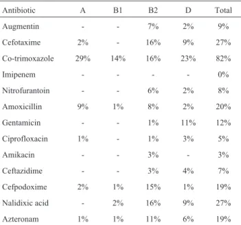
High-level resistance to azteronam (78%), co-trimoxa-zole (61%), cefpodoxime (48%) were found among UPEC while commensal E.coli isolates showed increased resis-tance to co-trimoxazole (82%), nalidixic acid (27%) and cefotaxime (27%). Resistance pattern of UPEC and commensalE. coliisolates were presented in Tables 1 and 2.
Multi-drug resistance which was defined as resis-tance to 3 or more classes or sub-classes of antibiotics (Canton and Ruiz-Garbajosa, 2011), was most commonly observed in UPEC (38%) compared with commensal E. coliisolates (22%).
Phylogenetic typing groups
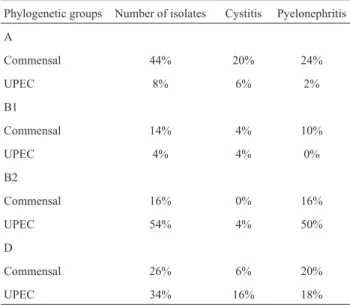
Phylogenetic groups A and D were commonly found among commensalE. coliisolates. However, UPEC iso-lates belonged to phylogenetic groups B2 and D,
predomi-Table 1- Antibiotic resistance pattern among phylogenetic groups in UropathogenicE.coliisolates from children with community acquired UTI.
Antibiotic A B1 B2 D Total
Augmentin - - 12% 2% 14%
Cefotaxime 3% 2% 39% 1% 45%
Co-trimoxazole - 1% 50% 10% 61%
Imipenem - - - - 0%
Nitrofurantoin - - 2% - 2%
Amoxicillin - - 16% - 16%
Gentamicin 2% - 19% 1% 22%
Ciprofloxacin - - 7% 1% 8%
Amikacin 1% 1% 5% 1% 8%
Ceftazidime - - 12% - 12%
Cefpodoxime 2% 6% 38% 2% 48%
Nalidixic acid - - 9% - 9%
Azteronam 1% - 48% 29% 78%
Table 2- Antibiotic resistance pattern among phylogenetic groups in commensalE.coliisolates from children with community acquired UTI.
Antibiotic A B1 B2 D Total
Augmentin - - 7% 2% 9%
Cefotaxime 2% - 16% 9% 27%
Co-trimoxazole 29% 14% 16% 23% 82%
Imipenem - - - - 0%
Nitrofurantoin - - 6% 2% 8%
Amoxicillin 9% 1% 8% 2% 20%
Gentamicin - - 1% 11% 12%
Ciprofloxacin 1% - 1% 3% 5%
Amikacin - - 3% - 3%
Ceftazidime - - 3% 4% 7%
Cefpodoxime 2% 1% 15% 1% 19%
Nalidixic acid - 2% 16% 9% 27%
nantly (Table 3). The results presented on Table 3 highlight a preliminary connection between pyelonephritis and phy-logenetic group B2 (p < 0.001).
Distribution of hemolytic activity and hlyD in UPEC and commensalE. coliisolate
hlyDwas detected in 26% of UPEC and 2% of com-mensal E. coli isolates, however, hemolytic activity was observed for 42% of UPEC and 6% of commensalE. coli
isolates (p < 0. 05) .
Distribution of intI1 in UPEC and commensalE. coli
isolates
intI1gene, which was significantly associated with pyelonephritis (22%) rather than cystitis (14%) (p < 0.05), was more frequently expressed in UPEC (24%) in compari-son with commensalE. coliisolates (12%).
Discussion
UTI is usually treated empirically without culture but it contributes for about 10-15% prolongation of hospital-ization due to the emergence of antimicrobial resistance among the causative bacteria, particularly UPEC isolates (Walter and Stamm, 2001). This may result in the spread of antibiotic resistant bacteria in the hospital and therefore, it has been suggested that more powerful antibiotics might better eliminate UPEC reservoirs and consequently reduce the incidence of chronic and recurrent UTIs among hospi-talized and outpatients (Kaperet al., 2004; Rodriguez-Siek
et al., 2005)
High incidence of co-trimoxazole resistance (61% for UPECs and 82% for commensalE. coliisolates) and of sus-ceptibility to imipenem(100% for both UPEC and com-mensal E. coliisolates) were detected. These data are in
agreement with the results of Farshadet al.(2008) forE. coli isolates obtained from children with community-acquired UTI. Thus, co-trimoxazole, which is a widely used for UTI treatment, has become nearly ineffective to treat UTI in this country.
In our study, different antibiotic resistance patterns were observed in UPEC compared with commensal iso-lates. Contrarily to the results of Alhajet al.(2007), lower resistance percentages to nalidixic acid (9%), amoxicillin (16%) and gentamicin (22%) was found among UPEC compared with commensal E.coli isolates. Nevertheless, resistance rates to ceftazidime (12%) and augmentin (14%) among UPEC isolates were in agreement with the studies of Limet al.(2009) with 47 nonrepeat E. coliisolates, col-lected from intensive care unit patients presented with UTI, in 5 public hospitals located in different areas of Malaysi. Consistent with Adegoke et al. (2011), our findings re-vealed that cefpodoxime and cefotaxime were less effective in UTI treatment than imipenem, nalidixic acid, ciprofloxacin, nitrofurantoin, augmentin and amikacin for all UPEC phylogenetic groups.
In a research by Morenoet al.(2006),E. coliisolates obtained from 150 patients presenting with acute uncom-plicated cystitis, acute pyelonephritis and urinary-source bacteraemia, revealed 21% and 18% resistance to quino-lones and fluoroquinoquino-lones, respectively. Recently, Shi-gemura et al. (2008) has reported the emergence of fluoroquinolone resistant E. coli responsible for UTI among patients attended at Kobe University Hospital, Ja-pan. In those studies a higher resistance to quinolones (27%) than to fluoroquinolones (5%) was observed among commensalE. coliisolates. However, they found that resis-tance to the two mentioned antibiotic classes was nearly the same among UPEC (9% and 8% respectively).
It should be considered that, in our study, resistance to amikacin in UPEC (8%) and commensalE. coliisolates (3%) was relatively lower, considering the 27% reported in a research conducted in Colombia by Villegaset al.(2004) onE. coliisolates obtained from hospitalized patients, in a study covering 62.3% of all general hospital beds in that country.
As previously noted, class 1 integrons were more prevalent than those of class 2 (Johnsonet al., 1998; Mu-hammadet al., 2011; Patti et al., 2008). Similar to a re-search by Colganet al.(2011), in our studyintI1gene was more frequently detected among UPEC than commensalE. coliisolates, which may contribute for the occurrence and transmission of MDR among UPEC isolates. Our results also showed that group B2 is the most frequent E. coli
phylogroup in UTI, as previously found (Johnson and Rus-so, 2002; Kovalevskaya, 2002; Mokadyet al., 2005). The UPEC isolates found in this studyprimarily belonged to one of two virulence groups (group B2 or D). Although a higher percentage of commensal isolates clustered into group A, a considerable proportion belonged to group D and this is
Table 3- Phylogenetic groups distribution of UPEC and fecalE.coli strains in patients with UTI.
Phylogenetic groups Number of isolates Cystitis Pyelonephritis
A
Commensal 44% 20% 24%
UPEC 8% 6% 2%
B1
Commensal 14% 4% 10%
UPEC 4% 4% 0%
B2
Commensal 16% 0% 16%
UPEC 54% 4% 50%
D
Commensal 26% 6% 20%
why a large proportion of commensal isolates were found to represent a potential human health threat, as well as the UPEC isolates (Burmanet al., 2003; Moulin-Schouleuret al., 2006).
Thus, our data indicate that group B2E. coliisolates are uncommon among commensal intestinal flora (16%); however, when present, they are highly virulent (Burmanet al., 2003; Moulin-Schouleur et al., 2006). In this study, only 42% of UPEC isolates had hemolytic activity, 26% of which carried hlyD gene. The relatively low percentage of
hlyDgene carriage rate, in the 100 UPEC isolates analyzed here, may be partially due to the relatively low percentage of B2 isolates (54%) detected in this study. Because B2 commensalE. coliisolates seem to have a privileged role in eliciting urinary tract infection, the intestinal normal flora would potentially act as a reservoir for developing UTI (Brangeret al., 2005). However, our findings challenge the “fecal urethral” pathway for the pathogenesis of UTI in children and instead support alternative routes of infection in this population (Johnsonet al., 2001a; Johnsonet al., 2001b).
Many studies have shown that urine isolates collec-tively differed dramatically from normal flora isolates with respect to phylogenetic background and virulence gene content profiles, suggesting an increased virulence poten-tial for the urine isolates (Clermontet al., 2000; Teraiet al., 2000; Vishalakshi, 2011). In fact, in our work, a consider-able difference between UPEC and commensalE. coli iso-lates was observed regarding their phylogenetic groups, presence of class 1 integron, carriage ofhly Dgene, hemo-lysin activity and resistance pattern.
Thus, we can conclude that some UPEC with differ-ent phylogenetic characteristics and virulence profiles are multiple drug resistant (MDR) isolates which make them a serious, challenging health problem. However it is reason-able to suppose that UPEC and commensalE. coliisolates might have similar fitness properties for adapting to an extraintestinal lifestyle, which, in turns, enable commensal
E. colito cause extraintestinal disease in humans as well as UPEC. As previously mentioned, commensalE. colimay potentially serves as a source or reservoir of virulence genes for human pathogenesis. Further research will be necessary to determine if commensalE. coliisolates can ac-tually overcome the hurdles necessary for human transmis-sion through the urethral route.
Acknowledgments
This work was supported bygrants from Faculty of Medical Sciences, Tarbiat Modares University, Tehran, Iran
Conflict of interest
Authors have no conflict of interest.
References
Adegoke Anthony A, Okoh Anthony I (2011) Prevalence, antibi-otic susceptibility profile and extended spectrum b -lacta-mase production among Escherichia coli from high vaginal swab (HVS). Afr J Pharm Pharacol 5:1287-1291.
Alhaj N, Mariana NS, Raha A, Ishak Z (2007) Prevalence of anti-biotic resistance among Escherichia coli from different sources in Malaysia. Res J Pharmacolo 1:44-49.
Blango MG, Mulvey MA (2010) Persistence of uropathogenic
Escherichia coli in the face of multiple antibiotics. Antimicrob Age Chemother 54:1855-1863.
Branger C, Zamfir O, Geoffroy S, Laurans G, Arlet G, Thien HV, Gouriou S, Picard B, Denamur E (2005) Genetic back-ground of Escherichia coli and extended-spectrum beta-lactamase type. Emerg Infect Dis11:54-61.
Burman WJ, Breese PE, Murray BE, Singh KY, Batal HA, Mac-kenzie TD, Ogle JW, Wilson ML, Reves RR, Mehler PS (2003) Conventional and molecular epidemiology of trime-thoprim-sulfamethoxazole resistance among urinary Esche-richia coliisolates. Am J Med 115:358-364.
Cantón R, Ruiz-Garbajosa P (2011) Co-resistance: an opportunity for the bacteria and resistance genes. Curr Opin Pharmacol 11:477-485.
Clermont O, Bonacorsi S, Bingen E (2000) Rapid and simple de-termination of the Escherichia coli phylogenetic group. Appl Environ Microbiol 66:4555-4558.
Colgan R, Williams M, Johnson JR (2011) Diagnosis and treat-ment of acute pyelonephritis in women. Am Fam Physician 84:519-526.
Dhakal BK, Kulesus RR, Mulvey MA (2008) Mechanisms and consequences of bladder cell invasion by uropathogenic
Escherichia coli. Eur J Clin Invest 38:2-11.
Farshad SH, Japoni A, Hosseini M (2008) Low distribution of integrons among multidrug resistantE.colistrains isolated from children with community -acquired urinary tract infec-tions in Shiraz, Iran. Pol j microbiol 57:193-198.
Forbes BA, Sahm DF, Weissfeld AS (2007) Bailey & Scott’s Di-agnostic Microbiology, Mosby Press.
Foxman B, Manning SD, Tallman P, Bauer R, Zhang L, Koopman JS, Gillespie B, Sobel JD, Marrs KF (2002) Uropathogenic
Escherichia coliAre More Likely than commensalE. colito be shared between heterosexual sex partners. Am J Epidemiol 156:1133-1140.
Johnson DE, Lockatell CV, Russell RG, Hebel JR, Island MD, Stapleton A, Stamm WE, Warren JW (1998) Comparison of
Escherichia colistrains recovered from human cystitis and pyelonephritis infections in transurethrally challenged mice. Infect Immun 66:3059-3065.
Johnson JR (1991) Virulence Factors inEscherichia coliUrinary Tract Infection. Clin Microbiol Rev 4:80-128.
Johnson JR, Delavari P, Kuskowski M, Stell AL (2001) Phylo-genetic distribution of extraintestinal virulence-associated traits inEscherichia coli. J Infect Dis 183:78-88.
Johnson JR, Stell A, Delavari P (2001) Canine feces as a reservoir of extraintestinal pathogenic Escherichia coli. Infect Im-mune 69:1306-14.
infec-tions in dogs and extraintestinal infecinfec-tions in humans. J In-fect Dis 183:897-906.
Johnson JR, O’Bryan TT, Sandberg T (2005) Phylogenetic and pathotypic comparison of concurrent urine and rectal Esche-richia coli isolatesfrom men with febrile urinary tract infec-tion. J Clin Microbiol 43:3895-3900.
Johnson JR, Russo TA (2002) UropathogenicEscherichia colias agents of diverse non- urinary tract extraintestinal infec-tions. J Infect Dis 186:859-864.
Kaper JB, Nataro JP, Mobley HL (2004) Pathogenic Escherichia coli. Nat Rev Microbiol 2:123-140.
Kovalevskaya NP (2002) Mobile Gene Cassettes and Integrons.
Mol Biolo 36:196-201.
Lim KT, Yasin R, Yeo CC, Puthucheary S, Thong KL (2009) Characterization of Multidrug Resistant ESBL-Producing
Escherichia coli Isolates from Hospitals in Malaysia. J Biomed Biotechno 2009:2009:165637.
Manges AR, Dietrich PS, Riley LW (2004) Multidrug-resistant
Escherichia coliclonal groups causing community-acquired pyelonephritis. Clin Infec Dis 38:329-334.
Mokady D, Gophna U, Ron EZ (2005) Extensive gene diversity in septicemic Escherichia coli strains. J Clin Microbiol 43:66-73.
Moreno E, Prats G, Sabate M, Perez T, Johnson JR, Andreu A (2006) Quinolone, fluoroquinolone and trimethoprim sulfamethoxazole resistance in relation to virulence determi-nants and phylogenetic background among uropathogenic Escherichia coli. J Antimicrob Chemother 57:204-211. Moreno E, Andreu A, Perez T, Sabatem M, Johnson JR, Prats G
(2006) Relationship betweenEscherichia colistrains caus-ing urinary tract infection in women and the dominant faecal flora of the same hosts. Epidemiol Infec 134:1015-1023. Moulin-Schouleur M, Schouler C, Tailliez P, Kao MR, Bree A,
Germon P, Oswald E, Mainil J, Blanco M, Blanco J (2006) Common virulence factors and genetic relationships be-tween O18: K1: H7Escherichia coliisolates of human and avian origin. J Clin Microbiol 44:3484-3492.
Muhammad I, Uzma M, Yasmin B, Mehmood Q, Habib B (2011) Prevalence of antimicrobial resistance and integrons in
Escherichia Colifrom Punjab, Pakistan. Braz J Microbiol 42:462-466.
Mulvey MA, Schilling JD, Martinez JJ, Hultgren SJ (2000) Bad bugs and beleaguered bladders: interplay between uropathogenic Escherichia coli and innate host defenses. Proc Nat Acad Sci USA 97:8829-8835.
Patti G, Mannini A, Balistreri M, Schito AM (2008) Virulence factors in urinary Escherichia coli strains: phylogenetic
background and quinolone and fluoroquinolone resistance. J Clin Microbiol 46:480-487.
Plos K, Connel H, Jodal U (1995) Intestinal carriage of P
fimbriatedEscherichia coliand the susceptibility to urinary tract infection in young children. J Infect Dis 171:625-631. Rodriguez-Siek KE, Giddings CW, Doetkott C, Johnson TJ,
Fakhr MK, Nolan LK (2005) Comparison of Escherichia coli isolates implicated in human urinary tract infection and avian colibacillosis. Microbiol 151:2097-2110.
Rodriguez-Siek KE, Giddings CW, Doetkott C, Johnson TJ, Nolan LK (2005) Characterizing the APEC pathotype. Vet Res 36:241-256.
Sabarinath A, Tiwari KP, Deallie C, Belot G, Vanpee G, Matthew V, Sharma R, Hariharan H (2011) Antimicrobial Resistance and Phylogenetic Groups of CommensalEscherichia Coli
Isolates from Healthy Pigs in Grenada. Web Med Central 42:1-10.
Sabate M, Moreno E, Perez T, Andreu A, Prats G (2006) Pathoge-nicity island markers in commensal and uropathogenic
Escherichia coliisolates. Clin Microbiol Infect 12:880-886. Schlager TA, Hendley JO, Bell AL , Whittam TS (2002) Clonal
Diversity ofEscherichia coliColonizing Stools and Urinary Tracts of Young Girls. Infect Immun. 70:1225-9.
Shigemura K, Arakawa S, Miura T, Nakano Y, Tanaka K, Fujisa-wa M (2008) Significance of fluoroquinolone resistant
Escherichia coliin urinary tract infections. Jpn J Infect Dis 61:226-228.
Sivick KE, Mobley HLT (2010) Waging war against uropathogenic Escherichia coli: winning back the urinary tract. Infect Immun 78:568-585.
Terai A, Ishitoya S, Mitsumori K, Ogawa O (2000) Molecular epi-demiological evidence for ascending urethral infection in acute bacterial prostatitis. J Urol 164:1945-1947.
Villegas MV, Correa A, Perez F, Miranda MC, Zuluaga T, Quinn JP (2004) Prevalence and characterization of extended-spectrumb-lactamases inKlebsiella pneumoniaeand
Esche-richia coli isolates from Colombian hospitals. Diag
Microbiol Infect Dis 49:217-222.
Vishalakshi B (2011) Detection of Virulence Markers of Uro-pathogenic Escherichia coli from Urinary Tract Infection, Karnataka, Banglore, 125 p (Department of Microbiology Mysore Medical College and Research Institute).
Walter E, Stamm MD (2001) An epidemic of urinary tract infec-tions. N Eng J Med 345:1055-1057.