Objective: To describe the clinical profile and treatment of Brazilian Guarani indigenous children aged less than five years hospitalized for acute lower respiratory infection (ALRI), living in villages in the states from Rio de Janeiro to Rio Grande do Sul. Methods: Of the 234 children, 23 were excluded (incomplete data). The analysis was conducted in 211 children. Data were extracted from charts by a form. Based on record of wheezing and x-ray findings, ALRI was classified as bacterial, viral and viral-bacterial. A bivariate analysis was conducted using multinomial regression. Results: Median age was 11 months. From the total sample, the ALRI cases were classified as viral (40.8%), bacterial (35.1%) and viral-bacterial (24.1%). It was verified that 53.1% of hospitalizations did not have clinical-radiological-laboratorial evidence to justify them. In the multinomial regression analysis, the comparison of bacterial and viral-bacterial showed the likelihood of having a cough was 3.1 times higher in the former (95%CI 1.11-8.70), whereas having chest retractions was 61.0% lower (OR 0.39, 95%CI 0.16-0.92). Comparing viral with viral-bacterial, the likelihood of being male was 2.2 times higher in the viral (95%CI 1.05-4.69), and of having tachypnea 58.0% lower (OR 0.42, 95%CI 0.19-0.92).
Conclusions: Higher proportion of viral processes was identified, as well as viral-bacterial co-infections. Coughing was a symptom indicative of bacterial infection, whereas chest retractions and tachypnea showed viral-bacterial ALRI. Part of the resolution of non-severe ALRI still occurs at hospital level; therefore, we concluded that health services need to implement their programs in order to improve indigenous primary care.
Keywords: Respiratory tract infections; Pneumonia; Indigenous population; Child.
Objetivo: Descrever o perfil clínico e o tratamento realizado nas crianças da etnia Guarani menores de cinco anos hospitalizadas por infecção respiratória aguda baixa (IRAB), residentes em aldeias nos estados do Rio de Janeiro ao Rio Grande do Sul.
Métodos: Das 234 crianças, 23 foram excluídas (dados incompletos), sendo analisadas 211. Os dados foram extraídos dos prontuários por meio de formulário. Com base no registro de sibilância e padrão radiológico, a IRAB foi classificada em: bacteriana, viral e viral-bacteriana. Foi utilizada regressão multinomial para análise bivariada. Resultados: A mediana de idade foi de 11 meses. Do total da amostra, os casos de IRAB foram assim distribuídos: viral (40,8%), bacteriana (35,1%) e viral-bacteriana (24,1%). Verificou-se que 53,1% das hospitalizações não possuíam evidências clínico-radiológico-laboratoriais que as justificassem. Na análise de regressão multinomial, ao comparar a IRAB bacteriana com a viral-bacteriana, a chance de ter tosse foi 3,1 vezes maior na primeira (intervalos de 95% de confiança — IC95% 1,11-8,70) e de ter tiragem 61,0% menor (Odds Ratio — OR 0,39, IC95% 0,16-0,92). Na comparação da IRAB viral com a viral-bacteriana, a chance de ser do sexo masculino foi 2,2 vezes maior na viral (IC95% 1,05-4,69) e de ter taquipneia, 58,0% menor (OR 0,42, IC95% 0,19-0,92) na mesma categoria.
Conclusões: Identificou-se maior proporção de processos virais do que processos bacterianos, bem como a presença de infecção viral-bacteriana. A tosse foi um sintoma indicativo de infecção bacteriana, enquanto a tiragem e a taquipneia apontaram infecção viral-bacteriana. Parte da resolubilidade da IRAB não grave ocorreu em âmbito hospitalar; portanto, propõe-se que os serviços priorizem ações que visem à melhoria da assistência à saúde indígena na atenção primária.
Palavras-chave: Infecções respiratórias; Pneumonia; População indígena; Criança.
ABSTRACT
RESUMO
*Corresponding author. E-mail: patigsouza40@gmail.com (P.G. Souza). aUniversidade Federal do Rio de Janeiro, Rio de Janeiro, RJ, Brazil. bFundação Oswaldo Cruz, Rio de Janeiro, RJ, Brazil.
Received on April 1st, 2017; accepted on June 22, 2017; available online on March 22, 2018.
ACUTE LOWER RESPIRATORY INFECTION IN
GUARANI INDIGENOUS CHILDREN, BRAZIL
Infecção respiratória aguda baixa em crianças indígenas Guarani, Brasil
Patricia Gomes de Souza
a,*, Andrey Moreira Cardoso
b, Clemax Couto Sant’Anna
a,
INTRODUCTION
At a global level, acute respiratory infections (ARIs) are the main cause of morbimortality among children. In developing countries, pneumonias — one of the main acute lower respi-ratory infections (ALRIs)1 — are in charge of 20 to 40% of hospitalizations of children aged less than 5 years, and of 20% of deaths in the same age group.2
In infant health, ALRIs are mainly represented by com-munity-acquired pneumonia (CAP)1 and acute viral bron-chiolitis (AVB).3 Even though most ALRIs are caused by a virus, the viral-bacterial infection is observed in more than one fourth of the hospitalized children.4 In 83 Guarani vil-lages, located in the South and Southeast of Brazil, the pro-portion of hospitalizations due to the same cause was 77.6% in children aged less than five years, and 83.4% in children aged less than one year.5 The high prevalence of wheezing found among Guarani children who were hospitalized by ALRI6 suggests high frequency of viral etiology,3 as well as of viral-bacterial infection.7
In developing countries, because of the difficulties to conduct laboratory tests to identify the viruses,8 the clini-cal-radiological criteria are the most used to distinguish the viral and the bacterial infections.1 It is necessary to know the profile of the ALRIs caused by different agents to formulate the treatment and reduce the unnecessary use of antiobiot-ics.9,10 This study aimed at describing the clinical profile and the treatment carried out with Guarani children hospitalized because of ALRI in the South and Southeast of Brazil, as well as at gathering evidence to assist the presumption of the eti-ology and the therapeutic decision-making in contexts with limited diagnostic resources.
METHOD
A cross-sectional study about the clinical profile and treatment of Guarani indigenous children aged less than five years hos-pitalized because of ALRI (2007-2008), living in 83 villages in the states of Rio de Janeiro down to Rio Grande do Sul. Indigenous primary health care is provided by a multidisci-plinary indigenous health team managed by the Indigenous Health Care Subsystem, component of the Unified Health System (SUS). Hospitalizations are referred to the SUS net-work outside the villages by the Reference and Counter-Reference System.11
The hospitalizations were recruited based on the surveil-lance system of a case-control study,12 and data extraction of the medical charts was conducted by using a standardized form. The methodological details are described in other articles.5,12
The variables are:
• Demographic and hospitalization seasons of the year: age group (0 to <2 months, 2 to 23 months and 24 to 59 months), sex, region of residence (South or Southeast) and seasons of the year (fall, winter, spring and summer).
• Clinics: hospital complexity in the National Record of Health Institutions, categorized in high and medium, based on the higher level of complexity in the hospital.
• Diagnostic hypothesis (DH) of the assistant physician used for the diagnostic classification of the study, based on the etiological hypotheses: CAP1=bacterial ALRI, AVB3=viral ALRI, and CAP+AVB = viral-bacterial ALRI.
Signs and symptoms assessed: cough; fever; dyspnea; tachypnea; rales; retractions; wheezes; signs of severity; simultaneous presence of other conditions, such as recur-rent wheezing — asthma or wheezing infant, cardiopathy and repetitive pneumonia. The classification of nutritional status was conducted based on “weight/age”, standardized in Z score: adequate weight for age (≥Z score -2 and ≤Z score +2) and low weight for age (<Z score -2). The clinical, labo-ratory and radiological evidence was used to indicate hospi-talization1,3. The laboratory evidence was based on leukocyte and rod count, anemia (hemoglobin <11 g/dL), peripheral oxygen saturation (SpO2) and blood culture. The radiolog-ical pattern was classified as:
• Viral: interstitial opacity, atelectasis, lung hyperinflation.
• Bacterial: alveolar opacity, condensation, pleural effu-sion, pneumatocele.1
• No description.
The treatment was assessed as: antibiotics — adequate dose for weight, adequate interval between doses, recommended duration, beginning of antibiotic therapy in relation to hos-pital admission (up to four hours, after four hours), change of antibiotics during hospital stay —, systemic and/or inhaled corticosteroids, nebulization with bronchodilator, oxygen ther-apy and adaptation of treatment.1,3 The following variables were also assessed: time of hospitalization (1-3 days, 4-6 days, 7-14 days and 15 days or more), re-hospitalization of the child because of the same episode (up to 14 days after discharge), complications and death. The variables with no record in the chart were defined as absent.
Based on the clinical-radiological criteria of the protocols,1,3 the diagnostic classification of the study was established as follows:
125
without description of the report or that the examina-tion was not taken.
• Viral ALRI: with record of wheezing and thoracic X-ray with at least one of the following findings: interstitial infiltrate, atelectasis, lung hyperinflation, rectification of costal arches, without description of the report or that the examination was not taken. Viral ALRI is also considered if there is no record of wheezing and tho-racic X-ray with at least one of the findings: intersti-tial infiltrate, atelectasis, lung hyperinflation or recti-fication of costal arches.
• Viral-bacterial ALRI: with record of wheezing and tho-racic X-ray, with at least one of the findings: condensa-tion, alveolar infiltrate, pleural effusion or pneumatocele.
The indication of hospitalization was assessed consider-ing age, clinical-radiological aspects and saturation of oxy-gen corresponding to the hospitalization criteria of CAP and AVB.1,3
The adequation of the treatment was analyzed based on the assistant physician’s DH, following the recommendations of the protocols.1,3 The indication of the following medications was assessed: prescription of antibiotics, use of empiric anti-biotics as the first choice, oxygen therapy, nebulization with bronchodilator and corticotherapy.
The data base and the statistical analysis were conducted using the software SPSS Statistics, version 21. In the descriptive analysis, mean±standard deviation (SD) and median were cal-culated for the continuous variables, and distribution of abso-lute and relative frequencies was used for categorical variables. The diagnostic classification of the study was considered as a dependent variable. The following step was the analysis of the distribution of variables of interest according to the categories of diagnostic classification of the study, using the chi-square test or Fisher’s exact test to verify the statistically significant differences in the distributions, considering a 5% significance level, besides the calculation of the respective 95% confidence interval (95%CI) values.
Afterwards, there was a bivariate analysis using the multino-mial logistic regression, estimating, with the Odds Ratio (OR), the association between variables, considering the viral-bacte-rial ALRI as the category of reference.
This sub-project is part of a case-control study about acute respiratory conditions affecting Guarani indigenous people.12 The general project was submitted to and approved by the Research Ethics Committee of National Public Health School at Fundação Oswaldo Cruz (Fiocruz) and by the National Research Ethics Commission.
RESULTS
This study included 234 children with ALRI, of which 23 were excluded due to incomplete data regarding clinical man-agement. The analysis was carried out with 211 children, with mean age of 11 months (zero to 58 months), and 75% of them were aged less than 21 months.
Of the total, 86 (40.8%) were classified with viral ALRI, 74 (35.1%), with bacterial ALRI, and 51 (24.1%), with viral-bacterial ALRI. There was higher frequency of hospital-ization among female participants (51.7%), those aged less than 24 months (79.6%), living in the Southeast (67.3%), in high complexity hospitals (70.1%), and in autumn (34.2%), as observed in Table 1. Gender was significantly associated with ALRI, and a higher proportion of boys presented with viral ALRI. The other variables were not associated with the type ot ALRI (Table 1).
CAP (167; 79.2%) was the most important cause of hos-pitalization. The diagnostic classification of the study was not compatible with the assistant physician’s DH in 54.5% of the cases; 53.1% of the hospitalizations did not have clinical, radiological or laboratory evidence to justify them (Table 2).
There was a record of comorbidity in 43.6% of the hos-pitalizations, mostly due to recurrent wheezing conditions. The adequate weight for age was verified in 71.6% of the 176 children with record of weight. Tachypnea, dyspnea, retrac-tion, fever, cough and rales presented frequencies ranging from 30.8% (retraction) to 80.1% (cough), as observed in Table 2, whereas wheezing was observed in 125 (59.2%) charts (data not tabulated). There was no seizure, sleepiness and grinding; The frequency of refusal to eat or drink was lower than 5%, so these data were excluded from the analysis.
ALRI was associated with the compatibility between the classification of the study and the physician’s DH, comorbid-ities, tachypnea, retraction and cough. The presence of com-patibility and cough was more common in bacterial ALRI, whereas retraction was less common in this group. Besides, the record of comorbidities and tachypnea was higher in viral-bac-terial ALRI (Table 2).
Two blood cultures were positive for Staphylococcus aureus — in both cases, it was the bacterial ALRI. Leukocytosis with left shift was registered in 50.8% of the children, and anemia, in 68.1% of the cases, however, without association with ALRI.
Antibiotics were prescribed in 206 (97.6%) hospitalizations, of which 20 (9.7%) had the physician’s DH of AVB. It was observed that 147 (71.4%) of the children started on antibiotic therapy in the first four hours after admission. Penicillin was the most used empiric antibiotics as the first choice (85.9%), followed by cephalosporins (12.1%). The prescribed dose of the antibiotic was considered adequate for the child’s weight in 52.3% of the hospitalizations (Table 3). The intravenous path was the most used one (86.4%), and the interval between doses was considered adequate in 94.2% of the prescriptions. The change of the first antibiotic took place in half of the hospitalizations, in average every 3.3 days. Also regarding the treatment, 201 (95.3%) patients used nebulization with bronchodilator; 24 (11.4%), inhaled corticosteroid; 27 (12.8%), oxygen therapy; and 145 (68.7%), systemic corticosteroid. A little more than one third of the children was offered treatment considered to be adequate based on the protocols.
The beginning of the antibiotic, its change, the cortico-steroid and the adaptation to treatment were associated with ALRI. More patients with bacterial ALRI started on antibiot-ics up to four hours after hospitalization, whereas the use of corticosteroid was lower in this group. On the other hand, the fact of changing antibiotics and having adequate treatment was more frequent in viral-bacterial ALRI (Table 3).
The mean time of hospital stay was 7.8±6.6 days, ranging from one to 63 days, considering that 8.5% of the hospital-izations lasted for less than three days, and 79.1%, for up to ten (Table 3). Eight children were re-hospitalized because of the same episode, two were transferred for intensive care, and one died (classified as viral-bacterial ALRI).
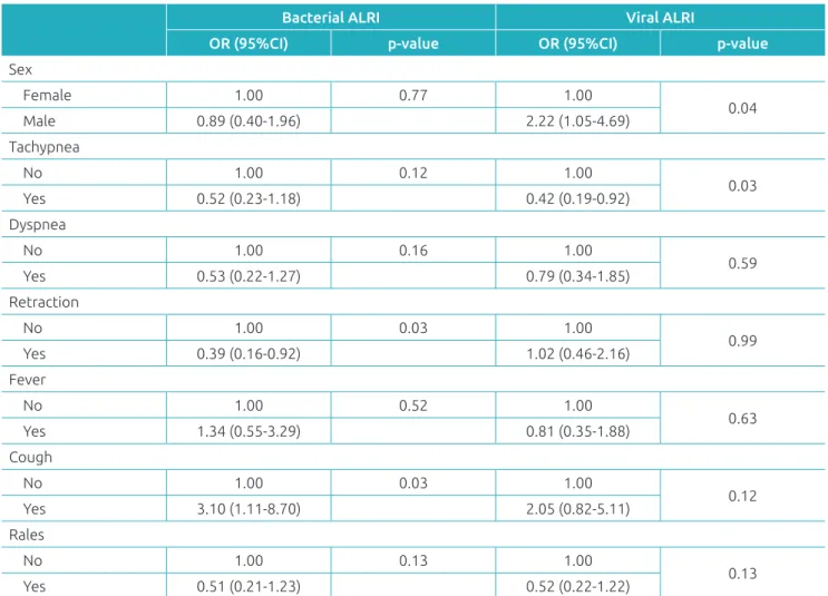
In the multinomial regression analysis, by comparing bac-terial ALRI with the viral-bacbac-terial ALRI, in the former the chances of cough were 3.1 times higher (95%CI 1.11-8.70), and retraction, 61.0% lower (OR 0.39, 95%CI 0.16-0.92). In the comparison between viral ALRI and viral-bacterial ALRI, in the former the chances of being male was 2.2 times higher (95%CI 1.05-4.69) and of having tachypnea, 58.0% lower (OR 0.42, 95%CI 0.19-0.92), as observed in Table 4.
Table 1 Frequency of hospitalization and proportion of acute lower respiratory infection, according to demographic
and climactic variables.
Hospitalization
due to ALRI Bacterial ALRI Viral ALRI Viral-bacterial ALRI p-value* n % n % 95%CI n % 95%CI n % 95%CI
Sex
Male 102 48.3 29 39.2 28.6-50.6 51 59.3 48.7-69.3 22 43.1 30.1-56.9
0.028 Female 109 51.7 45 60.8 49.4-71.4 35 40.7 30.7-51.3 29 56.9 43.1-69.9
Age (months)
0 to <2 14 6.6 6 8.1 3.4-16.1 7 8.1 3.6-15.4 1 2.0 0.1-9.3
0.290 2 to 23 154 73.0 49 66.2 54.9-76.3 63 73.3 63.2-81.8 42 82.3 70.1-91.0
24 to 59 43 20.4 19 25.7 16.7-36.5 16 18.6 11.4-27.9 8 15.7 7.6-27.6 Region of residence
South 69 32.7 19 25.7 16.7-36.5 32 37.2 27.5-47.8 18 35.3 23.2-49.1
0.271 Southeast 142 67.3 55 74.3 63.5-83.3 54 62.8 52.2-72.5 33 64.7 50.9-76.9
Season of hospitalization
Autumn 72 34.2 25 33.8 23.7-45.1 27 31.4 22.3-41.8 20 39.2 26.6-53.0
0.903
Winter 54 25.6 21 28.4 19.0-39.4 23 26.7 18.2-36.8 10 19.6 10.4-32.2
Spring 60 28.4 19 25.7 16.7-36.5 25 29.1 20.2-39.3 16 31.4 19.8-45.0 Summer 25 11.8 9 12.2 6.1-21.1 11 12.8 6.9-21.1 5 9.8 3.7-20.4 Hospital complexity
High 148 70.1 48 64.9 53.5-75.1 60 69.8 59.5-78.8 40 78.4 65.6-88.1
0.264
Medium 63 29.9 26 35.1 24.9-46.5 26 30.2 21.2-40.5 11 21.6 11.9-34.4
127
DISCUSSION
The disparity between the health of indigenous and non-in-digenous peoples is clear,13-15 and this has been attributed, for instance, to the poor socioeconomic conditions, the high load of infectious diseases, and the limitations in the continuity of their traditional means of subsistence.16
This study identified a higher proportion of presumed viral processes, and it points to the presence of viral-bacterial coinfec-tion. This seems to represent higher severity and worse therapeutic response, whereas the cases of non-combined viral or bacterial ALRI present a challenge to diagnosis and to the decision regarding the therapy. The study contributes with the subject not only because
Table 2 Frequency of hospitalization and proportion of acute lower respiratory infection, according to clinical variables.
Hospitalization
due to ALRI Bacterial ALRI Viral ALRI Viral-bacterial ALRI p-value* n % n % 95%CI n % 95%CI n % 95%CI
APDH compatible with SC
Yes 96 45.5 71 95.9 89.4-99.0 21 24.4 16.2-34.3 4 7.8 2.5-17.8
<0.001 No 115 54.5 3 4.1 1.0-10.6 65 75.6 65.7-83.8 47 92.2 82.2-97.5
Evidence for hospitalization
Yes 99 46.9 30 40.5 29.8-52.0 43 50.0 39.5-60.5 26 51.0 37.4-64.5
0.392 No 112 53.1 44 59.5 48.0-70.2 43 50.0 39.5-60.5 25 49.0 35.6-62.6
Presence of comorbidity
Yes 92 43.6 30 40.5 29.8-52.0 31 36.0 26.4-46.6 31 60.8 47.0-73.4
0.015 No 119 56.4 44 59.5 48.0-70.2 55 64.0 53.4-73.6 20 39.2 26.6-53.0
Weight/age
Adequate 66 37.5 21 37.5 25.6-50.7 29 38.7 28.2-50.0 16 35.6 22.7-50.2
0.919 Risk 60 34.1 17 30.4 19.4-43.3 27 36.0 25.8-47.3 16 35.6 22.7-50.2
Low weight 50 28.4 18 32.1 20.9-45.2 19 25.3 16.5-36.1 13 28.9 17.1-43.3 Tachypnea
Yes 110 52.1 34 45.9 34.9-57.4 41 47.7 37.3-58.2 35 68.6 55.0-80.2
0.025 No 101 47.9 40 54.1 42.7-65.1 45 52.3 41.8-62.7 16 31.4 19.8-45.0
Dyspnea
Yes 136 64.5 41 55.4 44.0-66.4 57 66.3 55.8-75.7 38 74.5 61.3-85.0
0.081 No 75 35.5 33 44.6 33.6-56.0 29 33.7 24.3-44.2 13 25.5 15.0-38.7
Retraction
Yes 65 30.8 14 18.9 11.2-29.0 31 36.0 26.4-46.6 20 39.2 26.6-53.0
0.021 No 146 69.2 60 81.1 71.0-88.8 55 64.0 53.4-73.6 31 60.8 47.0-73.4
Fever
Yes 141 66.8 54 73.0 62.0-82.2 55 64.0 53.4-73.6 32 62.7 48.9-75.1
0.374 No 70 33.2 20 27.0 17.9-38.0 31 36.0 26.4-46.6 19 37.3 24.9-51.1
Cough
Yes 169 80.1 65 87.8 78.9-93.9 69 80.2 70.8-87.6 35 68.6 55.0-80.2
0.030 No 42 19.9 9 12.2 6.1-21.1 17 19.8 12.4-29.2 16 31.4 19.8-45.0
Rales
Yes 144 68.2 47 63.5 52.1-73.9 56 65.1 54.6-74.6 41 80.4 67.8-89.6
0.099 No 67 31.8 27 36.5 26.1-47.9 30 34.9 25.4-45.4 10 19.6 10.4-32.2
of the knowledge of the clinical profile manifested by Guarani children hospitalized with ALRI, but especially for its proposal of a clinical-radiological classification of the etiological hypothesis.
ALRI is one of the main causes of hospitalization of indig-enous children in developed7,17 and developing5 countries. In the case of the Guarani children, our results indicate high frequency of hospitalization for non-severe ALRI (53.1%), with slight prevalence of viral ALRI in comparison to bacterial ALRI. Once these conditions are potentially treatable in primary health care,1,3 we observed that the indication for hospitaliza-tion is not always clinical, but it may result from factors such as the difficulty of the family regarding home care,1 language
limitations, difficulties to schedule appointments and to obtain medication, and the high turnover of professionals, as well as their insufficient skills to manage these cases.5
There were more hospitalizations caused by viral ALRI in male children aged less than 24 months, which is similar to stud-ies conducted with indigenous populations from New Zealand18 and the United States,19,20 as well as non-indigenous children.21 According to the literature, the household agglomeration and the exposure to the wood burning stove smoke were associated with higher risk of hospitalization due to AVB in indigenous children.12
Even though most ALRIs are viral,18 viral-bacterial infections were observed in approximately one fourth of the non-indigenous
Table 3 Frequency of hospitalization and proportion of acute lower respiratory infection, according to treatment
and outcome.
Hospitalization
due to ALRI Bacterial ALRI Viral ALRI Viral-bacterial ALRI p-value* n % n % 95%CI n % 95%CI n % 95%CI
ATB dose adequate for weight
Yes 90 52.3 30 52.6 39.7-65.3 36 50.7 39.2-62.2 24 54.5 39.8-68.7
0.921 No 82 47.7 27 47.4 34.7-60.3 35 49.3 37.8-60.8 20 45.5 31.3-60.2
Beginning of ATB in hospital
Up to 4 hours 147 71.4 63 85.1 75.6-91.9 52 63.4 52.6-73.3 32 64.0 50.1-76.4
0.005 After 4 hours 59 28.6 11 14.9 8.1-24.4 30 36.6 26.7-47.4 18 36.0 23.7-49.9
Adequate interval of ATB doses
Yes 194 94.2 72 97.3 91.4-99.5 77 93.9 87.0-97.7 45 90.0 79.2-96.2
0.222
No 12 5.8 2 2.7 0.5-8.6 5 6.1 2.3-13.0 5 10.0 3.8-20.8
Adequate duration of ATB
Yes 57 27.7 24 32.4 22.5-43.7 21 25.6 17.1-35.9 12 24.0 3.7-37.2
0.509 No 149 72.3 50 67.6 56.3-77.5 61 74.4 64.1-83.0 38 76.0 62.8-86.3
Change of ATB
Yes 103 50.0 33 44.6 33.6-56.0 37 45.1 34.6-56.0 33 66.0 52.1-78.1
0.034 No 103 50.0 41 55.4 44.0-66.4 45 54.9 44.0-65.4 17 34.0 21.9-47.9
Use of corticosteroid
Yes 145 68.7 36 48.6 37.4-60.0 68 79.1 69.5-86.7 41 80.4 67.8-89.6
<0.001 No 66 31.3 38 51.4 40.0-62.6 18 20.9 13.3-30.5 10 19.6 10.4-32.2
Adequate treatment
Yes 79 37.4 21 28.4 19.0-39.4 27 31.4 22.3-41.8 31 60.8 47.0-73.4
<0.001 No 132 62.6 53 71.6 60.6-81.0 59 68.6 58.2-77.7 20 39.2 26.6-53.0
Hospitalization days
1 to 3 29 13.7 9 12.2 6.1-21.1 17 19.8 12.4-29.2 3 5.9 1.5-15.2
0.167
4 to 6 87 41.3 33 44.6 33.6-56.0 36 41.8 31.8-52.5 18 35.3 23.2-49.1
129
hospitalized children,22,23 as in this study, in which the num-ber represented 24.1%. Studies with indigenous peoples in Australia7 reported the occurrence of viral-bacterial coinfec-tion in patients with ALRI.
Studies of CAP etiology in a non-indigenous population reported frequency of viral-bacterial infection in 30 to 66% of the cases.22,24 In the coinfections, it is common for the S.
pneu-moniae to be associated with the human rhinovirus10,23,25 or to the respiratory syncytial virus.10,23 In the cases reported here, this analysis was not conducted due to the absence of exam-inations for the research of respiratory viruses, besides the low request for blood culture.
Cough, retraction and tachypnea were associated with ALRI, confirming their importance for diagnosis.1,3 Cough was asso-ciated with bacterial ARLI, demonstrating to be a symptom that indicates bacterial infection,1 whereas retraction and tachy-pnea were associated with viral-bacterial ALRI. Once retrac-tion and tachypnea are also present in bacterial ALRI,1 the valorization of these clinical data added to wheezing would
help the clinical differential diagnosis between bacterial and viral-bacterial ALRI.1,3
Thoracic X-ray, blood test, blood culture and SpO2 are recommended for children with CAP who need hospitaliza-tion.1 Even though almost 80% of the hospitalizations were caused by CAP, it was observed that not all patients underwent these tests. This fact can reflect the operational difficulties in the health units, or the insufficient knowledge about the pro-tocols.1 The radiological pattern was mostly showing alveolar infiltrate, associated or not with pleural effusion and/or pneu-matocele. Considering the high frequency of wheezing in this population, suggesting the existence of bronchial obstruction and the possibility of these cases evolving to atelectasis, there is the possibility of radiological diagnostic error between the alveolar and the interstitial infiltrate and/or atelectasis, over-estimating the diagnosis of CAP.1
Children with suspicion of viral infection were on antibiot-ics, nebulization with bronchodilator and corticosteroids in a little discerning manner. Antibiotics and corticosteroids are not
Table 4 Acute lower respiratory infection and associated factors, according to the multinomial logistic regression.
Bacterial ALRI Viral ALRI
OR (95%CI) p-value OR (95%CI) p-value
Sex
Female 1.00 0.77 1.00
0.04
Male 0.89 (0.40-1.96) 2.22 (1.05-4.69)
Tachypnea
No 1.00 0.12 1.00
0.03
Yes 0.52 (0.23-1.18) 0.42 (0.19-0.92)
Dyspnea
No 1.00 0.16 1.00
0.59
Yes 0.53 (0.22-1.27) 0.79 (0.34-1.85)
Retraction
No 1.00 0.03 1.00
0.99
Yes 0.39 (0.16-0.92) 1.02 (0.46-2.16)
Fever
No 1.00 0.52 1.00
0.63
Yes 1.34 (0.55-3.29) 0.81 (0.35-1.88)
Cough
No 1.00 0.03 1.00
0.12
Yes 3.10 (1.11-8.70) 2.05 (0.82-5.11)
Rales
No 1.00 0.13 1.00
0.13
Yes 0.51 (0.21-1.23) 0.52 (0.22-1.22)
recommended for AVB, and the use of bronchodilators is con-troversial, used as therapeutic evidence and maintained in the presence of clinical response.3 This conduct may be attributed to the difficulty to distinguish bacterial and viral ALRI1,26 and to the absence of a routine medical team. The last hypothesis could explain the change of empiric antibiotics in half of the hospitalizations, despite its apparent initial adaptation.
The mean time of hospitalization was longer than in non-in-digenous children, in accordance with some studies,27 which leads to the conclusion that, in the absence of clinical sever-ity1 for hospital discharge, other factors should be taken into consideration. It is the case of malnutrition, which may lead to reduced immunity; living in indigenous areas of difficult access; lack of public vehicles addressed to the transportation of patients and lack of structure in primary health care to con-tinue the treatment.28
There were limitations inherent to a study based on second-ary data, such as incomplete records in charts, which prevented some analyses. This fact shows the need to improve the hospital records, in order to improve care and the offer of information about the use and quality of services, thus contributing with the evaluation of health policies, medical-care strategies and research.29 Besides, the classification of ALRI used in this study may be criticized, once it was carried out in an arbitrated man-ner, without the proper etiological proof of the cases.
This study suggests that part of the resolution of non-se-vere ALRI cases in Guarani children happens in the hospital environment. This model is opposed to the guidelines of the
National Policy of Indigenous Peoples Health Care11 and the National Policy of Children’s Health Care;30 both are based on integral health care, guided by principles that aim not only at curing diseases, but also at prevention and health promo-tion for children.
The proposal is that services addressed to these populations prioritize actions whose goal is to improve primary health care in the villages, reducing hospitalization rates. On the other hand, in the hospital environment, there is the need to create medical routines followed by continuous professional training, with good articulation between the levels of care, through an efficient reference and counter-reference system.
Besides, it is important to conduct studies that aim at ana-lyzing the identification of causal agents of ALRIs in indigenous populations, in order to subsidize adequate programs in child care.
ACKNOWLEDGEMENTS
The authors thank Professor Ronir Raggio Luiz (Bioestathistics NESC/UFRJ) for suggestions and help in the statistical planning.
Funding
Conselho Nacional de Desenvolvimento Científico e Tecnológico – Brasil. Edital MCT-CNPq/MS-SCTIE-DECIT nº 26/2006 (Processo nº 409677/2006-0).
Conflict of interests
The authors declare no conflict of interests
REFERENCES
1. Sociedade Brasileira de Pneumologia e Tisiologia. Diretrizes brasileiras em pneumonia adquirida na comunidade em pediatria - 2007. J Bras Pneumol. 2007;33:S31-50.
2. Wardlaw T, Johansson EW, Hodge M, World Health Organization, UNICEF. Pneumonia: the forgotten killer of children. Geneva: WHO; 2006.
3. Sociedade Brasileira de Pediatria. Diretrizes para o manejo da infecção causada pelo vírus sincicial respiratório (VSR). Rio de Janeiro: SBP; 2011.
4. Michelow IC, Olsen K, Lozano J, Rollins NK, Duffy LB, Ziegler T, et al. Epidemiology and clinical characteristics of community-acquired pneumonia in hospitalized children. Pediatrics. 2004;113:701-7.
5. Cardoso AM, Coimbra Jr. CE, Tavares FG. Hospital morbidity among Guarani indians in Southeastern and Southern Brazil. Rev Bras Epidemiol. 2010;13:21-34.
6. Souza PG, Cardoso AM, Sant’Anna CC. Prevalência de sibilância e fatores associados em crianças indígenas Guarani hospitalizadas por doença respiratória aguda no Sul e Sudeste do Brasil. Cad Saúde Pública. 2014;30:1427-37.
7. O’Grady KF, Chang AB. Lower respiratory in Australian i n d i g e n o u s c h i l d r e n . J Pa e d i a t r C h i l d H e a l t h . 2010;46:461-5.
8. Sant’Anna CC, D’Elia C. Bronquiolite. In: Benguigui Y, Antuñano FJL, Schumunis G, Yunes J, editors. Infecções respiratórias em crianças. Brasília: Organização Pan-Americana da Saúde/ Organização Mundial da Saúde; 1998. p.263-81.
9. Esposito S, Zampiero A, Terranova L, Ierardi V, Ascolese B, Daleno C, et al. Pneumococcal bacterial load colonization as a marker of mixed infection in children with alveolar community-acquired pneumonia and respiratory syncytial virus or rhinovirus infection. Pediatr Infect Dis J. 2013;32:1199-204.
10. Tsolia MN, Psarras S, Bossios A, Audi H, Paldanius M, Gourgiotis D, et al. Etiology of community-acquired pneumonia in hospitalized school-age children: evidence for high prevalence of viral infections. Clin Infect Dis. 2004;39:681-6.
131
© 2018 Sociedade de Pediatria de São Paulo. Published by Zeppelini Publishers. This is an open access article under the CC BY license (http://creativecommons.org/licenses/by/4.0/).
12. Cardoso AM, Coimbra Jr. CE, Werneck GL. Risk factors for hospital admission due to acute lower respiratory tract infection in Guarani indigenous children in Southern Brazil: a population-based case-control study. Trop Med Int Health. 2013;18:596-607.
13. Chang AB, Brown N, Toombs M, Marsh RL, Redding GJ. Lung disease in indigenous children. Paediatr Respir Rev. 2014;15:325-32.
14. Montenegro RA, Stephens C. Indigenous health in Latin America and the Caribbean. Lancet. 2006;367:1859-69.
15. Coimbra Jr. CE, Santos RV. Health, minorities and inequality: some webs of inter-relations, emphasizing indigenous peoples in Brazil. Ciênc Saúde Coletiva. 2000;5:125-32.
16. Gracey M, King M. Indigenous health part 1: determinants and disease patterns. Lancet. 2009;374:65-75.
17. Moore H, Burgner D, Carville K, Jacoby P, Richmond P, Lehmann D. Diverging trends for lower respiratory infections in non-Aboriginal and Aboriginal children. J Paediatr Child Health. 2007;43:451-7.
18. Grimwood K, Cohet C, Rich FJ, Cheng S, Wood C, Redshaw N, et al. Risk factors for respiratory syncytial virus bronchiolitis hospital admission in New Zealand. Epidemiol Infect. 2008;136:1333-41.
19. Holman RC, Curns AT, Cheek JE, Bresee JS, Singleton RJ, Carver K, et al. Respiratory syncytial virus hospitalizations among American Indian and Alaska Native Infants and the General United States Infant Population. Pediatrics. 2004;114:e437-44.
20. Lowther SA, Shay DK, Holman RC, Clarke MJ, Kaufman SF, Anderson LJ. Bronchiolitis-associated hospitalizations among American Indian and Alaska Native children. Pediatr Infect Dis J. 2000;19:11-7.
21. Alvarez AE, Marson FA, Bertuzzo CS, Arns CW, Ribeiro JD. Epidemiological and genetic characteristics associated with the severity of acute viral bronchiolitis by respiratory syncytial virus. J Pediatr (Rio J). 2013;89:531-43.
22. Cevey-Macherel M, Galetto-Lacour A, Gervaix A, Siegrist C, Bille J, Bescher-Ninet B, et al. Etiology of community-acquired pneumonia in hospitalized children based on WHO clinical guidelines. Eur J Pediatr. 2009;168:1429-36.
23. Juvén T, Mertsola J, Waris M, Leinonen M, Meurman O, Roivainen M, et al. Etiology of community-acquired pneumonia in 254 hospitalized children. Pediatr Infect Dis J. 2000;19:293-8.
24. Oliveira JR. Etiologia da pneumonia adquirida na comunidade em crianças hospitalizadas, com ênfase em derrame pleural [master’s thesis]. Bahia: Universidade Federal da Bahia; 2012.
25. Honkinen M, Lahti E, Österback R, Ruuskanen O, Waris M. Viruses and bacteria in sputum samples of children with community-acquired pneumonia. Clin Microbiol Infect. 2012;18:300-7.
26. Virkki R, Juven T, Rikalainen H, Svedström E, Mertsola J, Ruuskanen O. Differentiation of bacterial and viral pneumonia in children. Thorax. 2002;57:438-41.
27. Burgner D, Richmond P. The burden of pneumonia in children: an Australian perspective. Paediatr Respir Rev. 2005;6:94-100.
28. Brasil. Conselho Nacional de Secretários de Saúde. A integração da saúde indígena no SUS: uma proposta da Gestão Estadual. Nota Técnica. Brasília: CONASS; 2014.
29. Bittencourt SA, Camacho LA, Leal MC. O Sistema de informação hospitalar e sua aplicação na saúde coletiva. Cad Saúde Pública. 2006;22:19-30.