online | memorias.ioc.fiocruz.br
Adenoviruses C in non-hospitalized Mexican children older than five
years of age with acute respiratory infection
Dora P Rosete/1, María Eugenia Manjarrez1, Blanca L Barrón/+
Laboratorio de Virología, Escuela Nacional de Ciencias Biológicas, Instituto Politécnico Nacional, Carpio y Plan de Ayala s/n, 11340 México D.F., México 1Departamento de Virología, Instituto Nacional de Enfermedades Respiratorias, SSA, Calzada de Tlalpan 4502,
14080 México D.F., México
Adenoviruses (AdV) are commonly involved in acute respiratory infections (ARI), which cause high morbidity and mortality in children. AdV are grouped in six species (A-F), which are associated with a wide range of diseases. The aim of this study was to identify the AdV species infecting non-hospitalized Mexican children with ARI symp-toms, attending to the same school. For that, a PCR/RFLP assay was designed for a region of the hexon gene, which was chosen, based on the bioinformatical analysis of AdV genomes obtained from GenBank. A total of 100 children’s nasopharyngeal samples were collected from January to June, 2005, and used for viral isolation in A549 cells and PCR/RFLP analysis. Only 15 samples produced cytopathic effect, and in all of them AdV C was identified. AdV C was also identified in eight additional nasopharyngeal samples which were negative for viral isolation. In summary, this outpatient population showed a rate of AdV infection of 23%, and only AdV C was detected.
Key word: PCR/RFLP - adenovirus C - ARI - non-hospitalized children
Financial support: IPN, SIP (2004628, 20051208), INER, SSA + Corresponding author: [email protected]
Received 20 October 2007 Accepted 12 March 2008
Acute respiratory infections (ARI) represent one of the main health problems in children in developing countries. Although ARI are spread worldwide, they have smaller impact in industrialized countries than in developing ones. Whereas 1 to 3% of the deaths in chil-dren under five years of age in industrialized countries are due to pneumonia, in the developing countries this disease causes 10 to 25% of the deaths in children of the same age group. (Weissenbacher & Avila 1999). ARI are also one of the main reasons for health consultations and hospitalizations, and the main basis for drug prescrip-tions. In most cases, these drugs are unnecessary, poten-tially dangerous, and can stimulate bacterial resistance. Excessive and unnecessary use of medications signifi-cantly increases health care costs without benefiting the patient’s health (Benguigui et al. 1999).
The etiological agents of ARI encompass a wide variety of microorganisms, and viruses are the most frequently involved agents (Benguigui et al. 1999). Ad-enoviruses (AdV) are the third most common viral respi-ratory pathogen in children, after respirespi-ratory syncytial and parainfluenza viruses. AdV infections are common in all age groups, causing both hospital- and community-acquired epidemics (Benguigui et al. 1999). AdV con-stitute a large family of nonenveloped viruses containing a linear double-stranded DNA genome of approximately 36 kbp. To date, 51 different serotypes of human AdV have been classified into six species, named A, B, C, D,
E, and F (which have been associated to different clinical infections), based on erythrocyte agglutination assays and on DNA sequence homology. Respiratory infections have usually been associated with AdV species B, C, and E; whereas species F seem to be restricted to the enteric tract (Lee & Gruber 1999, Echavarria 2004, Fauquet et al. 2005, Wold & Horwitz 2007).
Diagnosis of AdV infections has typically relied on the detection of a cytopathic effect (CPE) in various cell lines (Mufson 2000). This method has been considered as the “gold standard” for AdV diagnosis. Although vi-ral isolation provides a sensitive method for diagnosis, it can take as long as three weeks to obtain results. Im-munofluorescence and other immunodiagnostic methods using direct antigen detection are faster than culture, but they lack sensitivity (Minnich & Ray 1980, Mahafzah & Landry 1989, Portillo & Cruz 2000). Recently polymerase chain reaction (PCR)-based assays have proven to be use-ful for AdV detection, showing higher specificity and sensitivity in comparison to the classic methods (Allard et al. 1990, Pring-Åkerblom & Adrian 1994, Alistair et al. 1996, Kajiwara et al. 1999, Xu et al. 2000, Avellón et al. 2001, Echavarria et al. 2001, Heim et al. 2003).
region of the hexon gene followed by DNA sequencing (Takeuchi et al. 1999, Sarantis et al. 2004), PCR ampli-fication followed by restriction endonuclease analysis, and multiplex PCR-enzyme hybridization methods with species-specific-oligonucleotide primers and probes (Gu et al. 2003, Pehler-Harrington et al. 2004, Echavarria et al. 2006).
There have been few studies about AdV infections in non-hospitalized children with mild respiratory infection (Freymuth et al. 1997), and in Mexico there is a lack of published data about the etiological agents causing ARI (Manjarrez et al. 2003, Cabello et al. 2006). For that rea-son, we investigated the presence of AdV involved in re-spiratory infections of non-hospitalized children, and in order to have an easy method for AdV screening in clinical samples and at the same time determine which AdV spe-cies were involved, a PCR followed by an enzymatic di-gestion using only one restriction enzyme was designed.
PATIENTS, MATERIALS AND METHODS
Viral propagation - Viral isolation and propagation were carried out according to the procedures described by Mufson (2000). Briefly, AdV reference strains: sero-type 5 and 6 (species C), serosero-type 4 (species E) used in this study were obtained from American Type Culture Collection. AdV serotype 41 (species F) isolated from a patient with gastrointestinal infection, was identified and serotyped as AdV 41 at the Instituto Nacional de Diagnóstico y Referencia, SSA, México. Clinical sam-ples: children older than five years of age (6 to 12 years old), attending to the same Elementary School in Mexico city were enrolled after the informed consent was ob-tained from their parents. A total of 100 nasopharyngeal aspirates were taken from January to June 2005, when children exhibited signs or symptoms of respiratory infection, within three days of the onset of the illness. Specimens were collected in transport medium (Levitz L-15, In Vitro, México D.F.) added with 0.5% of bo-vine albumin (Sigma-Aldrich) and antibiotics (penicil-lin/streptomycin 100 UI/ml and 100 μg/ml, Sigma), and sent to the laboratory. Samples were clarified by centrif-ugation for 5 min at 6,000g and transferred to cryovials and immediately frozen at -70°C.
AdV reference strains were propagated in A549 hu-man lung carcinoma cells grown in Eagle’s minimal es-sential medium added with 10% fetal bovine serum.
Clinical samples were inoculated by duplicate in A549 human lung carcinoma cells grown in four well Nunclon multidishes. Two wells were used as mock in-fected cell control. The cultures were incubated at 37ºC and daily scored for CPE. When CPE was observed, the monolayer was scraped and used for AdV detection by PCR. If the cultures did not show CPE after 21 days they were discarded.
DNA extraction - Viral genomic DNA was extracted from 500 µl of reference viral lysate, viral isolates and clinical samples using the QIAamp® Ultrasens™ Virus Kit (QIAGEN, cat 53704), following the manufacturer’s instructions.
Primers and PCR amplification - A set of primers was designed based on the in silico analysis of the AdV hexon gene. For that, AdV sequences obtained from GenBank (Table) were aligned to identify conserved regions by us-ing ClustalW (www.ebi.ac.uk/tools/clustalw). Forward and reverse primers were designed using the Primer3 software (Rozen & Skaletsky 2000). The specificity of each one of the primers was assessed using the BLAST algorithm available at the National Center for Biotech-nology Information website (Maglott et al. 2000). The sequences of the designed primers are shown below: For-ward 5´TCCTTCTTCAGAAACTTCCGCCATGAG3´, and reverse 5´TCGATGACGCCGCGGTGCGGCTG3´. The PCR was carried out using the Taq PCR core kit (QIAGEN, cat 201203), according to the manufacturer’s instructions. Amplification was performed in a thermo-cycler (TECHNE Genius) and PCR conditions were: an initial denaturing at 94ºC for 10 min and 35 cycles of 94ºC for 40 s, 60ºC for 40 s, 72ºC for 40 s, with a final elonga-tion at 72ºC for 7 min. PCR products were analyzed on a 1% agarose gel stained with ethidium bromide.
Amplicon restriction - The designed primers pre-dicted a 434 bp amplicon for all the AdV tested. Each amplicon sequences was analyzed using the Webcutter program version 2 (www.webcutter.com), in order to obtain a different restriction fragment length pattern (RFLP) for each AdV species, using only one restriction enzyme. It was found that the digestion of the predicted amplicons with HaeIII produced a different RFLP for each AdV species (Table). A sample of 10 μl of PCR-am -plified DNA product was incubated with 1U of HaeIII
TABLE
Expected and obtained RFLP for the adenoviruses (AdV) 434 bp amplicon digested with HaeIII
GenBank AdV Analyzed Expected RFLPa
Obtained RFLP Accession numbers Species serotypea (Size fragments, bp) Tested serotype (Size fragments, bp)
NC_001460 A 12 57, 122, 255 NT
NC_004001 B 11 70, 160, 204 NT
NC_001405 C 2 144, 290 5, 6 144, 290
NC_002067 D 17 70, 109, 118, 137 NT
NC_003266 E 4 12, 58, 70, 144, 150 4 12, 58, 70, 144, 150
NC_001454 F 40 87, 160, 187 41 70, 118, 246
restriction endonuclease (Invitrogen) for 3 h. After di-gestion, the products were analyzed by 12 % acrylam-ide gel electrophoresis, using the standard acrylamacrylam-ide monomer: N,N´-methylenebisacrylamide concentration (30:1), stained with ethidium bromide.
RESULTS
Adenovirus isolation in cell culture - Fifteen nasopha-ryngeal samples out of 100 samples of children with ARI showed AdV-like CPE, characterized by rounding and grapelike clustering of the swollen infected cells. These viral isolates were obtained from children in all age groups, and the number of viral isolates did not showed significative differences among the analyzed children age’s groups.
Adenovirus detection by PCR - The expected 434 bp amplicon from the hexon gene was obtained in all the AdV reference strains tested (Fig. 1). A similar amplicon was observed in the 15 viral isolates and in eight nasopha-ryngeal samples which did not produce CPE, although all the negative CPE nasopharyngeal samples were tested for AdV presence by using the designed PCR.
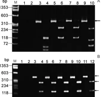
Typing of Adenovirus species by RFLP - The diges-tion of the amplicons from the AdV reference strains with HaeIII showed the expected RFLP for the serotypes 5 and 6 (species C), and for the serotype 4 (species E). The serotype 41 (species F) did not produce the expected RFLP, instead of that, three fragments of different size were obtained (70, 118, 246 bp), but this RFLP was clearly different from the RFLP of AdV C or E (Fig. 2A, Table).
Typing of Adenovirus from children with ARI - After restriction with HaeIII, all the 23 adenovirus amplicons (8 from the children’s samples and 15 viral isolates) showed the characteristic RFLP of AdV C (2 fragments of 144 and 290 bp) as shown in Fig. 2B.
Distribution of adenovirus according to children’s age and time of sampling - The higher number of stu-dents with symptoms of ARI was found on January, Feb-ruary and March, and the lowest number was found on June. AdV were only detected in children suffering ARI from January to April; being January the month with the highest number of cases of ARI positive for adeno-virus. The nasopharyngeal samples taken on May and
June were negative for adenovirus either by PCR or by isolation in cell culture (Fig. 3). There was no differ-ence on the rate of Adenovirus detection and the age of the children. The symptoms presented by children with acute respiratory infection were similar in both patients positive for adenovirus and in those negative.
DISCUSSION
Adenovirus infections occur worldwide in humans and are common in all age groups, causing both hospi-tal- and community-acquired epidemics. AdV probably account for 3% of the infections in civilian population. In children younger than five years old, they cause about 5% of upper tract respiratory infections, and are prob-ably responsible for about 10% of the pneumonias in childhood. Approximately by age 6, 95% of children are seropositive for AdV (D´Ambrosio et al. 1982).
In Mexican children suffering acute respiratory in-fections there have been very few studies analyzing the viral etiology for these infections. Furthermore, most of the studies have investigated respiratory syncytial viruses and recently metapneunovirus, especially in children during the first two years of life (Golubjatnikov et al. 1975, Tirado et al. 1995, Bustamante-Calvillo et al. 2001, Manjarrez et al. 2003, Noyola et al. 2005,
Ca-Fig. 1: adenoviruses (AdV) 434 bp amplicon obtained by polymerase chain reaction (PCR) using AdV reference strains. Agarose gel elec-trophoresis. Lanes - M: molecular size markers (ΦX174 HaeIII di -gest); 1: serotype 4 (species E); 2: serotype 5 (species C); 3: serotype 6 (species C); 4: serotype 41 (species F); 5: a nasopharyngeal sample; 6: negative control. The arrow indicates the 434 bp amplicon.
Fig. 2A: restriction fragment length polymorphism (RFLP) of the reference adenoviruses (AdV) strains amplicon digested with HaeIII. 12% polyacrylamide gel. Lanes - M: molecular size markers: ΦX174 HaeIII digested; 3, 5, 7, and 9: undigested; 4, 6, 8, and 10: digested with HaeIII. The arrow indicates the 434 bp amplicon; 1-2: nega-tive controls; 3-4: serotype 4 (species E); 5-6: serotype 5 (species C); 7-8: serotype 6 (species C); 9-10: serotype 41 (species F). B: RFLP of the AdV amplicon obtained from representative clinical isolates. Lanes - M: molecular size markers (ΦX174 HaeIII digest); 3, 5, 7, 9, 11: undigested amplicons; 4, 6, 8, 10, 12: amplicons digested with HaeIII; 1-2: negative controls; 3-4: positive control (serotype 5, AdV C); 5-6: viral isolate; 7-8: viral isolate; 9-10: viral isolate; 11-12: viral isolate. The arrows indicate the 434 bp amplicon; and the 144 and 290 bp fragments.
A
B
M
M
bello et al. 2006). Therefore the aim of this paper was to investigate the presence of AdV associated to ARI in non-hospitalized children older than five years old, as an example of adenovirus infection in a community, and at the same time to identify the AdV species involved in these infections. For that reason, children attending to the same Elementary School in Mexico city were en-rolled in this study, and nasopharyngeal samples were taken from each child when he or she had symptoms of acute respiratory infection. The period of sampling was during the winter-spring seasons (January-June, 2005). AdV were isolated from 15 nasopharyngeal samples out of 100 samples inoculated in A549 cells. These viral isolates were identified as AdV by PCR, since a frag-ment of 434 bp from the hexon gene was amplified. This amplicon was also obtained with the reference strains tested and had the expected size according to the bioin-formatical analysis performed with the GenBank AdV sequences. Besides the 15 viral isolates, other eight na-sopharyngeal samples negative for viral isolation in cell culture were positive for AdV by PCR detection. This finding agrees with previous studies which have shown that even though viral culture has been considered as the “gold standard” for AdV diagnosis, the PCR-based assays are more sensitive for this purpose (Allard et al. 1990, Pring-Åkerblom & Adrian 1994, Alistair et al. 1996, Kajiwara et al. 1999, Xu et al. 2000, Avellón et al. 2001, Echavarria et al. 2001, Heim et al. 2003).
The 23 AdV amplicons obtained from the children’s samples analyzed by restriction using only one restriction enzyme such as HaeIII produced the RFLP for AdV C. This RFLP showed the number and the size fragments expected according to the in silico analysis carried out with AdV C serotype 2, and was similar to the one ob-tained by digestion of the two AdV C reference strains tested (serotypes 5 and 6). These results suggested that the PCR/RFLP method described in this paper could be a useful screening approach for AdV detection and at the
same time for AdV species typing, using a single restric-tion enzyme, HaeIII, in contrast to others RFLP strate-gies which have been used to identify adenoviruses sero-types or species by using several restriction enzymes (Li et al. 1999, Elnifro et al. 2000, Allard et al. 2001, Ebner et al. 2006). However, to evaluate the capability of the PCR/RFLP described in this paper to detect the differ-ent AdV species is necessary to test more AdV reference strains and isolates, since sequencing of the AdV hexon gene has revealed a divergence at protein level of 0.5 to 25.4% among the 51 serotypes (Ebner et al. 2005a), and this variability may explain the unexpected RFLP found with the AdV serotype 41 (species F).
The finding of AdV C in all the children’s samples agrees with previous studies which have reported that AdV C is mostly associated to upper respiratory infec-tions in children (Echavarria et al. 2006). It is known that AdV C (serotypes Ad1, Ad2, Ad5, and Ad6) infect more than 80% of the human population early in life, causing roughly 5% of symptomatic upper respiratory infections and 15% of lower respiratory tract infections in children younger than five years. Besides that, AdV C can establish an asymptomatic infection in immuno-competent hosts characterized by intermittent viral ex-cretion in nasopharyngeal seex-cretions, and also display prolonged fecal excretion for months, and even years (Garnett et al. 2002, Wold & Horwitz 2007). However, there is not information if persistent infections also oc-cur in children like the population that was studied in this paper; therefore it would be important to investigate that, since persistent adenovirus DNA in the lungs has been postulated to be a cofactor in chronic obstructive pulmonary disease in adults (Matsuse et al. 1992).
AdV infections are usually more frequent in winter and spring, as it was found in this paper. The infections were distributed among the different age groups (6-12 years old) and no differences were found in the respira-tory symptoms between the students’ positive for adeno-virus and the students with ARI without AdV, implying that other pathogens may be involved in these respira-tory infections. In summary the set of primers used and the restriction of the amplicon with one enzyme allowed the detection and typing of the AdV C infecting children with ARI and attending to the same Elementary School, suggesting that AdV C is probably the most frequent adenovirus associated to upper respiratory infections in non-hospitalized patients as has been reported in hospi-talized children.
REFERENCES
Alistair HK, Jonsson M, Garwicz D, Kajon AE, Wermenbol AG, Verweij MW, De Jong JC 1996. Rapid subgenus identification of human adenoviruses isolates by a general PCR. J Clin Microbiol 34: 622-627.
Allard A, Albinsson B, Wadell G 2001. Rapid typing of human adeno-viruses by a general PCR combined with restriction endonuclease analysis. J Clin Microbiol39: 498-505.
Allard A, Girones R, Juto P, Wadell G 1990. Polymerase chain reac-tion for detecreac-tion of adenoviruses in stool samples. J Clin Micro-biol28: 2659-2667.
Avellón A, Pérez P, Aguilar JC, Ortiz de Lejarazu R, Echeverría JE Fig. 3: AdV detection in children with acute respiratory infections
2001. Rapid and sensitive diagnosis of human adenovirus infection by a generic polymerase chain reaction. J Med Virol92: 113-120.
Benguigui Y, López-Antuñano FJ, Schmunis G, Yunes J 1999. Re-spiratory infections in children, Pan American Health Organiza-tion, Washington DC, 453 pp.
Bustamante-Calvillo ME, Velásquez FR, Cabrera-Munoz L, Torres J, Gomez-Delgado A, Moreno JA, Muñoz-Hernandez O 2001. Molecular detection of respiratory syncytial virus in postmortem lung tissue samples from Mexican children deceased with pneu-monia. Pediatr Infect Dis J 20: 495-501.
Cabello C, Manjarrez ME, Olvera R, Villalba J, Valle l, Paramo I 2006. Frequency of virases associated with aute respiratory in-fections in children younger than five years of age at a locality of Mexico City. Mem Inst Oswaldo Cruz 101: 21-24.
Casas I, Avellon M, Mosquera M, Jabado O, Echevarria JE, Cam-pos RH, Rewers M, Pérez-Breña P, Lipkin WI, Palacios G 2005. Molecular identification of adenoviruses in clinical simples by analyzing a partial hexon genomic region. J Clin Microbiol43: 6176-6182.
D´Ambrosio E, Del Grosso N, Chica A, Midulla M 1982. Neutralizing antibodies against 33 human adenoviruses in normal children in Rome. J Hyg 89: 155-161.
Ebner K, Pinsker W, Lion T 2005a. Comparative analysis of the hexon gene in the entire spectrum of human adenovirus serotypes: phy-logenetic taxonomic and clinical implications. J Virol79: 12635-12642.
Ebner K, Raush M, Preuner S, Lion T 2006. Typing of human adeno-viruses in specimens from immunosuppressed patients by PCR-fragment length analysis and real-time quantitative PCR. J Clin Microbiol44: 2808-2815.
Ebner K, Soda M, Watringer F, Lion T 2005b. Molecular detection of the entire spectrum of human adenoviruses by a two-reaction RQ-PCR assay. J Clin Microbiol43: 3049-3053.
Echavarria M 2004. Adenoviruses. In A Zuckerman, J Banstavala, J Pattison, P Griffiths, B Schoub, Principles and practice of clinical virology, 5th ed., John Wiley & Sons Ltd, New York, p. 343-360.
Echavarria M, Forman M, van Tol MJ, Vossen JM, Charache P, Kroes AC 2001. Prediction of severe disseminated adenovirus infection by serum PCR. Lancet358: 384-385.
Echavarria M, Maldonado D, Elbert G, Videla C, Rappaport R, Car-ballal G 2006. Use of PCR to demonstrate presence of adenovirus species B, C or F as well as coinfection with adenovirus species in children with flu-like symptoms. J Clin Microbiol44: 625-627.
Elnifro EM, Cooper RT, Klapper PE, Bailey AS 2000. PCR and re-striction endonuclease analysis for rapid identification of human Adenovirus subgenera. J Clin Microbiol38: 2055-2061.
Fauquet CM, Mayo MA, Maniloff J, Desselberger U, Ball LA 2005. Family Adenoviridae. In CM Fauquet, MA Mayo, J Maniloff, U Desselberger, LA Ball (eds), Virus taxonomy, Classification and nomenclature of viruses, 8th Report of the International Com-mittee on the Taxonomy of viruses, Elsevier Academic Press, London, p. 213-228.
Freymuth F, Vabret A, Galateau-Salle F, Ferey J, Eugene G, Petitjean J, Gennetay E, Brouard J, Jokik M, Duhamel JF, Guillois B 1997. Detection of respiratory syncytial virus parainfluenza 3 adeno-virus and rhinoadeno-virus sequences in respiratory tract of infants by polymerase chain reaction and hybridization. Clin Diagn Virol
8: 31-40.
Garnett CT, Erdman D, Xu W, Gooding LR 2002. Prevalence and quantitation of species C adenovirus DNA in human mucosal lymphocytes. J Virol76: 10608-10616.
Golubjatnikov R, Allen VD, Olmos-Blancarte MP, Inhorn SL 1975. Serologic profile of children in a Mexican highland communi-ty: prevalence of complement-fixing antibodies to Mycoplasma pneumoniae respiratory syncytial virus and parainfluenza vi-ruses. Am J Epidemiol 101: 458-464.
Gu Z, Belzer SW, Gibson CS, Bankowski MJ, Hayden RT 2003. Mul-tiplex real-time PCR for quantitative detection of human adeno-virus. J Clin Microbiol41: 4636-4641.
Heim A, Ebnet C, Harste G, Pring-Åkerblom P 2003. Rapid and quantitative detection of human adenovirus DNA by real-time PCR. J Med Virol 70: 228-239.
Hierholzer JC 1973. Further subgrouping of the human adenoviruses by differential hemagglutination. J Infect Dis128: 541-550.
Hierholzer JC, Stone YO, Broderson JR 1991. Antigenic relationships among the 47 human adenoviruses in reference horse antisera.
Arch Virol 121: 179-197.
Kajiwara J, Hamasaki M, Mori R, Oniki S 1999. Detection and identi-fication of adenovirus from ophtalmological specimens by virus isolation and PCR. Jpn J Infec Dis52: 1-2.
Kidd AH, Jonsson M, Garwics D, Kjon AE, Wermenbol AG, Verweij MW, De Jong JC 1996. Rapid subgenus identification of hu-man adenovirus isolates by a general PCR. J Clin Microbiol 34: 622-627.
Lee SG, Gruber WC 1999. Adenoviruses. In R Dolin, PF Wright,
Viral Infections of the Respiratory tract, Marcel Dekker, New York, p. 223-251.
Li QG, Henningsson P, Juto F, Elgh F, Wadell G 1999. Use of restric-tion fragment analysis and sequencing of a serotype-specific re-gion to type adenovirus isolates. J Clin Microbiol37: 844-847.
Maglott DR, Katz KS, Sicotte H, Pruitt KD 2000. NCBI’s LocusLink and RefSeq. Nucleic Acids Res28: 126-128.
Mahafzah AM, Landry ML 1989. Evaluation of conventional culture centrifugation culture and immunofluorescent reagents for the rapid diagnosis of adenovirus infections. Diagn Microbiol Infect Dis12: 407-411.
Manjarrez ME, Rosete DP, Rincon M, Villalba J, Cravioto A, Cabrera R 2003. Comparative viral frequency in Mexican children under 5 years of age with and without upper respiratory symptoms. J Med Microbiol52: 579-583.
Matsuse T, Hayashi S, Kuwano K, Keunecke H, Jefferies WA, Hogg JC 1992. Latent adenoviral infection in the pathogenesis of chronic airways obstruction. Am Rev Respir Dis 146: 177-184.
Metzgar D, Osuna M, Yingst S, Rahka M, Earhart K, Elyan D, Es-mat H, Saad MD, Kajon A, Wu K, Grey G, Ryan MAK, Russell KL 2005. PCR analysis of Egyptian adenovirus isolates includ-ing identification of species serotypes and coinfections. J Clin Microbiol43: 5743-5762.
Minnich L, Ray CG 1980. Comparison of direct immunofluorescencent staining of clinical specimens for respiratory virus antigens with conventional isolation techniques. J Clin Microbiol12: 391-394.
Morfin FS, Dupuis-Girod S, Mundweiler S, Falcon D, Carrington D, Sedlacek P, Bierings M, Cetkovsky P, Kroes AC, van Tol MJ, Thouvenot D 2005. In vitro susceptibility of adenovirus to antivi-ral drugs is species-dependent. Antivir Ther10: 225-229.
Mufson M 2000. Respiratory viruses. In S Specter, L Hodinka, SA Young, Clinical virology manual, 3rd ed., ASM Press, Washing-ton, p. 235-251.
metapneu-movirus infections in Mexico: epidemiological and clinical char-acteristics. J Med Microbiol54: 969-974.
Pehler-Harrington K, Khanna M, Waters CR, Henrickson KJ 2004. Rapid detection and identification of human adenovirus species by adenoplex a multiplex PCR-enzyme hybridization assay. J Clin Microbiol42: 4072-4076.
Portillo CM, Cruz J 2000. Implementación del método rápido de di-agnóstico de virus por inmunofluorescencia en niños hospital-izados por infecciones respiratorias agudas. Arch Argent Pediatr 98: 99-102.
Pring-Åkerblom P, Adrian T 1994. Type and group-specific polymerase chain reaction for adenovirus detection. Res Virol145: 25-35.
Rosen I 1960. A hemagglutination-inhibition technique for typing of adenoviruses. Am J Hyg71: 120-128.
Rozen S, Skaletsky H 2000. Primer3 on the WWW for general users and for biologist programmers. In S Krawetz, S Misener, Bioin-formatics Methods and Protocols: Methods in Molecular Biol-ogy, Human Press, Totowa, p. 365-386.
Sarantis H, Johnson G, Brown M, Petric M, Tellier R 2004.
Compre-hensive detection and serotyping of human adenoviruses by PCR and sequencing. J Clin Microbiol42: 3963-3969.
Takeuchi S, Itoh N, Uchio E, Aoki K, Ohno S 1999. Serotyping of ad-enoviruses on conjuntival scraping by PCR and sequence analy-sis. J Clin Microbiol37: 1839-1845.
Tirado R, Sarmiento RE, Bustos J, Thompson O, Gomez B 1995. Occurrence of respiratory syncytial virus subtypes in Mexican infants with acute lower respiratory tract disease. Arch Med Res 26: 122-126.
Weissenbacher MC, Avila M 1999. Viruses as the cause of upper and lower ARI in children: General characteristics and diagnosis. In Y Benguigui, FJ López-Antuñano, G Schmunis, J Yunes, Respi-ratory infections in children, Pan American Health Organization, Washington DC, p. 87-104.
Wold WSM, Horwitz MS 2007. Adenoviruses. In DM Knipe, M How-ley, Virology, Wolters Kluwer Lippincott Williams & Wilkins, Philadelphia, p. 2395-2436.