Anaesthetic
aversion in
Zebrafish
(Danio rerio)
Jorge Miguel Custódio Martins Ferreira
Mestrado em Ecologia, Ambiente e Território
Departamento de Biologia 2016
Orientador
Todas as correções determinadas pelo júri, e só essas, foram efetuadas. O Presidente do Júri,
Agradecimentos
Queria começar esta secção por agradecer à Doutora Ana Valentim, uma orientadora incansável que desde o primeiro minuto em que agarrei esta tese esteve sempre lá pronta a ensinar-me tudo (e um pouco mais) que era necessário para conseguir chegar a bom porto e que nunca deixou de me propor desafios que sem dúvida me fizeram crescer e que com eles aprendi e levo valores e conhecimento que me acompanharão durante toda a vida numa jornada científica que apenas agora está a começar e que tenho a certeza que terá um caminho cruzado com o dela ao longo dos anos vindouros. Agradeço-lhe ainda a paciência que foi necessária ter com um orientando como eu em que perguntas nunca faltavam e que nenhuma ficou sem resposta. Foi uma sábia orientação que demonstra que em Portugal existem óptimos profissionais que não se deixam levar pelo pessimismo social e que mantêm a paixão pela Ciência acesa dentro deles.
Queria agradecer a todo o grupo Laboratory Animal Science por me terem acolhido entre vocês e me terem feito sentir como um de vocês. Todos vocês são óptimos colegas de trabalho. Não posso também deixar de agradecer à Doutora Anna Olsson por toda a flexibilidade que demonstrou e ainda por ser uma líder e não apenas uma chefe.
De seguida quero agradecer aos meus compinchas do Addiction Biology, pois no grupo ao lado encontrei um conjunto de amigos improváveis. Obrigado Joana e João por esta jornada conjunta, foi difícil, mas todos conseguimos juntos terminá-la. Obrigado ainda Renata por todos os conselhos e horas que passaste a ouvir queixumes e devaneios de quem está a fazer uma tese e tem poucas horas de sono, foste aquela amiga mais experiênciada neste mundo científico que por vezes não tinha respostas para os problemas mas tinha sempre uma piada para animar o espírito.
Quero agradecer agora aos meus pais, sem eles nada disto tinha sido possível. A eles agradeço toda a liberdade e independência que me deram e terem-me deixado crescer e tornar-me o que sou deixando-me tomar as minhas próprias decisões, tendo eu a certeza que no fundo existia um cunho deles que não me deixaria tomar as decisões erradas.
Não posso deixar de agradecer ao Professor Nuno Formigo, pelo seu claro amor ao ensino e pela qualidade cientifíca da sua mentoria e conselhos.
Quero agradecer aos meus colegas do MEAT, foram dois anos de luta e cá estamos todos. Adultos prontos a enfrentar o mundo e tudo o que eles nos tiver para atirar. Venham os desafios porque sabemos que estamos prontos, e se não sabemos é porque ainda não nos testaram, porque este grupo é sem dúvida uma esperança em que todos iremos brilhar no que decidirmos seguir. A todos vós um boa sorte e um obrigado.
Aos meus Tasqueiros, um grupo crescente de amigos que se torna família. A personificação da frase “os verdadeiros amigos encontram-se na faculdade”. A todos os que iniciam a jornada comigo, tenho-vos a felicitar e a agradecer a paciência, as longas horas de conversa na faculdade, laboratório ou numa qualquer mesa de um tasco por aí. Com vocês sorri, chorei, cresci e brilhei. Sei que fizemos isto todos juntos e que continuaremos a ser a irmandade que somos. Obrigado Talica por momentos como a Monumental e o Comboio. Obrigado Rémi, obrigado Luís, obrigado Rita, Nala, Ana, Tó, Leonardo, Mourão, Joana (a tasqueira desaparecida), Gun, Dida e aos pseudo tasqueiros Mónica, Cú e Ashley. A todos vós ergo o meu copo e vos saúdo. Venho buscar os que ficam e conhecer os que se juntam. Que o Tasco esteja sempre connosco meus irmãos.
A ti Nessie, porque o prometido é devido e as acções falam mais alto que as palavras, obrigado por as infindáveis horas em que me ouviste, em que me apoiaste e que me fizeste ver que as coisas só podiam melhorar e nunca piorar. Foste sem dúvida uma peça chave neste trabalho no que toca à sanidade mental e nunca me esquecerei de tudo o que fizeste e disseste. Levo no coração a amizade e dedicação incansável que demonstraste ter para comigo. A vida é feita de altos e baixos, mas no fim, depois de toda a tempestade, vem a bonança. Sei que de uma forma ou outra, estarás lá para ver o resto do meu percurso e eu o teu, obrigado do fundo do coração. Obrigado pequena Ecotona.
Aos meus companheiros de longa data, que sempre demonstraram algum interesse no meu trabalho mas que demonstraram um gigante interesse em fazer-me esquecer que existia stress na vida transportando-me para um mundo de fantasia,
felicidade e companheirismo agradeço-lhe as longas horas à volta da mesa a conversar. Obrigado Barros, João e Nandes.
Ao Pereira, porque os irmãos não precisam de ser de sangue e ainda porque te enquadras em demasiadas categorias, fica o agradecimento curto, porque a alguém como tu, cerca de 20 anos não se agradecem em meia dúzia de palavras, mas obrigado por tudo meu irmão.
À Maria Luís, pois mesmo distante, acabaste por estar lá nos momentos certos para me apoiar cumprindo sempre o papel que te propuseste desde sempre.
Às minhas afilhadas, por todo o carinho, orgulho e apoio que me proporcionaram neste tempo em que entraram na minha vida.
A ti Fatinha, por todo o carinho demonstrado, pelos sorrisos e perguntas. Sei que tenho tanto a aprender contigo como tu comigo e isso faz toda a diferença. Obrigado pelo apoio e dedicação e por demonstrares que é possível juntar coisas que à partida não parecem fazer parte uma da outra mas que se complementam. Obrigado minha Geógrafa Ecóloga.
Queria agradecer ainda à MLP pelo completo fascínio com que olhas para a natureza e por contigo aprender a olhá-la com outros olhos.
Obrigado Pipa, obrigado Xana e todas as restantes pessoas que no último ano surgiram e estiveram lá.
Obrigado aos meus irmãos d’A tua mãe. Estamos e estaremos sempre juntos, mais perto ou mais longe. Obrigado à restante família que a constitui.
Por fim, obrigado a todos aqueles que estiveram ou estão na minha vida, cada um de vocês contribuiu para isto. Se me esqueço de alguém, obrigado também a ti pelo que fizeste.
Este trabalho não é só meu, é de todos vocês e só me resta agradecer de novo a todos.
“You become responsible, forever, for what you have tamed.” The Little Prince - Antoine de Saint-Exupéry
Abstract
Although the interest in zebrafish has been rising in research, there are still refinements required to some procedures. The refinement of anaesthesia tends to be reduced to clinical efficacy, and animals’ welfare is often disregarded. Thus, we aim to study an anaesthetic protocol that is efficient and that induces no or less aversion in adult zebrafish, using a combination of Propofol/lidocaine and the most used anaesthetic in fish, MS222. This study was divided in two, where the first part aimed to assess which concentrations are equipotent, check the complete anaesthetic profile and clinical quality of the anaesthetics. The second part aims to assess the aversion of both anaesthetic protocols. Thirty-six mixed-sex AB zebrafish were randomly assigned to MS222 (150mg.L-1) and Propofol/lidocaine (5mg.L-1 of propofol combined with 150mg.L-1 of lidocaine) group. Aversion was tested in a conditioned place aversion task where each protocol was paired with a previously trained environment, and, afterwards, animals were tested again without the anaesthetic. The aversion degree was measured by the animals’ preference for the conditioned place. The present work contradicted other studies where MS222 is aversive, and showed that Propofol/lidocaine is also no/less aversive to zebrafish. Moreover, both provide a full anaesthesia and recovery in short time. Thus, both anaesthetic protocols seem to be recommended to use in adult zebrafish, and the new anaesthetic protocol presented has the advantage of being more practical to use and cheaper than MS222. Further experiments need to be made to refine this anaesthetic protocol and improve zebrafish welfare in different ages, strains, and experimental situations.
Sumário
Embora o interesse em peixe-zebra tenha aumentado na investigação científica, existem ainda alguns procedimentos que necessitam de refinamento. O refinamento da anestesia tem tendencialmente sido reduzido apenas à sua eficácia clinica e o bem-estar animal tem sido colocado em segundo plano. Assim, o nosso objectivo é estudar um protocolo anestésico para peixe-zebra que seja eficaz e que seja pouco ou não aversivo para o peixe-zebra adulto, usando uma combinação de Propofol/lidocaina e o anestésico mais comummente usado em peixe, MS222. Este estudo consiste em duas partes, sendo que a primeira tinha como objectivo perceber qual a concentração equipotente dos anestésicos, estudar a qualidade clínica e o perfil anestésico. A segunda parte tinha como objectivo verificar se estes anestésicos eram aversivos. Trinta e seis animais da estirpe AB de ambos os sexos foram aleatoriamente atribuidos por cada um dos dois grupos, MS222 (150mg.L-1) e Propofol/lidocaina (5mg.L-1 de propofol combinado com 150mg.L-1 de lidocaina). A aversão a estes anestésicos foi então testada com um teste de aversão condicionada a um local em que cada protocolo anestésico foi associado a um local previamente preferido, e de seguida os animais foram testados novamente sem o anestésico. O grau de aversão foi medido através da preferência demonstrada pelos animais ao local condicionado. Este estudo veio contradizer outros em que o MS222 é aversivo, mostrando que o Propofol/lidocaina é também não ou pouco aversivo para o peixe-zebra adulto. Para além disso, ambos os protocolos induziram uma anestesia completa e uma rápida recuperação. Deste modo, ambos os anestésicos são recomendáveis para o peixe-zebra. O protocolo novo aqui apresentado tem as vantagens de ser mais prático a nível de preparação e mais barato do que o MS222. São ainda necessários mais estudos para refinar este protocolo anestésico e melhorar o bem-estar do peixe-zebra de diferentes estirpes, idades e em diferentes situações experimentais.
Palavras-chave: Anestesia, Bem-estar animal, Peixe-zebra, MS222, Propofol, Lidocaina
Contents
Agradecimentos ... i Abstract ...v Sumário... vi Introduction ... 1 Zebrafish in nature ... 1 Zebrafish in science ... 3(Zebra)fish and anaesthesia ... 5
Anaesthetics ... 9
Zebrafish behaviour: aversion ... 12
Aims of the study ... 13
Material and Methods ... 15
Animals and housing ... 15
Reproduction and development ... 15
Study 1: Establishment of equipotent dose and clinical variables evaluation ... 17
Study 2: Aversion to anaesthetics ... 18
Conditioned place avoidance ... 19
Habituation ... 20
Training phase ... 20
Testing period ... 21
Data analysis ... 22
Results ... 24
Establishment of equipotent doses and clinical variables ... 24
Old fish pilot study ... 24
Young fish assay ... 29
Light conditions preference ... 31
Training ... 33
Testing ... 33
Discussion ... 38
Bibliography ... 44
Annex 1: Recipe for E3 medium ... 54
Annex 2: Old fish assay data... 55
Annex 3: Young fish assay data ... 57
Annex 4: CPA scheme ... 58
Introduction
The crescent interest in Zebrafish by the scientific community is evident in the publication of more than 2400 papers in the last year regarding this species; actually the number of publications quadruplicated in the last 15 years (Pubmed database; keyword: “zebrafish” in Title/Abstract) (Figure 1). It is then of utmost importance to address some methodologies used daily in research, such as anaesthesia, to fulfil gaps in knowledge, and achieve a complete insight of the animal’s needs to refine its use.
Zebrafish in nature
Fish represent the largest class of vertebrate animals with more than 31,000 known species. Weber estimates that over 3000 species have some kind of use, such as a protein and nutrient source both for humans and for other animals, as a recreational and commercial resource, as a companion or display pet, and for scientific purposes (Weber, 2011).
There are 6 families in the Cypriniformes order: Cyprinidae, Catostomidae, Gyrinocheilidae, Psilorhynchidae, Cobitidae, and Balitoridae (Nelson, 2006). The
Fig. 1: Number of publications referent to zebrafish during the past 15 years (keyword “zebrafish” in Title/Abstract according to Pubmed database).
Year N u mb er o f p u b li cat io n s
gender Danio of the Cyprinidae family is placed in the largest clade of freshwater fishes and in the second largest vertebrate animals family (Mayden et al., 2007; Nelson, 2006). This Order is a large group of freshwater fishes distributed throughout North America, Africa, and Eurasia (Mayden et al., 2007). Danio rerio was first described by Francis Hamilton, a surgeon of the British East India Company that was in West Bengal at the beginning of the 19th century. This species was included in a publication named
An Account of the Fishes Found in the River Ganges and its Branches in 1822 with
nine more species of the gender Danio (Spence & Gerlach, 2008).
Danio rerio, commonly referred also as zebrafish or zebra danio (Figure 2), is a
small cyprinid fish, native of the streams of South-eastern Himalayan region (Mayden et al., 2007), especially around the basins of the river Ganges and Brahmaputra, in the north-eastern India, Bangladesh and Nepal. There are some reports of zebrafish sightings in the Indus, Cauvery, Pennar, Godavari and Mahanadi river basins, in the Krishna river basin (Spence & Gerlach, 2008) and in the states of Rajasthan, Gujarat and Andra Pradesh (river basins draining into the Arabian Sea) as well as in northern Myanmar and Sri Lanka (Spence & Gerlach, 2008). Individuals of this species are normally encountered in shallow slow moving waterbodies with scarce aquatic vegetation and with gravel or silt as substract. This species is also often found in waterbodies near rice cultivations, being this association possibly related to the fertilizer used in agriculture that may promote the growth of zooplankton which is the main resource on their diet (Spence & Gerlach, 2008). This highly social species is characterized by a rapid growth rate, and it can grow to 35mm in nature (Spence & Gerlach, 2008) with a lifespan of approximately 3 years. Females have a protuberant genital papilla and appear to be larger than males with the last being more fusiform. It is common for zebrafish to achieve maturity after 75 days, possibly mating every two days after reaching it, and females can produce several hundred eggs in a single spawn (Spence & Gerlach, 2008).
Zebrafish in science
Fish has been a rising animal model with increasing popularity in the recent decades (Detolla et al., 1995; Jenkins et al., 2014) and in certain countries, being the third-most commonly used animal group in scientific experiments (Overturf, 2009). Thus, this increasing interest in fish demands a growth of knowledge in the following areas: aquatic animal medicine, as a support for veterinarians and aquaculture; companion animal medicine; conservation medicine; laboratory animal medicine; zoo and aquarium medicine; ecosystem health; restocking and protection of endangered species. This has been leading to a development of the aquatic medicine and a greater care of these animals. Methodologies need to be adapted to the unique characteristics of this paraphyletic group to improve science and decrease the impact that they may have on fish (Weber, 2011).
Since the 80’s, zebrafish (Danio rerio) has been used on laboratory and, as we have seen above, the number of papers with this animal quadrupled in the last decade (Valentim 2016), showing a clear importance of this animal model among scientists. This increase in popularity has economical and physiological reasons.
Danio rerio has been extensively used in research and analysis of the effects of
alcohol (R. Gerlai, Lahav, Guo, & Rosenthal, 2000), drug addiction (Darland & Dowling, 2001), learning and memory (Al-Imari & Gerlai, 2008; Pather & Gerlai, 2009), aggression (Robert Gerlai, 2003), social behaviour (Pather & Gerlai, 2009), fear, anxiety and aversion (Robert Gerlai, Fernandes, & Pereira, 2009; Speedie & Gerlai, 2008) have been successfully studied in this animal model. It has also been used as a model for human diseases, such as cancer (Lieschke & Currie, 2007), cardiovascular
diseases (Zon, 1999), immune system diseases (Lieschke & Currie, 2007) and tuberculosis (Cronan & Tobin, 2014). It is particularly interesting in toxicological, developmental (Lele & Krone, 1996), and regeneration studies (Poss, Keating, & Nechiporuk, 2003). Over 800 labs worldwide are now using zebrafish at daily basis in fundamental and applied research (ZFIN1) (Howe, Bradford, & Conlin, 2013) and there is an increasing interest in zebrafish as a model for understanding the genetic basis of behaviour (Gerlai et al., 2000; Gerlai, 2003; Guo, 2004; Spence & Gerlach, 2008).
Its strength as a model organism lies in that, as a vertebrate, it is more comparable to humans than invertebrate model species such as Drosophila
melanogaster (Barbazuk et al., 2000; Postlethwait & Talbot, 1997) or Caenorhabditis elegans, while, as a simple vertebrate, it is easier to perform genetic and embryonic
manipulations than in traditional biomedical model species such as mouse or rat in which such procedures are both complicated and costly (Hölttä-Vuori et al., 2010; Spence & Gerlach, 2008).
Thus, zebrafish are very cost-efficient, easy to breed and breeds all year round, has high fecundity (200–300 eggs per female per spawning every other day) (Gerlai et al., 2009), short lifecycle, small size allowing to be housed in large numbers in a relatively small space, being easily maintained and used in laboratory settings (Kalueff, Stewart, & Gerlai, 2014; Readman, Owen, Murrell, & Knowles, 2013). Plus, the zebrafish genome is also well characterized and fully sequenced, revealing a close parallel between mammals (even humans) and zebrafish regarding behaviour, genetics and physiology. Thus, zebrafish has been considered one of the primary model organisms in modern day genetics (Grunwald & Eisen, 2002). Adding to all these features, it also possesses rapid development and a relatively long lifespan, which makes them a useful model for various human brain disorders. External fertilization and transparency of embryos and larvae increase their value in genetic manipulations, protein tracking, developmental and toxicological studies. The availability of multiple zebrafish strains is another important aspect of this species, enabling studies of strain differences in brain function, behaviour, and drug responses (Stewart, Braubach, Spitsbergen, Gerlai, & Kalueff, 2014).
1 ZFIN: https://zfin.org/
In spite of this increasing use, this has not been accompanied by a full understanding of the animal welfare, and the best procedures to be performed, leading to the need of refinement of several techniques, as, for example, anaesthesia protocols.
(Zebra)fish and anaesthesia
Anaesthesia is required for mammals in research and clinical settings on a daily basis to avoid or decrease distress and pain associated with invasive procedures or procedures requiring immobility only. Thus, before addressing the characteristics of anaesthesia process, it is important to focus on a misconception that it is still believed by many. Do fish feel pain? Many address that they don’t, even though a review of the literature provides no compelling reason to consider fish any differently from other vertebrates. There are evidences that fish feel pain and can experience suffering as birds and mammals do. In fact, they may feel even more pain than human neonates and preterm babies (Braithwaite, 2010). Fish have been shown to possess nociceptors that are physiologically identical to those found in mammals, brain structures and opioid receptors necessary to feel pain, and a capacity to associate specific events with noxious and stressful stimuli (Sneddon, 2003, 2009). Stressors are responsible for the disturbance of the individuals’ normal physiology and general health, potentially leading to certain disorders. This disruption is dependent on the duration and intensity of the stress or stressors (Weber, 2011). Some of the most common stressors identified in fish include: changes in either chemical or physical water quality conditions (water temperature, pH, alkalinity, salinity, dissolved oxygen), accumulation of nitrogenous waste in water, other environmental pollution, handling, transport, excess of noise, poor/inadequate nutrition, overstocking, aggression, predation (environment), infectious diseases (parasitic, bacterial, viral, fungal), among others (Weber, 2011). Although there are few studies involving zebrafish (Sneddon, 2011), we should consider that they feel pain and stress until prove it wrong. And that’s why any invasive or potentially harmful procedure in zebrafish should be subject to monitoring and appropriate ethical review (Reed & Jennings, 2011). Indeed, this is now a legal requirement for fish in general from the moment they become an independent free feeding larva, as stated in
the European Directive 2010/63/EU on the protection of animals used for scientific purposes (European Parliament & Council, 2010).
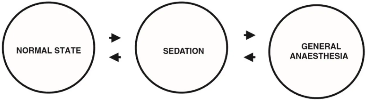
The word anaesthesia has a Greek derivation, meaning loss of sensation or insensibility; it has also been described as a reversible process of intoxication where the individual undergoes on a state of unconsciousness and can be awake without harm. This loss of sensation is a continuum as presented in Figure 3. Anaesthesia is preceded by sedation, a state of drowsiness with reduced sensory perception, but without a major loss of equilibrium or sensory perception (Ross, Ross, & Robb, 2000).
Anaesthetic agents are then normally used for several procedures in fish, such as surgery (e.g.: cardiac regeneration studies), fin clipping for genetic identification, and retrieval of blood samples in laboratory and field settings (Nickum, 1988; Trushenski et al., 2013). They can also be used to assess if the anaesthetic by itself can produce changes on a neurological, physiological and behavioural level. This evaluation may be important not only for the fish welfare, but it may also have clinical and toxicological implications. Anaesthetic drugs can also be used in overdose as euthanasia agents or at least as agents that induce loss of consciousness before another method of euthanasia is performed. Thus, almost all laboratory fish, some free range fish and a few aquaculture fish will have contact with anaesthetic agents at some point in their life, which translates into millions of animals per year exposed to anaesthetics (Readman et al., 2013). In fact, different types of anaesthetics have been used to aid in the capture, handling, artificial reproduction, and transport of fish as an anti-stress procedure in modern aquaculture (Roubach & Gomes, 2005).
Anaesthesia for fishes is an area where relatively little is known or formally reported, and has many practical difficulties. Further, the wide range of species and evolutionary context makes generalisations across fish species extremely problematic (Readman et al., 2013). It should also be noted that there is a large spectrum of areas, from research to field ecology, aquaculture and veterinary science that would benefit from better anaesthetic protocols.
The actual mode of action of anaesthetics in invertebrates and fish is poorly known but some agents appear to show an inverse relationship between the dose required to induce loss of consciousness and the animal position in the evolutionary tree. Therefore, to anaesthetize a fish, it may be necessary a larger dose of anaesthetic than it would be necessary to administer in a mammal. This phenomenon may be related with the evolution of molecular mechanisms and to an increase of active sites for certain compounds in higher vertebrates (Ross et al., 2000).
Although the difficulty to control anaesthesia depth, if the induction is slow, several stages may be detected. There are different scales to evaluate anaesthetic depth in fish, and Table 1 is an example of these scales.
In general, anaesthesia is fully achieved by the induction of three main components: hypnosis (loss of consciousness), analgesia (lack of sensation which also blunts autonomic reflexes) and muscle relaxation (Miller, Eriksson, & Fleisher, 2009).
With unconsciousness, amnesia may also be attained. For practical purposes, anaesthesia may be reduced to three obvious phases: induction, maintenance and recovery of anaesthesia (Ross et al., 2000). Induction is a phase where the animal is exposed to the anaesthetic agent, increase its activity (excitement phase), and lose consciousness/equilibrium. This is followed by the anaesthesia maintenance that involves extending the previous stage without harming the animal. If long duration maintenance is needed, the anaesthetic concentration must be decreased, and oxygenation provided. To recover from continuous anaesthesia, the anaesthetic administration is stopped and, after some time, the fish regain equilibrium in the water, returning later to its normal state. If the necessary precautions are not taken, and the anaesthesia lasted for too long, death may occur due to overdose, especially if the drug concentration is high. In order to avoid death after a procedure, general good care and cautious handling are essential (Ross et al., 2000).
Anaesthesia of small fish is normally induced by placing the fish into an anaesthetic solution that is absorbed through the gills and enters the arterial blood circulation, and the anaesthetics or their metabolites are excreted via the gills (Ross et al., 2000). The animal regains consciousness when it returns to fresh water. As this type of anaesthesia induces a massive absorption, extreme care must be taken to avoid overdosing or too deep stages of anaesthesia (Carter, Woodley, & Brown, 2011). The difficulty to control the anaesthetic depth in an anaesthetic bath is an animal welfare liability as it causes stress to the animal, risk of mortality and may be a variable that induces bias in the results between assays.
Another concern raised is, as anaesthesia is both used in the field and closed facilities, there is an associated risk of anaesthetics to become pollutants (Valcárcel et al., 2012). As these pharmaceuticals are synthetic chemicals, they are also classified as xenobiotics. Although they have a strong role and a beneficial effect on nowadays society, the environmental implications of the use of certain anaesthetics remains largely unknown and unconsidered, which raises some concerns. The polluting effects of these xenobiotics are often assessed in the ecotoxicology field, where the zebrafish embryo model is used (Busch et al., 2011).
Anaesthetics
According to the literature, a good anaesthetic for fish have several characteristics, such as high solubility in any matrix, high potency, high safety margin, induction of a complete recovery, the ability to be administered in various ways (immersion and injection) and to be able to induce a range of depths of anaesthesia (Readman et al., 2013; Thienpoint & Niemegeers, 1965). It was also described that the ideal anaesthetics induce “anaesthesia within 3 minutes or less, cause no toxicity to fish at treatment levels, present no mammalian safety problems, leave low tissue residues after a withdrawal time of 1 h or less, and be reasonable in cost” (Marking & Meyer, 1985).
The standard anaesthetic used nowadays for fish is tricaine methanesulfonate (TMS; also known as MS222). This is a local anaesthetic that acts as muscle relaxant and blocks sodium and to a lesser degree potassium currents in nerve membranes. It is currently the only anaesthetic approved for aquatic species by the US Food and Drug Administration (FDA) and the most widely used sedative and anaesthetic for zebrafish.
MS222 is available as a water-soluble powder that is acidic in solution, altering the pH to 5 or lower, which may cause irritation in the skin and mucous membranes of the fish, increasing the risk of acidosis on fish’s plasma. This should be avoided by making a buffered solution adding sodium bicarbonate or sodium hydroxide (Harper & Lawrence, 2012) or Tris buffer (Westerfield, 2000) to MS222. However, it appears that the stress is induced by the anaesthetic and not by the low pH of the water in channel catfish (Welker, Lim, Yildirim-Aksoy, & Klesius, 2007). Similarly, zebrafish immersed in buffered or unbuffered MS222 typically respond with tachyventilation before righting reflex, body, and opercular movements cease (Wilson, Bunte, & Carty, 2009). Occasionally, gill bleeding occurs, and some fish do not recover from anaesthesia (Matthews & Varga, 2012). This local anaesthetic is administered in a water bath, acting systemically as it is absorbed through the gills and skin of some fishes. It has been recently contested because of the possibility of inducing neuromuscular blockade instead of inducing loss of consciousness, causing stress. In addition, it may affect the hemodynamic equilibrium as it lowers the heart rate, increasing the risk of accidental death, especially in long duration anaesthesia (Huang et al., 2010; Readman et al.,
2013; Reed & Jennings, 2011). This accidental death can occur as, quoting the renowned toxicologist and zebrafish scholar von Hohenheim: “Poison is in everything, and nothing is without poison. The dosage makes it either a poison or a remedy”. The users pro-MS222 advocate that this potential mortality may be caused by a misused of the anaesthetic and more training is needed.
Therefore, the establishment of suitable anaesthetics is not easy, as it depends on the purpose of the experiment (type of procedure and the potential influence of the drug in the outcomes), the species of the fish, gender, its size and the stage of development, which may limit the use and efficiency of these products (Ribeiro et al., 2015). It is then necessary to take into account these variables and more different anaesthetic protocols should be carefully evaluated and characterized. In this sense, we intend to study a new anaesthetic protocol, the combination of propofol and lidocaine.
Propofol (Diprivan®, Rapinovet®, Propoflo®, Lipuro®) is 2,6-diisopropylphenol with sedative-hypnotic properties (Larijani & Gratz, 1989). It has been widely used as an intravenous anaesthetic drug in human patients (Andrews et al., 1997) and in veterinary practice (Gholipourkanani & Ahadizadeh, 2013). In contrast to MS222, propofol is a widely used anaesthetic in mammalian species (Oda, Bailey, Lewbart, Griffith, & Posner, 2014). Propofol is a short-acting, rapidly metabolized agent, which is characterized by a lack of cumulative effects and by a rapid recovery after its administration in effective doses or by continuous infusion (Gholipourkanani & Ahadizadeh, 2013). It provides a reliable, rapid and smooth induction of anaesthesia, adequate hypnosis, minimal suppression of vital organ functions and some analgesia at high doses during surgical interventions (Gholipourkanani & Ahadizadeh, 2013), although this last effect is highly debatable (Fassoulaki, 2011). Moreover, recovery is observed to be rapid, uncomplicated and complete. This substance has been used as an injectable to induce anaesthesia in reptiles, some fish species such as the Gulf of Mexico sturgeon (Acipenser oxyrinchus desotoi), in which MS222 isn’t effective (Fleming, Heard, Francis Floyd, & Riggs, 2003), and in spotted bamboo sharks (Chiloscyllium plagiosum) (Miller, Mitchell, & Heatley, 2005). Subanaesthetic concentrations of propofol might also be useful as a sedative for brief handling and transportation (Oda et al., 2014). However the efficacy and safety of any anaesthetic agent vary among species, life stages and environmental conditions, and more studies
are needed to take advantages of this anaesthetic in ornamental fish (Gholipourkanani & Ahadizadeh, 2013).
Lidocaine (2-(diethylamino)-N-(2, 6-dimethylphenyl) acetimide), also commonly known as Lignocaine or Xylocaine, is a widely used analgesic synthetized in 1943 that was used for many years as a local anaesthetic agent and it is also an antiarrhythmic drug (Collinsworth, Kalman, & Harrison, 1974) in mammals, inducing cardiac depressant effects. Nevertheless, it has been used in fish, and its analgesic and/ or anaesthetic effects have been demonstrated in zebrafish (Collymore, Tolwani, Lieggi, & Rasmussen, 2014), Algansea lacustris (Lopez, Mendoza, Ross, Rivera Lopez, & Orbe Mendoza, 1991), goldfish and Cyprinus carpio, especially if used in conjunction with sodium bicarbonate to stabilise the dose response (Carrasco, Sumano, & Navarro-Fierro, 1984; Feldman, Defrancisco, & Cascella, 1975). In a water bath, lidocaine is normally used in the hydrochloride form, because without the hydrochloride salt it is insoluble in water and only soluble in acetone or alcohol (Park, Park, Gil, Nam, & Kim, 2011). It is relatively cheap, easy to obtain and, in most fish, it has a good safety margin (Ross et al., 2000).
Recent research showed that propofol provokes a consistent analgesia in zebrafish only in high doses (Valentim, Félix, Carvalho, Diniz, & Antunes, 2016) which may increase the risk of overdose; high concentration of lidocaine in a water bath also increased zebrafish mortality (Collymore et al., 2014). Nevertheless, when propofol is combined with lidocaine both doses can be decreased (Valentim et al., 2016) inducing a balanced anaesthesia. The application of this concept leads to a safe protocol with analgesia and potentially fewer side effects. Propofol/lidocaine showed to be suitable for procedures needing a quick loss of consciousness and analgesia gain (e.g.: tail fin clipping), while MS222 is more adequate when a rapid recovery is essential (e.g.: to test an immediate effect of a substance administered intraperitoneally) (Valentim et al., 2016). Thus, this combination was already tested regarding clinical parameters (Valentim et al., 2016), but little is known about the impact on animal welfare.
A greater effort should be done to refine anaesthesia in fish regarding not only clinical effectiveness, but also animal welfare. Little has been done in the past to assess the possibility that these compounds may induce distress in fish (Readman et al., 2013).
Zebrafish behaviour: aversion
Fear is a functional emotion with a deep evolutionary origin, which let us understand that Earth has always been an environment filled with hazardous situations that the organisms had to surpass to stay alive. The instinct of staying alive consists in the goal of having offspring and perpetuating the genes. Hence, even the more basic forms of life have developed defence mechanisms and responses to deal with the daily threats that they had to face, as dangerous chemicals, weather changes, the presence of a predator or an aggressive conspecific (Öhman, 2008). There are many strategies/ behaviours to cope with these threats (Ahmed, Seguin, & Gerlai, 2011). Fish may freeze, hide or, alternatively, escape from the aversive environment, depending on the environmental circumstances (Ahmed et al., 2011). It is then well documented that fish manifest fear in several different ways (Domenici, 2010). Clinical signs of distress are observed in fish home-tanks, such as, body orientation and swimming, feeding, hiding, and spatial position, i.e. in the water column, near the water surface or in the bottom, near aeration or heater (Weber, 2011).
Fear and anxiety are overlapping, aversive, active states centred on a threat/ aversive stimulus. Both involve strong negative feelings that are often manifested physically. The signals expressed when animals face aversive stimuli may be similar between fear and anxiety, being difficult to phenotypically distinguish them. However, they are different in a psychological level, involving different brain pathways. Fear is a dread of imminent disaster and an urge to self-defence, primarily by fleeing from the hazard. Anxiety is denoted as an unpleasant feeling due to prediction of a possible hazardous situation happening. The best way to assess the degree of aversion and stress that a compound may induce in the individuals would be to ask the animal. As this is not literally possible, behaviour has been a widely used tool described in the literature to assess learning, anxiety, and aversion (Colwill, Raymond, Ferreira, & Escudero, 2005; Williams, White, & Messer, 2002; Xu, Scott-Scheiern, Kempker, & Simons, 2007) and it can help researchers and veterinarians to establish many nonspecific signs of distress in fish. In this study, we used the escape response, a key behaviour for the survival of this paraphyletic group, avoiding predation. The escape response to an aversive stimulus has attracted a lot of attention by the scientific
community in different areas of research such as, neurophysiology (Eaton, Lee, & Foreman, 2001), biomechanics (Wakeling, 2006), kinematics (Domenici & Blake, 1997), and behavioural ecology (Godin, 1997).
Our experiment was performed in a modified apparatus light/dark box. This is usually used to assess anxiety in rodents (Wong, Makowska, & Weary, 2013), and also in fish (Stewart, Braubach, Spitsbergen, Gerlai, & Kalueff, 2014). In the literature there are some contradictory results regarding the dark/light environment preference of adult zebrafish, as the preference may depend on light intensity and other stimuli present (Stephenson, Whitlock, & Partridge, 2011). Thus, we used the referred apparatus to test the conditioned place aversion, which relies on fleeing or avoiding an aversive environment where the escape behaviour and/or alteration of spatial occupation are the visual signs to understand the degree of avoidance. This paradigm has been used in rodents and fish, and it consists on pairing an aversive experience with a previously neutral/positive environment, resulting in the avoidance of the previously neutral/preferred place (Wong et al., 2013). The degree of avoidance is directly linked to the failed attempts to enter in the aversive side, the latency to enter in the previously preferred side and the time spent in each side (Readman et al., 2013). It is probable that aversion assessed in this test results from anxiety caused by the introduction of the potentially aversive stimulus in a neutral or preferred place.
Therefore, behavioural assessments help to understand the degree of aversion that individuals may suffer without the need of more invasive techniques, saving money and time.
Aims of the study
As referred, Propofol/lidocaine is a promising combination to be used in zebrafish. However, there is no knowledge regarding its effect on the animal welfare. Therefore, this experiment intends to provide evidences about the efficacy and welfare improvement of Propofol/lidocaine combination in comparison with the standard anaesthetic MS222 in adult zebrafish.
Thus, the main aim of the study is to establish the least aversive and more effective anaesthetic protocol to refine anaesthesia for adult zebrafish. The more specific aims are the following:
Study 1
- To define an equipotent dose for the combination Propofol/lidocaine and MS222;
- To study different clinical parameters (loss of consciousness, loss of response to a light or to a painful stimuli and anaesthesia
recovery);
Study 2
- To assess aversion of Propofol/lidocaine and MS222 in adult zebrafish using equipotent doses in the conditioned place aversion
test.
A new anaesthetic protocol, less or not aversive for adult zebrafish, will improve zebrafish welfare having less impact in the research output, and reducing the variability inherent to a poor animal welfare. Also, a clinical efficacy will increase the survival rate and reduce the costs. Therefore, the refinement of this combination can then lead to more efficient, cheaper, and robust scientific outcomes with less expense of animal suffering. This kind of study serves the purpose of continuing the work to assess new methodologies that can be used interdisciplinary being this study the start of a continuum needed to validate the use of a new protocol that can replace or be a valid option to the standard one if proven to be better, not only in laboratory, but also in field studies and aquaculture.
Material and Methods
All procedures were carried out under personal and project licenses approved by the National Competent Authority for animal research (Direção-Geral de Alimentação e Veterinária, Lisbon, Portugal). All experimental procedures were performed in accordance with the European Directive 2010/63/EU on the protection of animals used for scientific purposes, and its transposition to the Portuguese law, ‘Decreto Lei’ 113/2013.
Animals and housing
Ninety-nine adult mixed-sex AB zebrafish bred in the Animal Facility of i3S from progenitors bought to IGC (Lisbon) were used. They were maintained in groups in 3.5 L tanks (maximum of 5 fish per liter), in a 14h:10h light:dark cycle, in a recirculating water system connected to a central unit of water purification and controlled conditions (27 ± 0.5 °C, pH of 7 + 0.5, conductivity of 700-715 µS). Adult fish were fed three times a day with a commercial diet (GEMA micro 300 for adults and GEMA micro 75 for larvae) supplemented with artemia (after 21 days post-fertilization). In the study 2, animals were housed in pairs during the conditioned place avoidance test (description below), to reduce stress induced by social isolation. As a result, the experimental unit for all variables on this assay was the pair, as zebrafish is a highly social species and it is expected that individuals prefer to be housed with conspecifics (Collymore, Tolwani, & Rasmussen, 2015; Engeszer, Patterson, Rao, & Parichy, 2007). The same conditions of water and light:dark cycle were maintained in the conditioned place aversion apparatus.
Reproduction and development
In order to obtain a known and pure strain of adult zebrafish (AB), we had to establish this line by breeding new animals in the institute facility.
To obtain fertilised eggs in the laboratory, it is important to mimic some naturalistic conditions to allow the animals to express similar behaviour to the one that would be expressed in the wild. Thus, zebrafish in a 2:3 male:female ratio were placed in a commercially available 1L breeding tank (Figure 4). This tank has a plastic grid on the floor to avoid animals to reach the eggs and eat them.
Near the end of the light cycle, genders were separated with a transparent barrier throughout the night to increase the probability of reproduction when together. In the next morning, during the first hours of the light-cycle, the barrier was withdrawn, the level of water was lowered, and the breeding tank was tilted. This procedure is based on the zebrafish preference to reproduce in shallow waters at the first light hours of the day. After ~3 hours, the adults were placed in their home tank and the eggs were retrieved to a petri dish filled with E3 zebrafish embryo medium (annex 1). Then a screening was performed to retrieve debris and non-fertilized eggs. After 24 hours post-fertilization (hpf), the eggs were bleached in a solution of 0.037% by putting the
embryos sequentially and for 5 minutes in the following containers with: bleach, water, bleach, water and water. The eggs that were properly bleached returned to a petri dish filled with E3 medium. On the fifth day, animals were transferred to a bigger container, and on the sixth day they started being fed three times a day. Twenty-one days post-fertilization and during one week, the E3 medium was gradually replaced with water being then the animals transferred to the recirculating water system at the age of one month and housed like adult fish.
Study 1: Establishment of equipotent dose and clinical variables
evaluation
In this context, the term equipotent dose is the dose of anaesthetics that induce the same clinical sign at the same time, in this case, the loss of equilibrium. To obtain the equipotent dose of each anaesthetic administered in an anaesthetic bath, a pilot assay with three different doses of MS222 and Propofol/lidocaine combination was made using thirty-four 1.5 year old zebrafish (N= 34). To reduce the number of animals used in this pilot assay, they were re-used with an interval of ~3 weeks between each anaesthesia. These initial concentrations were chosen based on prior work of our research group (Valentim et al., 2016) and on the work of Wong (Wong, Von Keyserlingk, Richards, & Weary, 2014), and a curve dose-response for loss of equilibrium was drawn, and the equipotent dose chosen had the requirement to induce anaesthesia in less than a minute to 100 seconds. Zebrafish were randomly assigned to six different groups: a group anaesthetized with 150mg/L of MS222 (M150 group), n= 10; a group anaesthetized with 175mg/L of MS222 (M175 group), n= 10; a group anaesthetized with 200mg/L of MS222 (M200 group), n= 10; a group anaesthetized with 2.5mg/L of propofol combined with 150mg/L of lidocaine (C25 group), n=10; a group anaesthetized with 5.0mg/L of propofol combined with 150mg/L of lidocaine (C50 group), n=10 and a group anaesthetized with 7.5mg/L of propofol combined with 150mg/L of lidocaine (C75 group), n=10. Buffered MS222 solution was prepared by adding ethyl 3-aminobenzoate methanesulfonate (MS222) powder (Sigma-Aldrich, USA) to system water, making a stock solution of 10g/L buffered with sodium bicarbonate until pH reached 7.0. Propofol (Lipuro 2%, B. Braun Melsungen AG,
Germany) and lidocaine (2%, Braun, Queluz de Baixo, Barcarena, Portugal) did not require any previous preparation.
The anaesthetic water bath of 200 mL was always freshly prepared with the system water and put in a 1L tank. Zebrafish were placed in the prepared water bath and time to lose the equilibrium was registered when fish stayed more than 3 seconds in lateral or dorsal recumbency. After the equilibrium loss was achieved, the side of the fish was touched with a plastic pipette until it stopped reacting (loss of light stimulus). Afterwards, the caudal fin was gently pinched with forceps until the animal had no response to this painful stimulus. The response to light and painful stimuli was evaluated every 30 seconds. After not responding to a painful stimulus, the tested individuals were placed in another tank with clean water. Latency to move and time to recover the equilibrium were registered. Equilibrium was gained when fish stayed in the upright position more than 3 seconds. If the fish took more than 20 minutes to equilibrium loss or pain response loss, it was placed in the recovery tank, and eliminated from the analysis.
Animals were observed 1, 12 and 24 hours after equilibrium gain to make sure of their complete recovery and normal behaviour re-establishment, insuring zebrafish welfare.
Afterwards, a confirmation assay with 4-7-month old animals (N=24) was made to ensure that the equipotent dose was the same for these younger animals. This age span is more relevant for zebrafish used in research.
Study 2: Aversion to anaesthetics
For this study, we used 4-7 months old mixed-sex zebrafish (N=56), randomly paired and divided in 3 groups: HCl group (animals subjected to water with a pH of 3, induced by hydrochloric acid; n= 10), MS222 group (n=9) and Propofol/lidocaine group (n=9). The animals from the last two groups were subjected to the equipotent concentrations of these anaesthetics established in study 1. Hydrochloric acid at a pH of 3 is aversive to zebrafish (Readman et al., 2013), thus HCl group acts as a positive control validating this methodology.
Conditioned place avoidance
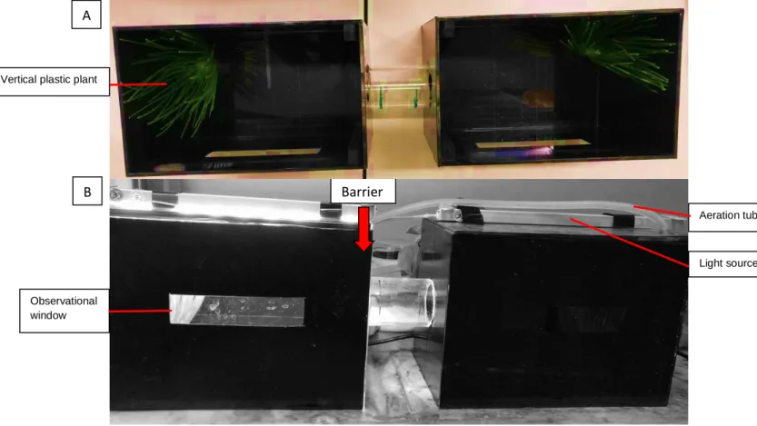
This task is based on Wong et al (2014) to study zebrafish aversion to the anaesthetics. This test is conducted in an apparatus that consists of two opaque aquariums linked by a tube that allows the fish to visit both sides (Figure 5). When a barrier is needed, it is placed in the illuminated side to restrain the animal from switching sides. As referred, in this test, zebrafish were housed in pairs. Each aquarium has a lid and each lid has a light source, but only one was on each given time. Plastic vertical plants were added as an environmental enrichment so the zebrafish could cope with pairing house, providing a place to hide. Plants were always placed in both side of the apparatus, in the opposite corner of the tube, near the back wall. Aeration was also provided and placed near the plant, as the system was closed (see scheme of the test in Annex 4).
Light source Aeration tube
Fig. 5 – Conditioned Place Aversion experimental apparatus top (A) and front view (B). Barrier
A
B Vertical plastic plant
Observational window
Habituation
This phase lasted 48 hours and allowed the animals to acclimatise to the apparatus. In this phase animals were fed 5 times a day in the side where they were found. Before feeding time, we recorded the movements of the animals for 10 minutes to assess in which side of the apparatus the individuals spend more time, i.e., their preference for the lightning conditions. In the end, the average of all trials showed that our animals spent more the lighted side. As, at this point, the animals were not tested; the word “control” corresponds to the 48 hours of habituation.
Training phase
In this phase, before feeding, animals were transferred to the illuminated side of the apparatus using a net; animals already at that side were also netted. Besides putting the animal in the right side for feeding, this procedure allowed zebrafish to be habituated to netting, minimising the stress of being handled. The animals stayed in the lighted side and could not escape to the dark side because of the divider placed in the lighted side. Then, light conditions were switched between sides, divider was removed, and food was placed in the current lighted side and animals were free to move and eat. Feeding animals in the lighted side will reinforce the preference established during habituation. Furthermore, the divider removal and the light switch will act as cues signalling the presence of food in the other side. This way, the animal is trained to pass the tube. This phase lasted for a maximum of 10 days or until the individuals reach the criterion of fully trained, i.e., when at least one animal from each pair entered 3 times in the lighted side in less than a minute and never took more than 2 minutes in the other trials in one day. Thus, during this period, latency to enter in the lighted side was recorded. The measures of the last day of training – Last training - were used to be compared with the testing period measures.
Testing period
Twenty-four hours after being fully trained, animals were video recorded for 15 minutes – Preanaesthesia. Then, each pair of fish was subjected to only one anaesthetic episode, wherein the concentration used was defined by the establishment of an equipotent dose (study 1). In the test period, a barrier was placed in the illuminated side between the tanks to avoid animals to pass to the non-illuminated side. In this side, the anaesthetic is diluted. Then, the light conditions were switched between sides, the barrier lifted and then the assay started with the food introduction in the illuminated side containing the anaesthetic. This trial ended when at least one of the individuals lost the equilibrium (assessed by observation), when 3 minutes after entering the side with anaesthetic elapsed (a safe measure of time to ensure that the animal in unconscious as the apparatus is opaque with only a small window and so it is not always possible to check the state of the animal) or when 15 minutes had passed.
This point of observation is called “Anaesthetic episode”. Afterwards, they are allowed to recover in a tank with clean water for at least 15 minutes, period after which the animals exhibited a normal swimming and behaviour. Then the animals were introduced again in the dark side of the experimental apparatus, with the barrier in this side to prevent animals to pass to the illuminated side for 5 minutes. The previous trial is then repeated without adding the anaesthetic to test if there is a conditioning regarding the side that previously had this compound (the previously preferred side)- Postanaesthesia. Time spent in each side, number of visits, attempts and latency to enter the lighted side were recorded to assess the aversion induced by each anaesthetic. Attempt is scored when fish enters the tunnel until 1 cm of the entrance, with head facing the tunnel, while a visit is considered when fish whole body enters in the aquarium.
All recordings were made with the camera placed facing the tunnel and later analysed manually using video and keyboard input recording software.
Prior to the test, a trial was made with food colouring to ensure that there was no diffusion of the anaesthetic to the other side of the apparatus. After 30 minutes, there was minimal diffusion to the opposite side, which was more than the duration of the trial, ensuring that there was no or only a negligible passage of the anaesthetics to the dark side of the apparatus during testing period.
Data analysis
First, data were analysed concerning normal distribution (Shapiro-Wilk test) and homogeneity of variances between groups (Levene’s test).
The time to lose equilibrium, reflexes and to recover from anaesthesia between different concentrations of the same anaesthetic protocol (three groups) and between anaesthetic protocols (comparisons between low, intermediate and high concentrations) can be evaluated by statistical tests that compare the means of a continuous dependent variable between unrelated groups. The same tests will also be used to compare the differences between anaesthetic protocols regarding the latency to pass to the illuminated tank, number of attempts and time spent in each tank in the conditioned place aversion test. If the assumptions normal distribution and homogeneity of variances between groups were fulfilled, a parametric test was used: Student’s t-test for comparisons between two unrelated groups or one-way ANOVA with Tukey as a posthoc test when three unrelated groups are evaluated. If the homogeneity of variances is not verified, a parametric test with corrections to this assumption violation will be used (Welch correction with Games-Howell as a posthoc test). When both assumptions are broken, the analysis was performed using a non-parametric test: Mann-Whitney U test for two unrelated samples and Kruskal-Wallis with the posthoc Dunn’s test for three unrelated samples.
The paired Student’s t-test compares the means of a continuous dependent variable between two related groups, while the Wilcoxon signed-rank test is the correspondent nonparametric test. These tests can be used to compare the differences in the time spent in the dark and illuminated side, in the preferred side before and after anaesthesia exposure, and to compare the number of attempts of the same animal in different points of observation within a treatment group. Friedman test is used to compare non-parametric data of more than two related groups, in this case, it was used to compare the latency of the same animal to enter in the lighted side between different points in time: the last day of training, anaesthetic episode and post-anaesthesia.
To study the anaesthetic protocols’ doses at which the majority of the animals lost equilibrium between 60 and 100 seconds, a scatter plot of the data was used.
All hypotheses were two-tailed tested and statistical significance was set at p≤ 0.05. Data was inserted in the Microsoft Excel™ 2010 (Microsoft Corporation, Redmond, WA, USA), and analysed using IBM SPSS™ 20 for Windows (SPSS Inc., Chicago, IL, USA). Prior to the beginning of the study, a sample size calculation2 was made using Wong’s results to ensure that our number of animals/pairs was enough to replicate the protocol (Wong et al., 2014)
Results
Detailed statistical results can be consulted in the annexes 2, 3 and 5.
Establishment of equipotent doses and clinical variables
Old fish pilot study
In our study, the equipotent doses were established as the doses of different anaesthetic protocols that induced a loss of ventral recumbency at the same time, between 60 and 100 seconds. Most of the animals that loss equilibrium at that range of time was treated with the high concentrations of both anaesthetics: C75 and M200 group (Figure 6). As both groups had low response variability, these concentrations were established as equipotent doses using 1.5 years zebrafish.
Regarding time to lose equilibrium, there were significant differences between concentrations in the Propofol/lidocaine combination groups (H (2) = 12.418, p= 0.002) where C75 animals lose the equilibrium faster than the ones from C25 group (p= 0.001). Regarding this variable, there are also significant differences between MS222 groups (H (2) = 11.980, p= 0.003), with M200 being quicker to lose equilibrium than the other groups (p= 0.036 and p= 0.003 for M175 and M150, respectively). Between anaesthetic protocols, M200 zebrafish lost the equilibrium faster than C25, and C75 was also faster than M150 (p= 0.002, p= 0.034, respectively) (Figure 6). However, there were no differences between all the other groups.
Fig. 6 - Time to lose equilibrium in each anaesthetic protocol. Data are presented as median [interquartile range]. C25, C50, and C75 - groups anaesthetized with 2.5mg/L, 50mg/L or 75mg/L of propofol, respectively, combined with 150mg/L of lidocaine; M150, M175, M200 - groups anaesthetized with 150mg/L, 175mg/L or 200mg/L of MS222, respectively. * for comparisons with C25 (p= 0.001) and M150 (p= 0.034); # for comparisons with M150 (p= 0.003), M175 (p= 0.036), and C25 (p= 0.002). Comparisons between groups using Kruskal-Wallis with Dunn’s test.
Regarding time to lose reaction to a light touch, there were significant differences between concentrations in the Propofol/lidocaine combination groups (F (2, 12.58) = 6.752, ppropofol/lidocaine= 0.010) where C75 animals lost reaction to this stimulus more quickly than C25 group (p= 0.049). In the animals anaesthetized with MS222 (F (2, 15.18) = 10.503, p= 0.001), M200 group presented a quicker loss of response to light stimulus than M150 (p= 0.004) and M175 (p= 0.042). Also, C75 animals lost the reaction to touch faster than M150 (p= 0.015) (Figure 7). No other difference between anaesthetic protocols or doses was detected.
Regarding time to lose the response to a painful stimulus, there were no significant differences between concentrations in the Propofol/lidocaine combination groups (F (2, 14.31) = 2.853, ppropofol/lidocaine= 0.091), but there were significant differences in the MS222 groups (F (2, 14.74) = 8.445, pms222= 0.004) where M200 zebrafish loss response to pain more quickly than the other MS222 groups (p= 0.015
Fig. 7 - Time to lose reaction to touch in each anaesthetic protocol. Data are presented as median [interquartile range]. C25, C50, and C75 - groups anaesthetized with 2.5mg/L, 5.0mg/L or 7.5mg/L of propofol, respectively, combined with 150mg/L of lidocaine; M150, M175, M200 - groups anaesthetized with 150mg/L, 175mg/L or 200mg/L of MS222, respectively. # for comparisons with C25 (p= 0.049) and M150 (p= 0.015); * for comparisons with M150 (p= 0.004), and M175 (p= 0.042). Comparisons between groups using Welch correction with Games-Howell’s as a posthoc test.
and p= 0.046 for M150 and M175, respectively). Also, M200 group lost response to this stimulus faster than C25 (p= 0.044), and C50 (p= 0.031) groups (Figure 8). Thus, 200mg/L of MS222 induced the loss of painful stimulus faster than the other anaesthetic protocol, except when 7.5mg/L of propofol combined with 150mg/L of lidocaine was used. No other significant differences were found.
During anaesthesia recovery, the animal’s latency to move is not different between the Propofol/lidocaine combination groups (H (2) = 5.058, ppropofol/lidocaine= 0.08) (Figure 9), nor between the MS222 groups (H (2) = 1.925, pms222= 0.382). However, Propofol/lidocaine groups took more time to start moving than MS222 groups, being data from C25, and C75 groups different from all MS222 groups (p≤ 0.012); C50 behaved differently from M150 (p= 0.011) and 175 (p= 0.010) (Figure 9). Concerning different anaesthetics, C50 and M200 are the only groups with similar latency to move after anaesthesia.
Fig. 8 - Time to lose reaction to a painful stimulus in each anaesthetic protocol. Data are presented as median [interquartile range]. C25, C50, and C75 - groups anaesthetized with 2.5mg/L, 5.0mg/L or 7.5mg/L of propofol, respectively, combined with 150mg/L of lidocaine; M150, M175, M200 - groups anaesthetized with 150mg/L, 175mg/L or 200mg/L of MS222, respectively. * for comparisons with M150 (p= 0.015), with M175 (p= 0.046), with C50 (p= 0.031) and with C25 (p= 0.044). Comparisons between groups using Welch correction with Games-Howell’s as a posthoc test.
Regarding time to regain equilibrium after anaesthesia, there were also no significant differences between concentrations in the Propofol/lidocaine combination groups (H (2) = 2.990, ppropofol/lidocaine= 0.224), nor in the MS222 groups (H (2) = 5.656, pms222= 0.059). However, all the MS222 protocols induced a quicker recovery compared with all the Propofol/lidocaine protocols (p≤ 0.025) (Figure 10).
Fig. 9 - Time to zebrafish start moving after each anaesthetic protocol. Data is presented as median [interquartile range]. C25, C50, and C75 - groups anaesthetized with 2.5mg/L, 5.0mg/L or 7.5mg/L of propofol, respectively, combined with 150mg/L of lidocaine; M150, M175, M200 - groups anaesthetized
with 150mg/L, 175mg/L or 200mg/L of MS222, respectively. * for comparisons with M150 (pC25< 0.001,
pC75= 0.001), M175 (pC25 < 0.001, pC75= 0.001), and M200 (pC25= 0.003, pC75= 0.012) ,# for comparisons
with M150 (p= 0.011),and M175 (p= 0.010). Comparisons between groups using Kruskal-Wallis with Dunn’s test.
Young fish assay
In order to confirm the equipotent doses showed by the previously pilot study, a new assay was performed with younger zebrafish, 4-7 months old, an age closer to the most used in research. Animals of that age were also going to be used in the Conditioned place aversion test. The equipotent dose of MS222 chosen for older animals (200mg/L), induce mortality in younger zebrafish with an inferior weight. Thus, this experiment showed that young animals need lower doses compared with the ones used in older zebrafish to achieve the same effects at a safe and efficient level. The dose of Propofol/lidocaine previously chosen did not show to be unsafe to younger zebrafish, but it needed to be adapted to the new equipotent dose of MS222 regarding the time to equilibrium loss.
Fig. 10 - Time to regain equilibrium after each anaesthetic protocol. Data are presented as median [interquartile range]. C25, C50, and C75 - groups anaesthetized with 2.5mg/L, 5.0mg/L or 7.5mg/L of propofol, respectively, combined with 150mg/L of lidocaine; M150, M175, M200 - groups anaesthetized
with 150mg/L, 175mg/L or 200mg/L of MS222, respectively. * for comparisons with M150 (pC25= 0.001, pC50
and C75= 0.011, ), M175 (p< 0.001) and M200 (pC25= 0.003, pC50 and C75= 0.025). Comparisons between groups
using Kruskal-Wallis with Dunn’s test.
Regarding the clinical variables presented in figure 11, the time to regain equilibrium during anaesthesia recovery is the only difference detected between the two anaesthetic protocols used. Animals anaesthetized with 175 mg/L of MS222 gained equilibrium faster than animals anaesthetized with a combination of 5 mg/L of propofol with 150 mg/L of lidocaine (U>0.001, p< 0.001). As there was no difference in time to lose equilibrium, these doses are equipotent for these zebrafish, inducing equilibrium loss in a range of 60-100 seconds for the majority of the animals. Compiling all data from both the pilot and the young fish assay, most of the animals anaesthetized with 175 mg/L of MS222 and 5 mg/L of propofol combined with 150 mg/L of lidocaine also lost equilibrium between 60 and 100 seconds (Figure 12).
Fig. 11 – Time to achieve a certain anaesthetic endpoint using MS222 or propofol/ lidocaine: equilibrium (Eq) loss, loss of response to a light touch (Touch loss) and to a painful stimulus (Pain loss), start moving (mov) during anaesthesia recovery, and equilibrium (eq) gain. MS222 group: animals anaesthetized with 175mg/L of MS222; Propofol/lidocaine group: animals anaesthetized with 5mg/L of propofol combined with
150mg/L of lidocaine. Data presented as median+ interquartile range. *p< 0.0001 for comparisons between
Conditioned place aversion
Light conditions preference
The assessment of the preference of side with different light conditions was made analysing the time spent on each side of the apparatus during the habituation phase. Regarding this, there was a significant difference within each group (t (7) = 4.185, pMS222 = 0.004, t (5) =4.382 pPropofol/lidocaine= 0.007, t (9) =2.533 pHCl = 0.032) (Figure 14), where pairs of zebrafish spent more time in the lighted side. Thus, they were trained to pass the tube to be fed in the lighted side. However, after training and before the anaesthetic episode, the same analysis was performed and there was no significant difference within each group in the preanaesthesia period (pMS222 = 0.401, pPropofol/lidocaine = 0.491, pHCl = 0.494). As there was no difference at the preanaesthesia Fig. 12 – Scatter plot of the time taken to lose equilibrium in both protocols regarding all data acquired from older and younger zebrafish to establish equipotent doses. MS222 group: animals anaesthetized with 175mg/L of MS222; Propofol/lidocaine group: animals anaesthetized with 5mg/L of propofol combined with 150mg/L of lidocaine. Each dot (square or rhombus) represents an animal. The grey area represents the 60 to 100 seconds time frame that most animals took to lose equilibrium.
T im e t o l o s e e q u il ib ri u m i n s e c o n d s


![Fig. 6 - Time to lose equilibrium in each anaesthetic protocol. Data are presented as median [interquartile range]](https://thumb-eu.123doks.com/thumbv2/123dok_br/19176218.943308/35.893.152.654.219.614/time-equilibrium-anaesthetic-protocol-data-presented-median-interquartile.webp)
![Fig. 7 - Time to lose reaction to touch in each anaesthetic protocol. Data are presented as median [interquartile range]](https://thumb-eu.123doks.com/thumbv2/123dok_br/19176218.943308/36.893.177.669.455.870/time-reaction-touch-anaesthetic-protocol-presented-median-interquartile.webp)

![Fig. 9 - Time to zebrafish start moving after each anaesthetic protocol. Data is presented as median [interquartile range]](https://thumb-eu.123doks.com/thumbv2/123dok_br/19176218.943308/38.893.207.709.182.591/time-zebrafish-moving-anaesthetic-protocol-presented-median-interquartile.webp)
