Behavioural effects of anaesthetics in mice
–
risk of
cognitive alterations
Ana Maria Marques Valentim
Tese de doutoramento em Ciências Veterinárias
Ana Maria Marques Valentim
Behavioural effects of anaesthetics in mice- risk of cognitive
alterations
Tese de Candidatura ao grau de Doutor em
Ciências Veterinárias
submetida ao Instituto de
Ciências
Biomédicas
Abel
Salazar
da
Universidade do Porto.
Orientador
–
Doutor Luís Miguel Antunes
Categoria
–
Professor Associado
Afiliação
–
Universidade de Trás-os-Montes e
Alto Douro
Co-orientador
–
Doutora Anna Olsson
Categoria
–
Investigadora Associada
Afiliação
–
Instituto de Biologia Molecular e
Celular
Co-orientador
–
Professor Doutor Pedro
Moradas-Ferreira
Categoria
–
Professor Catedrático
The outcomes of this thesis have been published:
In conference proceedings:
Valentim A., Di Giminiani P., Ribeiro P., Olsson A., Antunes L. (2008). Depth of
Anesthesia Effects on Cognition – a Battery of Behavioral Tests. Proceedings of the American Society of Anesthesiologists Annual meeting 2008; 109, A323
Valentim A., Olsson I.A., Antunes L.M. (2010). Ketamine/Midazolam Do Not
Interfere with Consolidation and Recall of a Spatial Task in Mice. Proceedings of the American Society of Anesthesiologists Annual meeting 2010, 16-20 of October, San Diego, California, USA, A275
Valentim A., Olsson I.A., Antunes L.M. (2011) Influence of anesthetic
ketamine/midazolam combination in spatial and motor learning. Proceedings of the American Society of Anesthesiologists Annual meeting 2011, 15-19 of October, Chicago, Illinois, USA, A1679
International journals:
Valentim A. M.; Alves H. C.; Silva A. M.; Olsson I. A. S.;. Antunes L. M (2008)
Effects of depth of isoflurane anaesthesia on a cognition task in mice. British Journal of Anaesthesia 101: 434-435.
Valentim A., Di Giminiani P., Ribeiro P., Rodrigues P., Olsson I. A., Antunes L.
i
Acknowledgements
Four years of discoveries, disappointments, hard work, happy moments, excitement, sharing knowledge, hopes and ideas… It seems difficult to mention all the people that deserve to be in here, but I am going to try!
First of all I would like to thank the Fundação para a Ciência e Tecnologia and FEDER for the financial support through a personal grant (SFRH/BD/36121/2007) and two project grants: “Refinements in laboratory animal anaesthesia: behavioural and physiological measures” (POCTI/CVT/59056/2004) and “Effects of ketamine anaesthesia in animal models used on research” (FCOMP-01-0124-FEDER-009497, PTDC/CVT/099022/2008).
To my supervisor, Luís Antunes, for giving me independence but also for being there to discuss, thank you. To Anna Olsson, my co-supervisor, and PI of the research group where I belong, thank you for all the listening, support and comprehension in sad moments, for all the good judgment and knowledge. To Prof. Pedro Moradas-Ferreira, thanks for all your comments, and perspectives and for accepting being my co-supervisor in a theme slightly out of your main area of research.
In UTAD, thanks to the technical support of Lígia Lourenço, and the collaboration of Dr. Paula Rodrigues in the histological methodology.
ii practicability to solve problems, thanks. To Dr Pedro Amorim, for all his enthusiasm, for making me feel more confident, for all the support, a big thank you!
Thank you also Kathy Murphy, and Amy Taylor from Oxford for your constant availability to help me in endless e-mails! In general, thanks to the Rodent Behaviour group in Oxford for receiving me in their group meeting, for listen, and for giving me precious advices.
I would like to thank many more friends. To my dearest friends and colleagues of Biology of Coimbra, that were always there for me, thank you, you are really important to me! And yes, I am finally really tired (they will understand). To my friends in Tondela, for the support and nice weekends.
For the people that I met during this journey that are so important to me: Alice, Carla, thanks for receiving me so well and for being there even in the saddest moments; Ana Castro, Nadja, thanks for all the care; Marina, thanks for your unexpected friendship; Margarida, for your strength, thank you; Susana, for your priceless help, listening, support, thanks for everything; Sónia, your support, and advices made me stronger, thank you.
To Ticha, Rita, Samira, Marisa, thanks for your friendship, far or close, you are part of my life, part of who I was and am. To my best friend Ângela, for listening, for being there, for existing, for being a FRIEND, because we are always together, thank you!
To my love Pedro, with all ups and downs, I thank you for all the support, your gags, and crazy stories just to make me feel better, and even for helping me with your programmer skills! Thank you because I really don‟t know where I would be without you…
To my brother, for telling me how proud he is, even not really understanding how science works, for showing me that having fun is also important, thank you!
iii
Abstract
Anaesthesia is compulsory during surgery and other invasive procedures that are part of everyday clinical practice, and in current experimental research using animal models. Anaesthesia acts as a reversible intoxication and depression of the central nervous system by interfering with the metabolism and the electrophysiology of the brain. The mechanism by which anaesthesia affects brain functions and the duration of its effects are not fully understood. In humans, particularly in the elderly, the post-operative cognitive dysfunction phenomenon is problematic, because it may last for months. In rodents, the effects of anaesthesia reported in the literature are not consistent, as it has been shown to compromise cognition and cell viability, to enhance cognition in young animals or to cause no alterations. The possible effects of anaesthesia on cognition in humans and other animals depend on several factors, such as age of the individual, type and dose of the anaesthetic used, acute or chronic administration, duration of anaesthesia, type of cognitive function, species, and genetic background.
In addition to the potential clinical implications of anaesthesia, its interference with behaviour may induce artefacts in the data generated from animal behavioural studies, thus contributing to disparity in results or even erroneous conclusions.
Evidences regarding the effects of anaesthesia on cognition are described in clinical contexts, in which the distinction between the effects of anaesthesia alone and the effects of surgery and all its variables is not possible. Hence, this project aimed to study the real effects of anaesthesia alone using experimental situations.
iv Four studies were performed: two using isoflurane (chapter 3, sections 3.1 and 3.2) and two using the ketamine/midazolam combination (chapters 4 and 5).
In chapter 3, we describe the use of different concentrations of isoflurane that were studied in a battery of behavioural tasks, using an opaque radial arm maze, an open- field arena, T-maze spontaneous alternation, and a water maze. Volatile agents are excellent to study the depth of anaesthesia, as expired gases can be easily monitored using a gas analyser, without invasive procedures. Two studies are reported in this chapter: 3.1 and 3.2. In study 3.1, we assessed the learning ability of animals treated with low (1%) and high (2%) concentrations of isoflurane. The study presented in section 3.2 included one additional concentration (1.5%), thus allowing the establishment of a dose-effect curve. In the latter study, we also assessed working memory and reference memory using a spatial task, and neurodegeneration.
In chapter 4, we describe the first approach to study the ketamine/midazolam combination, in which we used the minimum dose that causes the loss of the righting reflex (40 mg.kg-1/10 mg.kg-1
)
. The effect of a spatial task (appetitive y-maze) on differentprocesses of memory - acquisition, consolidation and recall - was evaluated. General cell death was also assessed by hematoxylin-eosin staining.
In the study described in chapter 5, we used a higher concentration of ketamine/midazolam (75 mg.kg-1/10 mg.kg-1
)
, as ketamine was described to have adose-dependent effect on cognition. Acquisition of a task is the most labile process of memory; thus, acquisition of a motor task (rotarod) and a demanding spatial task (radial arm maze) were studied.
Studies performed using isoflurane (chapter 3) showed that low isoflurane concentration (1%) caused learning impairment only in spatial tasks. In support of the finding that low concentrations of this volatile affected spatial memory, a higher number of cells expressing caspase-3 was detected at lower concentrations of isoflurane in the region CA1 of the hippocampus. The impairment detected in the water maze was not found 5 days post-anaesthesia; thus, the effect was transient.
vii
Resumo
A anestesia é essencial para a realização de cirurgia ou outros procedimentos invasivos que fazem parte da rotina clínica ou da investigação. A anestesia actua como uma intoxicação e depressão reversível do sistema nervoso central, interferindo com o metabolismo e electrofisiologia do cérebro. Os mecanismos pelos quais a anestesia afecta as funções cerebrais e a duração desse efeito não são completamente compreendidos. A disfunção cognitiva pós-operativa encontrada em humanos, especialmente em idosos, tem sido descrita como problemática, pois pode durar meses. Na literatura, a descrição dos efeitos da anestesia em roedores não é consistente, uma vez que têm sido descritos efeitos nefastos na memória e na viabilidade celular, outros estudo revelam uma melhoria na memória em animais jovens e há até estudos que mostram a anestesia como sendo inócua a este respeito. Os potenciais efeitos da anestesia na memória de humanos e de outros animais dependem de vários factores como a idade do indivíduo, o tipo e dose de anestésico usado, administração aguda ou crónica, duração da anestesia, tipo de função cognitiva, espécie e até do fundo genético.
Para além das implicações clínicas que a anestesia pode ter, a sua interferência no comportamento animal pode induzir artefactos nos resultados de projectos de investigação com animais, onde esta variável é estudada, contribuindo para a disparidade de resultados ou mesmo conclusões erróneas.
Muitas das evidências de que a anestesia influencia a memória foram encontradas em contextos clínicos, onde não é possível separar os efeitos causados apenas pela anestesia e os efeitos causados pela cirurgia e todas as suas variáveis. Assim, este projecto visa estudar o efeito real da anestesia sozinha recorrendo a situações experimentais.
viii e humanos. Os murganhos usados foram testados em diferentes tarefas comportamentais usadas para avaliar diferentes tipos de memória e regiões cerebrais.
Foram realizados quatro estudos: dois usando isoflurano (capítulo 3- secção 3.1 e 3.2) e dois usando a combinação cetamina/midazolam (capítulo 4 e 5).
No capítulo 3, foi descrito o estudo de diferentes concentrações de isoflurano numa bateria de testes comportamentais usando um labirinto de braços radial opaco, arena em campo aberto, alternância espontânea num labirinto em T e labirinto de água. Os agentes voláteis são excelentes para estudar profundidade anestésica, uma vez que o gás expirado pode ser facilmente medido por um analisador de gases, evitando procedimentos invasivos. Este capítulo contém dois estudos: 3.1 e 3.2. No estudo 3.1, animais tratados com baixa (1%) e alta (2%) concentração de isoflurano foram testados com vista a avaliar a sua capacidade de aprendizagem. No estudo da secção 3.2, foi acrescentada uma concentração de isoflurano (1.5%) para que uma curva dose-efeito pudesse ser traçada. Neste segundo estudo também se avaliou a memória do trabalho e de referência numa tarefa espacial e a neurodegeneração por apoptose.
No capítulo 4 encontra-se a descrição da primeira abordagem ao estudo da combinação cetamina/midazolam que consistiu em escolher a dose mínima necessária para os animais perderem o reflexo de se endireitarem (40mg.kg-1/10 mg.kg-1). Neste estudo, foram avaliados os efeitos desta combinação nos diferentes processos de memória - aquisição, consolidação e evocação de uma tarefa espacial (labirinto em Y apetitivo). A técnica de coloração hematoxilina-eosina foi usada para avaliar a morte celular em geral.
Comparativamente com o estudo anterior, o estudo reportado no capítulo 5 analisou uma dose maior de cetamina/midazolam (75mg.kg-1/10 mg.kg-1), uma vez que a cetamina foi descrita como tendo um efeito dependente da dose na funcionalidade da memória. Como a aquisição de uma tarefa é considerado o processo mais lábil da memória, este estudo avaliou a aquisição de uma tarefa motora (rotarod) e de uma tarefa espacial exigente (labirinto de braços radial).
ix isoflurano. O défice observado na tarefa do labirinto de água não foi encontrado 5 dias após a anestesia, revelando que este efeito é transitório.
Ao resultados do estudo descrito no capítulo 4, não revelaram nenhum efeito nefasto da dose sedativa da combinação cetamina/midazolam sobre a aquisição, consolidação ou evocação de uma tarefa espacial. Este resultado é apoiado pelo igual número de células mortas detectadas nos cérebros tratados com a combinação e nos cérebros controlo. Mesmo com uma dose mais elevada (capítulo 5), esta combinação não interferiu no processo de aquisição da tarefa espacial labirinto de braços radial, da tarefa motora, nem interferiu na memória do trabalho.
xi
Index
Acknowledgements ... i
Abstract ... iii
Resumo ... vii
Index ... xi
Abbreviations ... xv
Outline of thesis ... 1
1. General Introduction ... 3
1.1. The use of laboratory animals: 3R‟s ... 5
1.2. Neuroanatomy ... 6
1.3. Memory ... 8
1.3.1. Memory according to contents ... 9
1.3.2. Memory according to duration ...11
1.3.3. Memory according to its nature ...12
1.3.4. Long term potentiation and hippocampus ...12
1.3.5. Hippocampus and spatial memory ...13
1.4. Anaesthesia ...14
1.4.1. Neurotransmitters ...15
1.4.1.1. GABA receptors ...17
1.4.1.2. Glutamate receptors ...18
1.4.1.2.1. NMDA receptors ...18
1.4.2. Anaesthetics ...20
1.4.2.1. Isoflurane ...20
Mechanism of action and clinical effects ...21
Uses and clinical implications ...22
1.4.2.2. Ketamine ...23
Mechanism of action and clinical effects ...23
Uses and clinical implications ...24
1.4.2.3. Midazolam ...25
Mechanism of action and clinical effects ...25
Uses and clinical implications ...27
1.4.2.4. Ketamine/midazolam combination ...28
Uses and clinical implications ...28
1.5. Anaesthesia vs. Memory...29
1.5.1. Age ...30
xv
Abbreviations
AMPA - α-amino-3-hydroxyl-5-methyl-4-isoxazole-propionate ANOVA – analysis of variance
ATP – adenosine triphosphate bpm – beats per minute brpm – breaths per minute C3E – caspase-3 expression Ca2+ - calcium ion
CA –cornu ammonis cm – centimetre
CNS – central nervous system DNA – deoxyribonucleic acid GABA –γ-Aminobutyric Acid
GABAA receptor– subtype A of the γ-Aminobutyric Acid receptor
GABAB receptor - subtype B of the γ-Aminobutyric Acid receptor
h – hour(s)
HE – hematoxylin-eosin
IF1% - group of animals anaesthetized with 1% of isoflurane IF1.5% - group of animals anaesthetized with 1.5% of isoflurane IF2% - group of animals anaesthetized with 2% of isoflurane ip. - intraperitoneal
K+ - potassium ion
kg – kilogram
K/M - group of animals treated with the combination ketamine/midazolam LTD – long-term depression
LTP – long-term potentiation m – metre
xvi MAC – minimum alveolar concentration
mg – milligram
Mg2+ - magnesium ion
min. – minute(s) Na+ - sodium ion
NMDA - N-methyl-D-asparticacid PCB – post-choice baiting
PCP - phencyclidine PND – post-natal day
POCD – post-operative cognitive dysfunction RAM – radial arm maze
rpm – rotations per minute s – second(s)
SD – standard deviation
1
Outline of thesis
In the General Introduction chapter some contents are explained. First, a number of issues that need to be taken into consideration in research with animals are addressed in section 1.1, “the use of laboratory animals: 3R‟s”. Thereafter, as it is important to recognize the structures in which anaesthetics may act, a section on “Neuroanatomy” is included. “Memory” and “Anaesthesia” are the following topics, and the relation between these topics is explored in the section “Memory vs. Anaesthesia”. Finally, the issue “Measuring anaesthetic effects” allows a better understanding of the methodologies latter described to evaluate the effects of anaesthetics on memory function in mice.
In chapter 2, the general material and methods with the sub-sections animals and husbandry, food restrictions schedule and reward, anaesthesia procedures, behavioural tasks, histopathological and immunohistochemial techniques are described. Chapters 3-5 report the experimental studies:
- Chapter 3 reports two studies about the effects of different concentrations of isoflurane on memory. The study from the section 3.2 had one more concentration of isoflurane compared with study from section 3.1, allowing the assessment of a curve-dose effect.
- Chapter 4 describes the effects of a sedative dose of ketamine/midazolam combination in acquisition, consolidation and/or recall of a spatial task.
- Chapter 5 describes a study that investigated the effects of a light anaesthetic dose of ketamine/midazolam in the acquisition of a motor task and of a demanding spatial task.
3
1. General Introduction
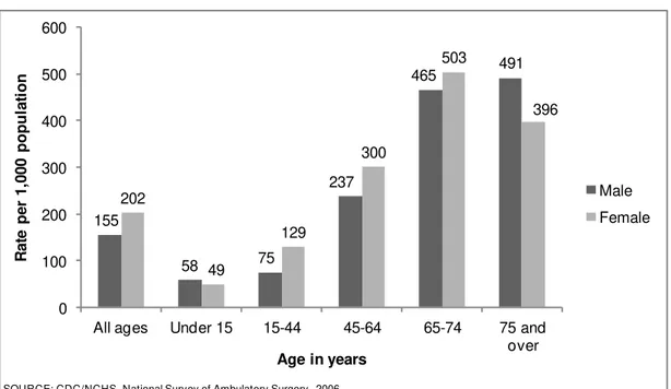
It is estimated that every year 234.2 millions of surgeries in humans are performed around the world. This number may even be underestimated, given the number of unregistered interventions (Weiser et al. 2008). Several procedures are now done on middle-aged and senior patients for diagnostics under sedation or light anaesthesia which are probably not taken into account. The National Health Statistics Reports estimated that in 2006 the most frequently performed procedures on ambulatory surgery, in the United States of America, included endoscopy of large intestine (5.7 million), endoscopy of small intestine (3.5 million), extraction of lens (3.1 million), injection of agent into spinal canal (2.0 million), and insertion of prosthetic lens (2.6 million) (Cullen et al. 2009). The age groups in need of more interventions were the middle-aged and senior patients (fig. 1). These procedures, along with many other invasive techniques that are part of everyday clinical practice, is nowadays inconceivable to be performed without anaesthesia.
Fig. 1: Rate of ambulatory surgical procedures per 1,000 of the population of the United States of
America by age and sex, in 2006 (from National Health Statistics Reports, number 11).
Regarding pre-clinical research using laboratory animals, a statistical report of procedures revealed that 33% of those were carried out under anaesthesia in the United Kingdom in 2009; the majority of the animals used were mice and fish (Home Office,
155 58 75 237 465 491 202 49 129 300 503 396 0 100 200 300 400 500 600
All ages Under 15 15-44 45-64 65-74 75 and
over R a te pe r 1 ,0 0 0 po pu la ti on
Age in years
Male Female
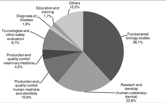
4 2010). Anaesthesia is a requirement for several procedures in neurobiological research, such as the implementation of monitoring devices (electrodes, arterial catheters, intracranial pressure monitors), neurotoxin administration, induction of traumatic brain injury and surgery, amongst others. Although there is no information on the proportion of animals being anaesthetized, official statistics of the European Union commission (European Commission, 2010) reported that, of the 12 million vertebrates(59,3% of which were mice) used in research in 2008, more than 60% had been used in research and development for human medicine, veterinary medicine, dentistry and in fundamental biological studies (fig. 2), many of which are estimated to have required the use of anaesthetics.
Fig. 1: Representation of the percentage of animals used in research in 2008 according with their
use (from Report from the Commission to the Council and the European Parliament, 2010).
Anaesthesia needs to be considered as a variable that potentially influences research results, in particular when animals are subsequently studied in behavioural/cognitive tests. Hence, this project focuses on the impact of anaesthesia on normal animal behaviour, which is essential to obtain reliable data from research projects using animals.
There are several studies about cognitive impairments detected after surgery and anaesthesia in the elderly and middle-age humans (Monk et al. 2008). In rodents, anaesthesia has been reported to cause cognitive dysfunctions to old (Culley et al. 2004a) and infant animals (Fredriksson et al. 2004). In this scenario, little is known about the
5 impact of anaesthesia in adulthood, which is the reason to use adult animals in this project. Although the younger and older brain may be more prone to injury, this does not mean that adults are safe from anaesthesia side-effects. To study the effects of anaesthesia alone, other variables, such as surgery, were excluded. Only anaesthesia was performed on these animals. Two different anaesthetic protocols often used in laboratory animals practice were evaluated: the volatile hypnotic anaesthetic isoflurane, and an injectable combination of ketamine (a dissociative) with midazolam (a benzodiazepine). Because isoflurane is a volatile anaesthetic, its concentration is easy to monitor in the expired air by a gas analyser, allowing the study of different depths of anaesthesia. Ketamine is probably the most commonly used injectable anaesthetic in laboratory rodents, and midazolam was chosen because of its rapid onset of action and recovery, being administered through an extra-vascular route with no harm for the tissues in the site of injection (Dundee et al. 1984).
1.1. The
use of laboratory animals: 3R’s
The general aim of this work is to study the impact of anaesthesia in the results of research projects using animals. Thus, the guiding principles for the use of animals in research are of extreme importance.
Those guidelines were first introduced by Russell and Burch in 1959 (Russell et al. 1992, special edition), who proposed a series of procedures so that animals in research could be treated in a more “humane” way, the 3R‟s politic, consisting in the principles of Replacement, Reduction and Refinement. First, the researcher has to be sure that the experiment cannot be done without animals, and instead be performed using alternative scientific methods involving non-sentient material such as molecular, computer, and mathematical models - Replacement. Russell and Burch also referred to the relative
Replacement where animals with lower complexity of nervous system or animal tissues are used. Then the researcher has to reduce the number of animals necessary, through a good experimental design, a proper statistical method and analysis (Festing et al. 2002), or choose the number of animals based on a preliminary study (Sherwin et al. 2003) -
Reduction. However care should be taken not to use so few animals that results become
inconclusive, and in the end, those animals were used in vain (Flecknell 2002). After efforts have been made to Replace and Reduce, researchers should apply Refinement,
6 during the life of the animals involved, and which enhances their wellbeing‟‟ (Buchanan-Smith et al. 2005). Refinement can be obtained with the improvement of husbandry conditions, refinement in the experimental procedures such as the use of anaesthesia and analgesic techniques, maintenance of daily routines, avoidance of single housing in social species, well trained operators/ researchers/ technicians, among other issues (Sherwin et al. 2003). This way the animal behaves as naturally as possible, reducing the data variability and achieving robust results on the experiment, which also reduces the number of animals needed.
The implementation of the 3R‟s is essential before beginning an experiment with animals since, besides being part of the legislation (Directive 2010/63/EU of the European parliament on the protection of animals used for scientific purposes), the 3R‟s are an ethical and scientific approach to be followed in research.
In this work, it is important to find an anaesthetic procedure that does not cause memory dysfunction. Thus, researchers can use that method to avoid the animal suffering in the experimental situation, refining the experiment and improving animal welfare.
1.2. Neuroanatomy
The aim of this section is to provide a brief notion about the location and structure of the main brain regions referred to throughout the thesis.
7
Fig. 1.2.1: Comparative basic neuroanatomy of human and mouse. The top image represents the
surface of the brain of each species and the bottom figure is a coronal section. The midbrain is not presented since it is located rostral to the pons. (figure adapted from Holmes & Cryan (2005) Nature Reviews Drug Discovery 4, 775-790).
The hippocampal formation is also of great importance for this project since it is strongly related with memory and several behavioural tasks. The hippocampus is formed by three subfields termed Cornu Ammonis (CA): CA1, CA2 and CA3; besides these regions, the hippocampal formation also comprises the dentate gyrus, and parahippocampal gyrus (subiculum and entorhinal cortex). The hippocampal formation presents three different cell layers: a molecular (scattered small nervous cells), multiforme (polymorphic nervous cells), and granular (in dentate gyrus) or pyramidal (in CA regions) stratum (El Falougy et al. 2008).
8
Fig. 1.2.2: Anatomy of the hippocampal formation in the human brain (a) and in the rat brain (b).
CA- Cornu Ammonis; DG- dentate gyrus (figure a from
http://en.wikipedia.org/wiki/File:Hippocampus_%28brain%29.jpg, original image donated by http://members.optusnet.com.au/changelingp/ Frank Gaillard Designs; figure b from http://quizlet.com/5076293/memory-systems-58-flash-cards/).
1.3. Memory
9 that of the patient named H.M., who had his hippocampus removed, as well as amygdala and part of his parahippocampal gyrus on both sides of the brain in order to reduce chronic seizures (Milner 1972). H.M. lost the capacity to form new long-term memories, although he remembered all events that had happened prior to the surgery, and his short-term memory was perfectly intact. Hence, memory involves several regions of the brain and different types of memory are stored in different neural systems (Kandel et al. 1995). Indeed, current behavioural and biological studies have shown that learning and memory are not a unitary process of mind, but rather distinct processes, composed of multiple systems with different principles and neuroanatomy (Squire 2004).
The classification of memory types differ between authors. In this work, the classification of Izquierdo will be used. According to Izquierdo (Izquierdo et al. 1999), memories can be classified according to their: content (declarative/ explicit, and nondeclarative/ procedural/ implicit), duration (short-term memory, and long-term memory), and nature which is divided in archival (short-term memory, long-term memory), and transient, moment-to-moment memory (working memory).
1.3.1. Memory according to contents
10 Fig. 1.3.1: The organization of different types of memory according to their content.
11
Fig. 1.3.2: Simplified anatomical framework for declarative and nondeclarative memory. parahipp.
region- parahippocampal region; HPA axis- hypothalamic-pituitary-adrenal axis. (from handouts of the Introductory Course in Neuroscience, lecture Learning and Memory by David P. Wolfer, MD in 15.03.2010; http://www.neuroscience.uzh.ch/education/handouts/wolfer_fs10.pdf).
1.3.2. Memory according to duration
12 (Bailey et al. 1996; McGaugh et al. 2000). The hippocampus, entorhinal and parietal cortex are crucial for short and long term memory, while the anterolateral prefrontal cortex and amygdala are described to be involved only in long term memory (Izquierdo et al. 1999).
1.3.3. Memory according to its nature
As previously mentioned, long and short term memories are a type of archival memory which is preserved offline for later use, while working memory is a type of transient memory. Another term found in the literature associated with long and short term memory is reference memory. Working memory involves the retention of information for short periods (seconds to a maximum of a few minutes) referring to a moment-to-moment memory for specific details in a particular situation (Hodges 1996; Izquierdo et al. 1999). Working memory is primarily dependent on the electrical activity of cells of the prefrontal cortex; it persists only as long as this electrical activity persists. The regions with a main role in this type of on-line system are the anterolateral prefrontal cortex, the hippocampus, entorhinal and parietal cortices (Izquierdo et al. 1999).
1.3.4. Long term potentiation and hippocampus
The amazing ability of the brain to receive inputs and to translate them into memories that can last for decades has been ascribed to the efficacy and plasticity of synaptic communication. It has been shown that repetitive activation of excitatory synapses in the hippocampus causes an increase in synaptic strength, helping the brain “to learn” and this could last for hours or days (Malenka et al. 1999). This process is called Long Term Potentiation (LTP) and it has been identified as the substrate to learning and memory processes (Myhrer 2003). Long term potentiation occurs in several regions of the mammalian brain (Malenka et al. 1999), but the most studied pathways of LTP are located in the hippocampus.
13
Fig. 1.3.3: Anatomical representation of the hippocampus and the pathways of long-term
potentiation. The signal passes from the entorhinal cortex (not shown) to the granule cells of the hilus of the dentate gyrus using the perforant path fibres; the axons from granule cells form the mossy fibre pathway that goes to the pyramidal cells of the CA3 regions. These cells are divided in two branches, the Schaffer collateral pathway – that connects the CA3 to the pyramidal cells of the CA1 region – and the commissural fibres that project to the contralateral hippocampus via the corpus callosum; the commissural fibres are not represented in this figure. (from http://thebrain.mcgill.ca/flash/a/a_07/a_07_cl/a_07_cl_tra/a_07_cl_tra.html).
1.3.5. Hippocampus and spatial memory
In the 1970s a ground-breaking theory proposed by O‟Keefe and Nadel postulated that hippocampal function is essential for the formation of cognitive maps. This occurs after the observation of place cells in the hippocampus, i.e. cells that become active only when the animal is located in a specific part of the environment (O'Keefe et al. 1971). This place cells were found mainly in the hippocampus but they can also be located in other regions of the brain, such as the frontal cortex, amygdala, and parahippocampal cortex (Bohbot et al. 2004). If each cell is consigned to a specific location, the firing pattern of the cells can form a very precise cognitive map about the position of the animal at any given time – cognitive mapping theory. Jeffery and O‟Keefe (Jeffery et al. 1999) showed that hippocampal cells respond firstly to visual inputs, especially the place cells located in CA1 region.
14 in the learning of maze tasks (Bannerman et al. 2001; Broadbent et al. 2004). There is also a growing body of evidence that the hippocampal formation in humans contributes to the encoding and retrieval of spatial information, for example through the observation that these processes are impaired in patients with hippocampal injuries (Good 2002). MRI studies also showed that healthy participants using a spatial strategy to solve a place-learning task have a significant activation of the hippocampus, contrarily to the individuals that chose a non-spatial strategy (Bohbot et al. 2004). Furthermore, Maguire and colleagues (Maguire et al. 2000) reported that the posterior hippocampus of experienced official London taxi drivers was significantly larger relative to control subjects, and that this volume was positively correlated with the years of experience. This reveals that the dependence on spatial navigational skills and training may induce morphological changes in the brain.
In general, the outcome from both human and animal studies concerning navigation tasks propose that the hippocampus has a crucial role in processing spatial information, such as the information about the position of certain visual cues related with the location of the individual.
1.4. Anaesthesia
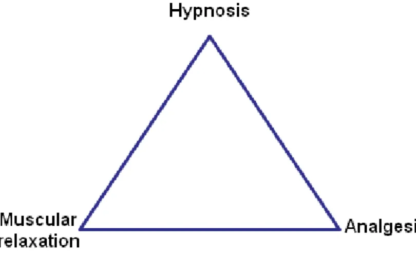
Although general anaesthetics were first used more than 150 years ago (Horine 1946), their mechanisms of action remain not fully understood. The concept of adequate anaesthesia was described in 1950 by Rees and Gray (Rees et al. 1950) as being a triad of components (fig. 1.4.1): hypnosis (lack of awareness), muscular relaxation, and analgesia (pain relief).
15 Lately, amnesia (inability to recall events) and suppression of stress response were also referred as important elements (Schneider et al. 1997). Since a sole drug does not seem to achieve all these effects with a reasonable safety margin, several drugs are used in combination. This is the concept of balanced anaesthesia (Tonner 2005), which postulates that low concentrations of drugs administered in combination act synergistically to produce the desired effects, while reducing the risk of side-effects such as hemodynamic instability (Arras et al. 2001; Alves et al. 2007).
These different actions indicate that anaesthetics have different CNS target locations to mediate specific effects. These targets are mainly proteins such as ion channels and neurotransmitter receptors that have different sensitivity and distribution in the central nervous system (Hemmings et al. 2005). For example, analgesia and immobility may be provoked by a blockage at the spinal cord level, but hypnosis and amnesia are effects only caused by interference in the brain (Villars et al. 2004). The action of anaesthetics towards ion channels regulates membrane potentials and synaptic transmission through neurotransmitter molecules. Hence, anaesthetics alter neuronal activity by hyperpolarization of neurons, increasing inhibition and/or decreasing excitation (Wakasugi et al. 1999; Alkire et al. 2009). This alteration in the action potential of the neurons blocks the elaborate paths of conscious perception within the central nervous system (Villars et al. 2004; Heinke et al. 2005). Neuroimaging (Peltier et al. 2005) and electroencephalogram (John et al. 2001) studies support that this blockage decreases brain activity and produces behavioural changes at anaesthetic concentrations, with impaired corticocortical and thalamocortical connectivity as well as electrical uncoupling of diverse brain regions. So, anaesthetics may cause loss of consciousness and amnesia by disrupting the interaction between brain areas and by the specific suppression of neuronal activity, reducing the integration and the amount of information processed (Heinke et al. 2005).
1.4.1. Neurotransmitters
16 postsynaptic receptor causing depolarization or hyperpolarization of the postsynaptic membrane (fig. 1.4.2). The effect of the binding described depends on the affinity of the molecule to the receptor and the receptor density in the brain regions (Heiss et al. 2006).
Glutamate and GABA are the major excitatory and inhibitory neurotransmitters of the central nervous system, respectively.
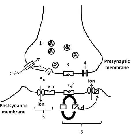
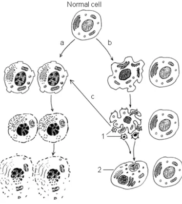
Fig. 1.4.2: Schematic representation of the synaptic transmission. 1- Neurotransmitter molecules
are stored in vesicles. 2- The action potential at the presynaptic neuron causes the entry of calcium (Ca2+) ions through voltage-activated calcium channels, leading vesicles to fuse with the presynaptic membrane and the release of their neurotransmitters into the synapse. 3- the presynaptic receptor or autoreceptor may bind to the neurotransmitters causing a negative feedback. 4- Neurotransmitters may also be re-uptaken through the presynaptic membrane. Neurotransmitters released in the synapse may bind to post-synaptic receptors causing a direct (5) or an indirect (6) opening of an ion-channel. 6- The neurotransmitter binding activates a second-messenger (triangle) that when attached to the ion-channel causes it to open. The opening of the ion-gated channel allows the correspondent ion to enter or leave the cell, depolarizing or hyperpolarizing the postsynaptic membrane.
+
Ca
2+ion
ion
1
2
3
4
5
6
Presynaptic
membrane
17
1.4.1.1.
GABA receptors
There are at least two subtypes of GABA receptors, GABAA and GABAB; no role in
anaesthesia is currently known for the GABAB receptor (Villars et al. 2004). The GABAA
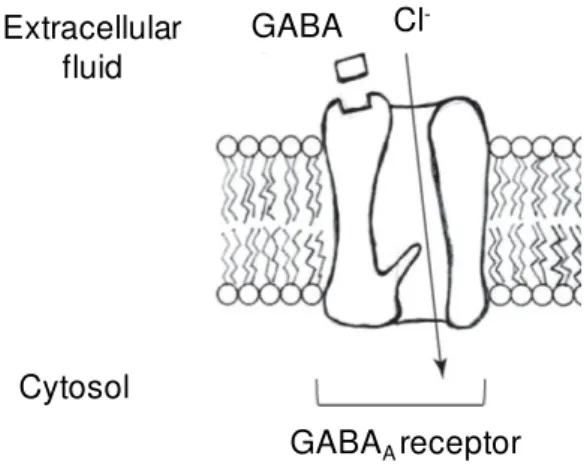
receptor has an inhibitory action due to its connection with a chloride ion channel; the entry of chloride ions in the cell makes the membrane less likely to depolarize (fig. 1.4.3).
Fig. 1.4.3: Representation of the GABAA receptor in the cell membrane. The binding of the γ
-Aminobutyric Acid (GABA), allows the opening of the ion chloride (Cl-) channel and the consequent
entry of this ion in the cell, hyperpolarizing it (from Villares et al (2004) AANA J, 72 (3) 197-205).
GABAA receptors consist of several protein subunits. Some subunits are
expressed almost throughout the brain, whereas the localization of most subunits is more narrowly confined. Anaesthetics have different affinities to the GABAA receptors
depending on the subunits configuration in these receptors (Uusi-Oukari et al. 2010). Aside from the GABA ligand, this receptor has binding sites for benzodiazepines (such as midazolam), barbiturates, steroids and volatile anaesthetics (such as isoflurane) (Harris et al. 1995; Jenkins et al. 2001). The neurotransmitter GABA are presented in 30-40% of all the synapses but its concentration is highest in the substantia nigra and globus pallidus nuclei of the basal ganglia, followed by the hypothalamus, the periaqueductal grey matter and the hippocampus (van der Zeyden et al. 2008).
The binding of benzodiazepines and most inhaled anaesthetics to GABAA receptor
increases the frequency of channel opening, enhancing the inhibitory effect of GABA on both synaptic and extrasynaptic GABAA receptors (Hemmings et al. 2005), which may
disturb memory function (Evans et al. 1996). This is of crucial relevance for this project, since isoflurane and midazolam, two GABA agonists agents, were studied in the present work.
Extracellular fluid
Cytosol
GABAAreceptor
Cl
18
1.4.1.2.
Glutamate receptors
Excitatory neurotransmitters bind to glutamate receptors causing an increase of flow of positive ions into the cell through the opening of ion channels. Glutamate receptors can be divided into ionotropic and metabotropic glutamate receptors. In the ionotropic receptor, the ion channel is activated when glutamate binds, while in the metabotropic receptor the activation is caused indirectly by a signalling cascade that involves G proteins. The ionotropic receptors are the α-amino-3-hydroxyl-5-methyl-4-isoxazole-propionate kainite (AMPA), and the N-Methyl-D-aspartic acid (NMDA) receptors. The name of the receptors is given according to one of the binding substances. Thus the designation of the NMDA receptor is due to the discovery that this receptor binds to the synthetic substance NMDA, not present in biological systems. (Jorgensen et al. 1995; Palmada et al. 1998). NMDA receptor is of particular interest in this work, since ketamine, one the anaesthetics studied, binds to it.
1.4.1.2.1.
NMDA receptors
19 Fig. 1.4.4: Schematic presentation of the NMDA receptor structure and its binding sites for the
different substances. Mg2+- magnesium ion; Zn2+- zinc ion; PCP- phencyclidine (adapted from
http://www.erowid.org/chemicals/dxm/faq/dxm_neuropharm.shtml).
Phencyclidine and ketamine only bind to the NMDA receptor when its channel is fully open, since the binding site of these substances is in the interior of the NMDA receptor channel. The channel is open just when glutamate or aspartate and glycine bind to the receptor (fig. 1.4.5.a). This opening only allows the passage of potassium and sodium ions, since magnesium partially blocks the channel (fig. 1.4.5. a). When the cell potential rises enough to release magnesium ions, calcium enters through the fully open NMDA channel (fig. 1.4.3. b). Inside the cell, calcium activates a series of reactions that improve the strength of the synapse, enhancing LTP, and so memory processes. This mechanism is associative since a strong activation of one set of synapses depolarizes adjacent regions of the dendritic tree, activating LTP in a large scale (Kandel et al. 1995; Malenka et al. 1999). Apart from this entry of calcium, LTP may also require activation of other metabotropic glutamate receptors and the generation of diffusible intercellular messengers (Bear et al. 1994). When postsynaptic calcium increases but do not reach the threshold for LTP, it can generate either a short-term potentiation, during 5-20 minutes, or a long-term depression (LTD), that decreases synaptic strength for a long time. This diversity of mechanisms may give increased flexibility to neural circuits involved in information processing and storage (Malenka et al. 1999).
Zn2+
Mg2+
Mg2+
Glutamate or
Aspartate Glycine
Polyamine PCP Cytosol
20
Fig. 1.4.5: NMDA receptor activation. a) The combined binding of glutamate (Glu) and glycine (Gly)
causes the partial opening of the channel allowing the passage of sodium (Na+) and potassium (K+) ions. This ion passage leads to the cell`s potential to rise and so, as represented in b), magnesium (Mg) is released allowing the entry of the ion calcium (Ca2+) into the cell (adapted from http://www.erowid.org/chemicals/dxm/faq/ dxm_neuropharm.shtml).
1.4.2. Anaesthetics
The activity of the neurotransmitter system may be modulated by anaesthetic agents.
According to the administration route, general anaesthetics may be classified in injectable and volatile agents. Anaesthetics are often administered in combination, since each one provides different actions to achieve a properly balanced anaesthesia (fig. 1.4.1). According to their main effects, agents used to cause anaesthesia may be classified as hypnotics, dissociatives, tranquilizers/ sedatives, muscular relaxants, and analgesics (Flecknell 2009).
In this project, two different anaesthetic protocols which drugs have different mechanisms of action were studied to have a broader view of the effects of anaesthesia in memory: isoflurane was chosen as the volatile agent and ketamine/midazolam as the injectable combination. The injections were administered intraperitoneally, since this is the route of administration routinely performed in laboratory mice. Intraperitoneal (ip.) injection is practical and triggers a rapid onset of action of the drug administered (Flecknell 2009).
1.4.2.1. Isoflurane
Isoflurane is a halogenated volatile drug (2-chloro-2-(difluoromethoxy)-1,1,1-trifluoro-ethane) routinely used in laboratory animal and in clinical anaesthesia. Although it is still
b)
a)
Cytosol Extracellular
fluid
Na+, K+ Mg
Gly
Na+, K+
Ca2+
Glu Glu
21 used in humans, it is gradually being replaced by sevoflurane (Gadani et al. 2011). Isoflurane is a hypnotic agent commercially available as a liquid that is placed in a vaporizer where it is mixed with oxygen to be administered in the breathing air, avoiding hypoxia. In the case of small animals, this mixture can be delivered into a chamber to induce anaesthesia, and then animals are put into a facial mask or intubated with an endotracheal tube for maintenance of anaesthesia. In large animals, the induction of anaesthesia is achieved with a face mask (Flecknell 2009). In humans, volatile anaesthesia is delivered by a facial mask for induction, followed by intubation for maintenance. The anaesthetic is inhaled into the lungs, where it rapidly diffuses to the bloodstream. Due to its high lipid solubility, it leaves the circulation and enters the brain inducing anaesthesia. At the brain level, isoflurane reaches an equilibrium when the concentration of anaesthetic measured in the exhaled air reflects alveolar gas concentrations, which are equivalent to the central nervous system concentrations (Hemmings et al. 2005). Thus, the rapid elimination of the major part of isoflurane by respiration confers safety to this anaesthetic and allows a rapid recovery avoiding toxic effects on liver and/or kidney. Only less than 0.2% is thought to be excreted by the urinary system in the form of inorganic fluoride and trifluoroacetic acid (Eger 1981).
Mechanism of action and clinical effects
The mechanism of action of volatile anaesthetics is still not well understood, and several theories have been brought forward. In the late 19th century, Meyer-Overton described the “lipid hypothesis” which stated that anaesthesia occurred when a sufficient number of volatile molecules were dissolved in the lipid cell membrane (Miller et al. 1967). However, the fact that optic isomers of inhalational anaesthetics, such as isoflurane and enflurane, do not have the same potency, contradicts this theory. Furthermore, this theory does not explain how anaesthesia occurs (Mashour et al. 2005). Hence, Mullins extended the “lipid hypothesis” to the “critical volume hypothesis”, proposing that inhalational anaesthetics bind to the membrane increasing the volume of a hydrophobic region within the cell membrane. This would subsequently distort channels required for sodium ion flux that leads to the development of action potentials necessary for synaptic transmission (Miller et al. 1973). In the end of the 20th century, the work of Franks and Lieb contradicted
the previous theory showing that lipids are not required for the action of volatile agents (Franks et al. 1994). Hence, other hypothesis called “protein hypothesis” appeared, showing that inhalational anaesthetics act on a variety of proteins, especially in ion channels, such as potassium and calcium channels, glycine, GABAA and nicotinic
22 the membrane action potential and propagating synaptic transmission (Rudolph et al. 2004). Probably, the mechanism of action of volatile anaesthetics is a combinations of the theories and hypothesis described, acting by a non-specific mechanism (lipophilic membrane site) and/or by specific mechanisms (protein sites) (Stanley et al. 1999).
It is widely described that isoflurane binds to the allosteric sites on the GABAA receptor
complex, increasing the conductance of ion chloride (fig. 1.4.3) and leading to the hyperpolarizing of the membrane. Isoflurane has also a minor effect on the NMDA receptor antagonist, inhibiting glutamate channels, and glutamate release (Hemmings et al. 2005; Alkire et al. 2009). Furthermore, high concentrations of isoflurane (as well as of the related substances halothane and enflurane) inhibit the receptors of substance P, a neuropeptide that modulates nociceptive transmission within the spinal cord. This could be the process leading to the reduction of the nociceptive response associated with these agents (Minami et al. 2002).
As a hypnotic, isoflurane changes individuals‟ state from consciousness to unconsciousness. This can be detected in data from the electroencephalogram of anaesthetized primates with high concentrations of isoflurane, which present lower amplitude from the posterior portion of the brain compared with the amplitude from the anterior portion that became more active (Clark et al. 1973; Tinker et al. 1977).
Uses and clinical implications
In both humans and other animals, high concentrations of isoflurane induce unconsciousness, amnesia, muscle relaxation, blunting of autonomic reflexes and immobility, allowing the maintenance of anaesthesia (Mashour et al. 2005; Flecknell 2009). However, in order to reduce concentrations and minimize side-effects, isoflurane is often used in association with an injectable agent for induction and maintenance of anaesthesia; analgesics may be used to increase analgesia in surgical procedures. Isoflurane alone can also be used at low concentrations to provide light anaesthesia for minor procedures not requiring unconsciousness (Antognini et al. 1997).
23 In conclusion, the main advantages of isoflurane anaesthesia are its relative safety and the quick recovery from its effects, with almost no toxicity associated (Eger 1981). Furthermore, it is easy to measure its concentration in the expired air (Hemmings et al. 2005), making the use of isoflurane a practical protocol when depth of anaesthesia is an important factor to control.
1.4.2.2. Ketamine
The ketamine molecule (2(2-chlorophenyl)-2-(methylamino)-cyclohexanone) has two enantiomers: S(+)ketamine and R(-)ketamine, and it is commercially available in a racemic mixture of an equal proportion of the two enantiomers. S(+)ketamine is an analgesic 2-3 times more potent than R(-)ketamine (Hirota et al. 1996).
Ketamine can be administered intravenously or intramuscularly in large animals. In rodents, the intraperitoneal route is very common and practical. The major metabolic pathway of ketamine involves its N-demethylation in norketamine by the hepatic microsomal system. Norketamine is an active metabolite with one third of the anaesthetic potency of ketamine. Norketamine is later solubilised by hydroxylation and excreted in the urine and, to a lesser extent, in the faeces (Adams et al. 1981). The active metabolites do not prolong the dissociative effects of ketamine (Reich et al. 1989) but may contribute to some of its pharmacological effects, such as analgesia (Bolze et al. 1998). Dissociative anaesthesia is “a CNS state characterized by catalepsy, analgesia and altered consciousness” (Muir et al. 2007).
Mechanism of action and clinical effects
Ketamine is a noncompetitive NMDA antagonist, which depresses the synaptic transmission of the excitatory neurotransmitter glutamate by blocking the NMDA receptors postsynaptically (fig. 1.4.4 and 1.4.5). Ketamine also interacts with nicotinic and muscarinic cholinergic, monoaminergic and opioid receptors, and with voltage-dependent ion channels, such as the calcium channel, and, at higher doses, it also blocks the sodium channels (Kohrs et al. 1998; Karmarkar et al. 2010). All these mechanisms contribute to the anaesthetic actions of ketamine.
24 electrophysiological inhibition of the thalamocortical pathways and a stimulation of the limbic systems (Kohrs et al. 1998; Bergman 1999). This capacity to disrupt thalamo-cortical gating of external and internal information to the cortex may lead to the development of hallucinations, emergence phenomena and schizophrenia symptoms, especially when low doses of ketamine are administered (Copeland et al. 2005a). Ketamine also induces amnesia, analgesia, and muscular rigidity. The analgesic effect is described to be mediated by the opiate receptors in the spinal cord, brain, and peripheral sites, while the neuromuscular rigidity is caused by the interaction between ketamine and muscarinic receptors in a dose-dependent manner (Haas et al. 1992). Inhibition of the sodium channel provides a mild local anaesthetic effect (Kohrs et al. 1998). In rodents, low doses of ketamine induce hyperactivity that is dependent of the changes in dopamine activity (Giuliani et al. 1997).
Uses and clinical implications
Ketamine is a relatively safe analgesic and short-acting anaesthetic agent (Bergman 1999). It is currently used in paediatrics, in critically ill patients, in the treatment of refractory status epilepticus (Borowicz et al. 2003), in asthmatic individuals (Haas et al. 1992; Bergman 1999), and in pain management (Schmid et al. 1999). Furthermore, in high doses and in combination with other drugs, ketamine is widely used in veterinary practice and in laboratory animal research (Maddison et al. 2008). It is also used as a drug of abuse (Morgan et al. 2004b).
Ketamine is the only intravenous anaesthetic that stimulates the cardiovascular system in a dose-dependent way, by the increase of heart rate, systemic and pulmonary arterial and vascular resistance (Haas et al. 1992).
25 to the superior salivatory nucleus (Reich et al. 1989; Bergman 1999). This effect seems to be dose-dependent (Morse et al. 2001). This anaesthetic may also cause an increase in the cerebral blood flow, oxygen consumption, and intracranial pressure (Copeland et al. 2005a). Cerebral vasodilation may be caused by the blockade of calcium channels (Kohrs et al. 1998).
In general, the use of ketamine is adequate when analgesia and amnesia are required for short periods of time without depressing the cardiovascular and respiratory system, maintaining hemodynamic stability. That is one of the reasons for the use of ketamine in situations where doctors have to work alone and with limited equipment for drug delivery and patients‟ monitoring (Copeland et al. 2005a).
1.4.2.3. Midazolam
Midazolam is an imidazobenzodiazepine (8-chloro-6-(2-fluorophenyl)-1-methyl-4H-imidazo[1,5-a][1,4]benzodiazepine), more potent and with a shorter action compared with other benzodiazepines. These properties are conferred by its imidazole ring, responsible for the stability and rapid metabolism of midazolam (Midtling 1987). In a pH environment lower than 4, the imidazole ring opens and the water solubility is increased with no need of solvents. At a physiological pH, the ring closes and midazolam becomes very lipophilic, which explains its rapid onset of action (Nordt et al. 1997). Other benzodiapepines, such as diazepam, require organic solvents which may not be well tolerated in the injection site (Dundee et al. 1984).
Midazolam can be administered orally, intravenously or intramuscularly. In laboratory animals it is more often delivered by a subcutaneous or intraperitoneal injection.
Midazolam is metabolized by hepatic microsomal oxidative mechanisms in α -hydroxymethylmidazolam (major metabolite), 4-hydroxymidazolam, and α,4 -hydroxymethylmidazolam (Pieri 1983). Only the major metabolite has an active role in midazolam effects contributing with 10% of the biological activity of the midazolam (Riss et al. 2008). In the end, α-hydroxymethylmidazolam is conjugated in glucuronide (Dundee et al. 1984) and excreted by the urine. Less than 0.03% of midazolam administered is excreted in the form of intact drug (Midtling 1987)
.
Mechanism of action and clinical effects
26 spinal cord and brain stem or to one of the allosteric binding sites of the GABAA receptor
in the brain. When midazolam binds to GABAA receptor, it facilitates GABA binding and
enhances GABA effects by increasing the cavity of chloride channel and its frequency of openings (fig.1.4.2). These effects cause membrane hyperpolarization and subsequent reduction in membrane excitability inducing neuronal inhibition. Hence, midazolam decreases the synaptic transmission, interfering with the encoding of information – the benzodiazepines‟ characteristic amnesic effect (Park et al. 2004). The occurrence and duration of anterograde amnesia depends on the dose of midazolam (Midtling 1987). The GABA uptake and its consequent accumulation caused by midazolam induce the sedative effects (Nordt et al. 1997). The binding of midazolam to the GABA receptor may also confer anticonvulsant activity. It is not fully clarified if the mild analgesic properties of midazolam are conferred by its binding to the GABA receptor or if it is associated with the sedative effect and to a possible central suppression of pain perception (Reves et al. 1985; Midtling 1987). Furthermore, after a stressful event, midazolam decreases the level of cathecholamines – stress-associated hormones – in the plasma, probably also through the GABA receptor (Pieri 1983; Crozier et al. 1987) (fig.1.4.6).
27
Fig. 1.4.6: The general effects of benzodiazepines in the presynaptic terminals of their
binding sites: GABA and glycine receptors. The benzodiazepine effects in different regions of the CNS are shortly described in bold. 1- Binding site of the neurotransmitter GABA; 2- Benzodiazepine allosteric binding site in the GABA receptor; 3- Binding site of the neurotransmitter glycine where benzodiazepine also binds. (Adapted from Richter. Anesthesiology (1981); 54: 66-76).
Uses and clinical implications
Midazolam has been used as a hypnotic-sedative agent, and, in combination with other drugs (ex.: volatile anaesthetics, opioids or ketamine), as an induction agent for general anaesthesia or for maintenance of anaesthesia. It is also used in the treatment of status epilepticus and tetanus, due to its anticonvulsant properties. Midazolam causes reliable amnesia, which is important for reducing incidence and severity of emergence agitation and to forget the unpleasant or painful procedures. It can also be used for premedication, sedation, as well as for diagnostic and therapeutic procedures such as treating anxiety, for dentistry and endoscopic practice (Dundee et al. 1984; Reves et al. 1985).
Cortex
Spinal cord Brain stem
Benzodiazepines enhanced inhibitory actions of GABA
Benzodiazepines mimics inhibitory actions of glycine Anticonvulsant
action
Amnesic and sedative action
Muscular
relaxation
Anxiolytic action
1 2
3
Motor circuits in the brain
Benzodiazepines
2 1
28 Midazolam causes minimal changes in the cardio-respiratory stability, being suitable for the elderly and cardiac patients (Dundee et al. 1984). However, a slight heart rate increase can be seen in high sedative doses (Dundee et al. 1984; Reves et al. 1985). Also in high midazolam doses, mild respiratory depression may occur, probably related with a depression in the central nervous system rather than related with respiratory mechanisms (Reves et al. 1985).
In conclusion, midazolam is almost free of side-effects. Its water solubility when injected, and short duration of action are also characteristics that promote its use.
1.4.2.4.
Ketamine/midazolam combinationAs previously referred to, there are advantages in using anaesthetic combinations in order to reduce the amount of drugs necessary to produce the desired effect (fig.1.4.1). The combination of agents with different clinical effects is common. In this case ketamine induces loss of righting reflex in rodents with mild analgesia, aided by midazolam that causes sedation, amnesia, and also muscular relaxation. In laboratory animals, it is very important to combine ketamine – which in itself causes muscle rigidity - with an agent that induces muscular relaxation (Maddison et al. 2008). In humans, the combination ketamine/ midazolam has an additive effect on hypnosis, but may not have any effect on the analgesic component (Hong et al. 1993). The majority of injectable combinations produce synergetic effects but, in this case, the additive effect may be the result of different mechanisms of action: ketamine acts mainly on the NMDA receptors, and midazolam on the benzodiazepine binding site in GABA and/or in glycine receptors (Schüttler et al. 2008). Therefore, a combination does not simply provoke the effects of the individual drugs, rather it has to be faced as an unique substance with its own properties (Short et al. 1992).
Uses and clinical implications
29 muscular rigidity provoked by ketamine, avoiding a potential poor recovery. Midazolam combined with ketamine avoids the recall of unpleasant effects and of unpredicted awareness during anaesthesia due to its amnesic properties (Maddison et al. 2008).
High doses of midazolam reduce the ketamine requirements for induction of anaesthesia, and so it may reduce the dose-related psychedelic effects (Bowdle et al. 1998). Furthermore, midazolam can also attenuate the cardiostimulatory responses of ketamine, conferring hemodynamic stability (White 1982; Toft et al. 1987; Reich et al. 1989). Most studies showed ketamine/midazolam as a cardiorespiratory stable combination (Morse et al. 2001).
1.5. Anaesthesia vs. Memory
Anaesthesia acts as a reversible intoxication and depresses the activity of the central nervous system interfering with the biochemistry and the electrophysiology of the brain (Alkire et al. 2009). General anaesthetics are highly liposoluble, penetrating the membrane and into organelles where they interact with cellular constituents. The reversibility of this action was thought to be dictated by the pharmacokinetic profiles of anaesthetic uptake and elimination. However, several studies showed that anaesthetics may affect gene expression, protein synthesis, cellular functions, among other effects which may interfere negatively with cognitive processes and persist after the drug is eliminated (Perouansky et al. 2009). The mechanism by which anaesthesia affects brain functions is not fully comprehended. How long these effects can last is also not known.
30 risk factors in healthy patients for these conditions. Likewise, Monk and colleagues found that higher age and less education are risk factors for POCD 3 months after surgery; in this study duration of anaesthesia was not a risk factor to POCD (Monk et al. 2008). Contradictory results could come from differences in the methodology to evaluate cognitive dysfunction such as in the tests used, interval between sessions, analysis of endpoints, statistical methods, and the definition of neuropsychological deficits (Rasmussen et al. 2001). A study in middle aged patients reported early POCD 7 days after surgery, from which the patients showed to recover when evaluated 3 months later; POCD persisted in only 11% of the patients (Johnson et al. 2002) and it was not statistical different from control group. In this younger age group, there was no association between duration of anaesthesia or educational level with early POCD. All these studies point to POCD at 3 months after non-cardiac surgery as primarily related to older age.
The balance between the effects of anaesthesia and the effects of surgery in the development of POCD is not clear. Moreover, the possible effects of anaesthesia on human and animal memory processes may depend on several factors. Age of the individual, type of anaesthetic, concentration of the anaesthetic, regime of administration, duration of anaesthesia, type of cognitive function, species and even genetic background are factors to take into consideration.
1.5.1. Age
31 characterized by some deranged mechanisms, such as the oxidative phosphorylation, the scavenging of free radicals through vitamins C, E and melatonin, mutations, and manifestation of silent genes (Cottrell 2008), that make it difficult for the brain to recover from an insult. Few studies are available on the evaluation of the effects of anaesthesia on memory of adult individuals.
1.5.2. Types of anaesthetic agents
Different types of anaesthetics, at clinically effective concentrations, act in different neurotransmitter systems and brain regions (Jevtovic-Todorovic et al. 2003). Thus, anaesthetics have different pathways of action that can lead to different impacts on behaviour and on cellular viability. For example, in rats, the neurotoxicity of nitric oxide was higher than that of ketamine in young adult brains (Jevtovic-Todorovic et al. 2005), but lower compared with high concentration of isoflurane (Jevtovic-Todorovic et al. 2003), while propofol produced no spatial memory impairment in aged rats (Lee et al. 2008). Furthermore, different anaesthetics have different electrophysiological effects in humans and animals (Alkire et al. 1997; Alkire et al. 1999; Antunes et al. 2003). Recent findings indicate that drugs acting by either inhibition via GABAA receptors or by decreasing
excitation through NMDA receptors induce widespread neuronal apoptosis (Jevtovic-Todorovic et al. 2003; Fredriksson et al. 2007; Bianchi et al. 2008) which may have serious implication on learning and memory processes.