Universidade de Lisboa
Faculdade de Medicina de Lisboa
Characterization of the affective behavioral effects of
chronic adolescent exposure to the cannabinoid
receptor agonist HU-210 in female rats
Jorge Miguel Farinha Ferreira
Orientador | Professora Doutora Ana Maria Ferreira de Sousa Sebastião
Dissertação especialmente elaborada para obtenção do grau de
Mestre em Neurociências
Todas as afirmações efectuadas no presente documento são da exclusiva responsabilidade
do seu autor, não cabendo qualquer responsabilidade à Faculdade de Medicina de Lisboa
pelos conteúdos nele apresentados.
A impressão desta dissertação foi aprovada pelo Conselho Científico da Faculdade de
Medicina de Lisboa em reunião de 19 de Fevereiro 2019
i
Agradecimentos
Quando decidi enveredar por este caminho, a minha ideia do que seria escrever uma dissertação era de que se trataria de um projecto inerentemente individual: a minha dissertação, o meu trabalho, a minha carreira. Aliás, estaria a mentir se não dissesse que essa foi uma das características que mais me atraiu acerca desta aventura. É por isso que agora, pouco mais de um ano depois de ter começado este trabalho, me encontro positivamente surpreendido com o quão errado estava. Apesar de ser o meu nome que está na capa desta dissertação, dizer que este trabalho é apenas meu não seria nada senão falso: este trabalho é tanto meu quanto das inúmeras pessoas que me ajudaram – directa ou indirectamente – ao longo dele, e sem as quais não o teria conseguido levar a bom porto. E embora tenha de agradecer a todos os membros do laboratório da Professora Ana Sebastião, pois cada um contribui da sua maneira única para fazer do laboratório o quão especial é, algumas pessoas merecem um destaque especial, pelos papeis que tomaram na minha vida.
Acima de tudo tenho de agradecer à minha orientadora, a Professora Ana Sebastião, por não só me ter recebido no seu laboratório, como por me ter aceite enquanto mestrando, e me ter dado a liberdade para fazer este trabalho algo mais meu. Mais ainda, não existem agradecimentos suficientes pelas oportunidades que me deu ao longo deste ano, e pelo tempo e disponibilidade que teve para discutir o projecto. Espero ter sido um bom mestrando e ter superado as suas expectativas para este trabalho.
Aos Mizeses devo, também, um obrigado colectivo – por me aturarem diariamente – e individual: ao João Gomes, por me ajudar com os blots, pelas conversas e momentos de brincadeira na nossa sala, pela variada banda sonora diária (não sei como te lembras de tantas músicas) e por me dar a perspectiva de que, se calhar, “isto” – seja lá o que “isto” for, em qualquer dia particular – não é o fim do mundo. À Rita Belo, lorde das bolhas, pela energia incontrolável, pelas conversas de café e os comentários aleatórios que nunca falham em me fazer rir, pelos conselhos científicos (e não só), e por ser uma das pessoas mais únicas que já tive o gosto de conhecer. À Sara Tanqueiro e à Catarina Beatriz, os meus putos, por me fazerem rir diariamente, por me darem conversa quando estou aborrecido, e sem as quais os dias no laboratório seriam muito menos felizes – isto sem vocês não era a mesma coisa. À Catarina Lourenço, por ter sempre uma resposta e um auxilio prontos às minhas dúvidas básicas, e me fazer companhia – com conversas sempre interessantes – durante tantos fins de semana no laboratório. E à Jéssica Rosa, que começou isto comigo,
ii
e com quem as conversas são uma constante caixa de boas surpresas, sem as quais a nossa sala não seria o que é. Devo também um enorme obrigado à líder dos Mizeses, a Professora Maria José Diógenes, pela ajuda crucial com a obtenção do THC, e por não só juntar à sua volta um grupo de tão bons alunos, como por ser um modelo de entusiasmo e curiosidade incansáveis (e, já agora, por me tentar casar).
À Nádia Raquel, minha dama do sinal e vítima favorita, não só pela ajuda com as dissecções mas, mais que isso, por ser a (pequena) pessoa fantástica que é: pela paciência para lidar com as minhas partidas constantes, por me fazer rir com as suas odisseias de electrofisiologia, pelas expressões hilariantes que faz quando eu digo as coisas absurdas que digo, pelos muitos cigarros que lhe cravei e, acima de tudo, pela companhia diária, quer dentro, quer fora do laboratório. És pequenina e resmungona, mas eu gosto de ti assim. Já agora, já que aqui estás, queres ir lá abaixo?
Ao Francisco Mouro, não só por me ter ensinado e ajudado a fazer comportamento, mas também pelas conversas acerca dos testes e do que estamos mesmo a fazer quando testamos animais, e por ter tido a disponibilidade para rever o capitulo de metodologia desta dissertação – este trabalho é melhor pela tua ajuda.
Aos Xapellis, mas em especial a três de vós: ao Rui Rodrigues, por ser o melhor público possível e se rir mesmo das minhas piores piadas, e pelas conversas quer sobre canabinóides, quer sobre a vida. À Marta Alonso, por ser uma das pessoas mais doces e simpáticas que já tive o prazer de conhecer, e cujo entusiasmo, boa disposição e sentido de humor melhoram qualquer dia, mesmo quando esse dia é um sábado ou domingo de Agosto passado no laboratório. E à Sara Raquel Paulo, por me ensinar a pipetar (e não me deixar esquecer disso), por ter a paciência e disponibilidade para me ajudar e ensinar a fazer imunos, por ainda não me ter batido apesar de eu lhe dar razões mais que suficientes para o fazer, e por – apesar de às vezes não nos entendermos – ser uma das melhores pessoas que conheci nos últimos tempos (tenta não ficar com o ego muito cheio).
À Sandra Vaz pela disponibilidade, paciência, encorajamento que me deu enquanto me ensinava a dissecar, e por me ter ajudado nas dissecções de uma das experiências. Mais ainda, pelo papel crucial que desempenha no laboratório, e por toda a ajuda que nos presta a todos diariamente. E à Joana Gomes, estudante da Sandra, por ser uma das minhas vitimas e ter um excelente sentido de humor acerca disso, pela companhia no tabagismo, e por me deixar lembrar-lhe diariamente que a vida é sofrimento.
iii
À Varél… Valéria Martins e à “Outra” (Beatriz Pereira), por terem sido as minhas underlings emprestadas, que tanto me ajudaram a fazer handling e a correr testes de comportamento. Mais que isso, por serem duas miúdas espectaculares, que trouxeram imensa animação, energia e “boa procrastinação” aos meus dias no laboratório, e que – por isso – fizeram dos meses que cá estiveram uma das minhas alturas favoritas. Podem ter radicalizado os meus ratos contra mim, e estar lá longe na Suécia, mas eu perdoo-vos e não me esqueço de vocês.
À Alexandra Maralhas, sem a qual eu ainda estaria à espera do meu primeiro frasco de HU-210, e que tornou – pela sua dedicação e trabalho incansável – este, e outros projectos futuros, possíveis. Mais ainda, um enorme obrigado pelo teu interesse e entusiasmo pelo meu trabalho.
À Alexandra Botelho e à Cristina Varandas, por toda a ajuda que me deram ao longo deste processo, e por me darem conversa quando eu estava a evitar ir fazer alguma coisa que não queria fazer. A vossa dedicação aos alunos e ao vosso trabalho é admirável.
Tenho também de agradecer ao Professor Paulo Ventura, da Faculdade de Psicologia da Universidade de Lisboa, o responsável por eu ter começado a fazer investigação, e com quem partilho a minha mais longa colaboração profissional. Agradeço-lhe não só por isso, mas por ser um modelo de como fazer ciência, de como há sempre mais por fazer e por descobrir, e por me ter dado – e continuar a dar – imensas oportunidades. Mais ainda, devo-lhe um agradecimento por ser no contexto do seu grupo que conheci e criei amizade com todo um conjunto de pessoas fantásticas – o João Delgado, o José Carlos Guerreiro, o Bruno Faustino e o António Fernandes – com os quais é um prazer, um orgulho, e uma honra, trabalhar. Um obrigado a todos vós.
Aos meus dois melhores amigos de longa data, as minhas fofinhas, o André Marques e o Carlos Santos, não há muito que seja necessário dizer. Mas isso nunca me impediu de falar, portanto aqui vai: obrigado pelos mais de 10 anos de amizade, pelas histórias e momentos de riso, por não me mandarem calar quando não falo de mais nada senão de trabalho, e por continuarem a ser as duas pessoas com quem posso falar de tudo e com os quais sei que posso contar seja para o que for. Aconteça o que acontecer, isto é para sempre, e se precisarem de um alibi para alguma eventualidade, sabem a quem ligar.
Não poderia, em boa consciência, deixar também de agradecer aos animais que deram a vida por este trabalho. Espero ter sido um bom dono, e ter feito a vossa curta vida o mais confortável possível. Num
iv
mundo ideal ter-vos-ia trazido a todas para casa comigo, e peço desculpa por não o poder ter feito. E, igualmente, não poderia não agradecer ao animal sem a qual a minha vida seria muito menos feliz: a Nina, o melhor cão que alguém poderia querer. Não há festinhas suficientes para te agradecer, mas eu tento na mesma.
Termino com um obrigado à minha família, mas em especial às duas pessoas sem as quais esta minha aventura não teria sido possível. Ao meu avô Quim e à minha avó Minda, que nunca me deram nada senão amor e aceitação incondicional, e que trago comigo no coração todos os dias. Se, neste último ano, não estive tão presente nas vossas vidas quanto gostaria, foi porque quis que o voto de confiança que fizeram em mim não fosse desperdiçado, e espero – acima de tudo – ter-vos deixado orgulhosos do vosso primeiro neto. Não há um dia em que não me sinta grato pela vossa ajuda, e por vos ter na minha vida.
Palavras, por muitas que sejam, não são o suficiente para agradecer tudo o que tenho para agradecer a todos vós. Resta-me tentar que, não só estas palavras, mas as minhas acções, vos mostrem o quanto gosto de todos vocês, e o quão sortudo me sinto ser por vos ter na minha vida. A todos, sem excepção, o mais profundo e sincero obrigado.
v
“There is no easy way from the earth to the stars.”
-
Lucius Annaeus Seneca
“The mind adapts and converts to its own purposes the obstacle to our acting. The impediment
to action advances action. What stands in the way becomes the way.”
-
vii
Scientific Production
During the year during which the current work was developed I was invited to participate in the writing of several review articles, two of which have since been published:
Ferreira, M.F.*, Castanheira, L. Sebastião, A.M., Telles-Correia, D. (2018). Depression assessment in clinical trials and pre-clinical tests: a critical review. Current Topics in Medicinal
Chemistry, 18(19), 1677-1703
Castanheira, L., Ferreira, M.F.*, Sebastião, A.M., Telles-Correia, D. (2018). Anxiety assessment in pre-clinical tests and in clinical trials: a critical review. Current Topics in Medicinal Chemistry, 18(19), 1656-1676
Rodrigues, R.S., Lourenço, D., Paulo S.L., Mateus, J., Moreira, J.B., Ferreira, M.F., Mouro, F.M., Ribeiro, F.F., Sebastião, A.M., Xapelli, S. (in review). Endocannabinoid actions on neural stem cells: implications for physiopathology. Molecules.
Furthermore, the scientific content of the present dissertation, has been presented in poster sessions at several national and international conferences, and constitutes the majority of a manuscript currently in preparation for publication:
Ferreira M.F., Paulo S.L., Fonseca-Gomes J., Rei N., Vaz S.H., Mouro F.M., Sebastião A.M. Of
rats, cannabinoids, and the blues: the unexpected short- and long-term effects of chronic adolescent HU-210 exposure on affective behavior.
viii
Table of Contents
Agradecimentos ... i
Scientific Production ... vii
Table of Contents ... viii
List of Figures ... x
List of Tables ... xiii
List of Abbreviations ... xiv
Resumo ... 1
Abstract ... 5
Chapter 1 – Introduction ... 8
1 – Cannabinoid Use and Abuse Worldwide... 8
2 – The Endocannabinoid System ... 10
3 – Adolescence and Chronic Cannabinoid Abuse ... 23
4 – HU-210 ... 41
5 – Reasoning, Aim and Organization of the Present Work... 43
Chapter 2 – Behavioral Methodologies ... 45
1 – Elevated Plus Maze ... 47
2 – Open Field Test ... 50
3 – Social Interaction Test ... 53
4 – Modified Forced Swim Test ... 54
5 – Sucrose Preference Test ... 57
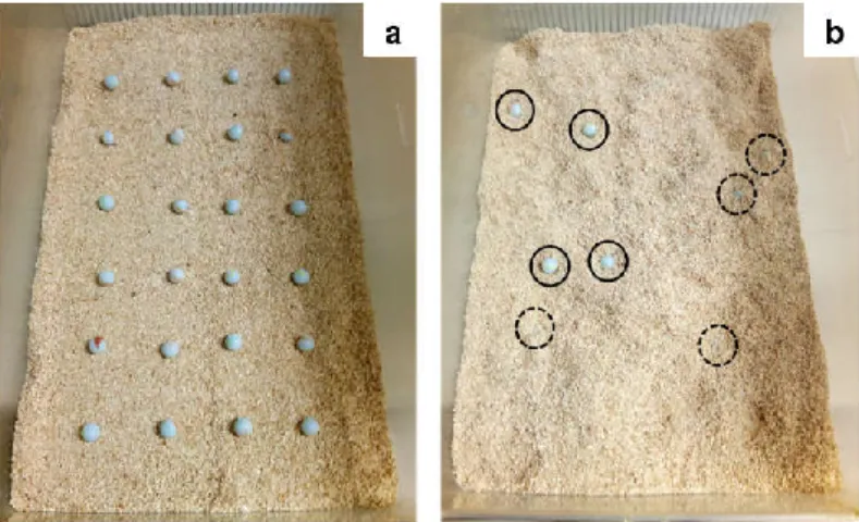
6 – Marble Burying Test... 58
Chapter 3 - Experiment 1 ... 62
1 – Rationale ... 62
2 – Methods ... 63
3 – Results ... 69
ix
Chapter 4 – Experiment 2 ... 78 1 – Rationale ... 78 2 – Methods ... 78 3 – Results ... 84 4 – Discussion ... 90 Chapter 5 – Experiment 3 ... 94 1 – Rationale ... 94 2 – Methods ... 95 3 – Results ... 98 4 – Discussion ... 101 Chapter 6 – Experiment 4 ... 104 1 – Rationale ... 104 2 – Methods ... 104 3 – Results ... 106 4 – Discussion ... 109Chapter 7 – General Discussion, Future Perspectives, and Conclusions ... 111
1 – General Discussion ... 111
2 – Future Perspectives ... 123
3 – Conclusions ... 127
x
List of Figures
Fig 1.1 – Schematic representation of the endocannabinoid system. ... 11
Fig 1.2 - Molecular structures of anandamide and 2-arachidonoyl-glycerol. ... 12
Fig. 1.3 – Molecular structures of the four CBRAs most used in research. ... 29
Fig. 2.1 – Schematic representation of the elevated plus maze. ... 49
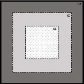
Fig. 2.2 – Schematic representation of the open field test. ... 52
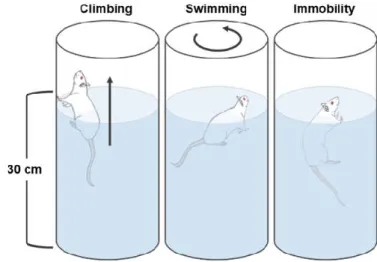
Fig. 2.3 – Schematic representation of the modified forced swim test. ... 56
Fig. 2.4 – Representative pictures of the marble burying test. ... 60
Fig. 3.1 – Timeline of drug administration. ... 64
Fig. 3.2 – Chronogram of behavioral experiments performed. ... 65
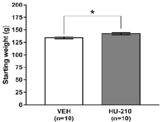
Fig. 3.3 – Animal weights at the start of the experiment (PND 35). ... 69
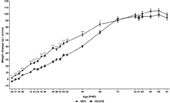
Fig. 3.4 – Change in animal weight relative to PND 35 over the course of the experiment. ... 70
Fig. 3.5 – Chronic adolescent exposure to HU-210 did not persistently alter adult anxiety-like behavior in the EPM after a 27-day drug washout. ... 70
Fig. 3.6 – Chronic adolescent exposure to HU-210 did not persistently alter adult anxiety-like behavior in the OFT after a 27-day drug washout. ... 71
Fig. 3.7 – Chronic adolescent exposure to HU-210 did not persistently alter adult locomotor activity in the OFT after a 27-day drug washout. ... 71
Fig. 3.8 – Chronic adolescent exposure to HU-210 did not persistently alter adult social behavior in the SIT after a 27-day drug washout. ... 72
Fig. 3.9 – Chronic adolescent exposure to HU-210 led to persistent alterations of stress-coping behavior in the mFST after a 27-day drug washout. ... 72
Fig. 3.10 – Chronic adolescent exposure to HU-210 did not persistently alter sucrose intake after a 27-day drug washout. ... 73
Fig. 3.11 – Chronic adolescent exposure to HU-210 did not persistently alter relative preference for sucrose after a 27-day drug washout. ... 74
Fig. 3.12 – Chronic adolescent exposure to HU-210 did not persistently alter adult anxiety-like behavior in the MBT after a 27-day drug washout. ... 74
xi
Fig. 4.1 – Timeline of drug administration. ... 79 Fig. 4.2 – Chronogram of behavioral experiments performed. ... 80 Fig. 4.3 – Change in animal weight relative to PND 28 over the course of the experiment. ... 84 Fig. 4.4 – Chronic adolescent exposure to HU-210 did not persistently alter adult anxiety-like behavior in the EPM after a 30-day drug washout. ... 85 Fig. 4.5 – Chronic adolescent exposure to HU-210 did not persistently alter adult anxiety-like behavior in the OFT after a 30-day drug washout. ... 85 Fig. 4.6 – Chronic adolescent exposure to HU-210 did not persistently alter adult locomotor activity in the OFT after a 30-day drug washout. ... 86 Fig. 4.7 – Chronic adolescent exposure to HU-210 did not persistently alter adult social behavior in the SIT after a 30-day drug washout. ... 86 Fig. 4.8 – Chronic adolescent exposure to HU-210 did not persistently alter stress-coping behavior in the mFST after a 30-day drug washout. ... 87 Fig. 4.9 – Chronic adolescent exposure to HU-210 did not persistently alter sucrose intake after a 30-day drug washout. ... 88 Fig. 4.10 – Chronic adolescent exposure to HU-210 did not persistently alter relative preference for sucrose after a 30-day drug washout. ... 88 Fig. 4.11 – Chronic adolescent exposure to HU-210 did not persistently alter adult anxiety-like behavior in the MBT after a 30-day drug washout. ... 89 Fig. 4.12 – Chronic adolescent exposure to HU-210 did not persistently alter CB1R protein levels, in regions involved in affective functioning, after a 30-day drug washout. ... 89 Fig. 5.1 – Chronogram of drug administration and behavioral testing. ... 96 Fig. 5.2 – Change in animal weight relative to PND 28 over the course of the experiment. ... 98 Fig. 5.3 – Chronic adolescent exposure to HU-210 did not alter anxiety-like behavior or locomotor activity in the OFT 24-hours after the last drug administration. ... 99 Fig. 5.4 – Chronic adolescent exposure to HU-210 altered stress-coping behavior in the mFST 24-hours after the last drug administration. ... 100
xii
Fig. 5.5 – Chronic adolescent exposure to HU-210 selectively altered the levels of CB1R protein, in regions involved in affective functioning, 24-hours after the last drug administration. ... 101 Fig. 6.1 – Chronogram of drug administration and behavioral testing. ... 105 Fig. 6.2 – Change in animal weight relative to PND 28 over the course of the experiment. ... 107 Fig. 6.3 – Chronic adolescent exposure to HU-210 did not alter anxiety-like behavior in the EPM 24-hours after the last drug administration. ... 108 Fig. 6.4 – Chronic adolescent exposure to HU-210 altered sucrose intake and preference 24 to 96 hours after the last drug administration. ... 109
xiii
List of Tables
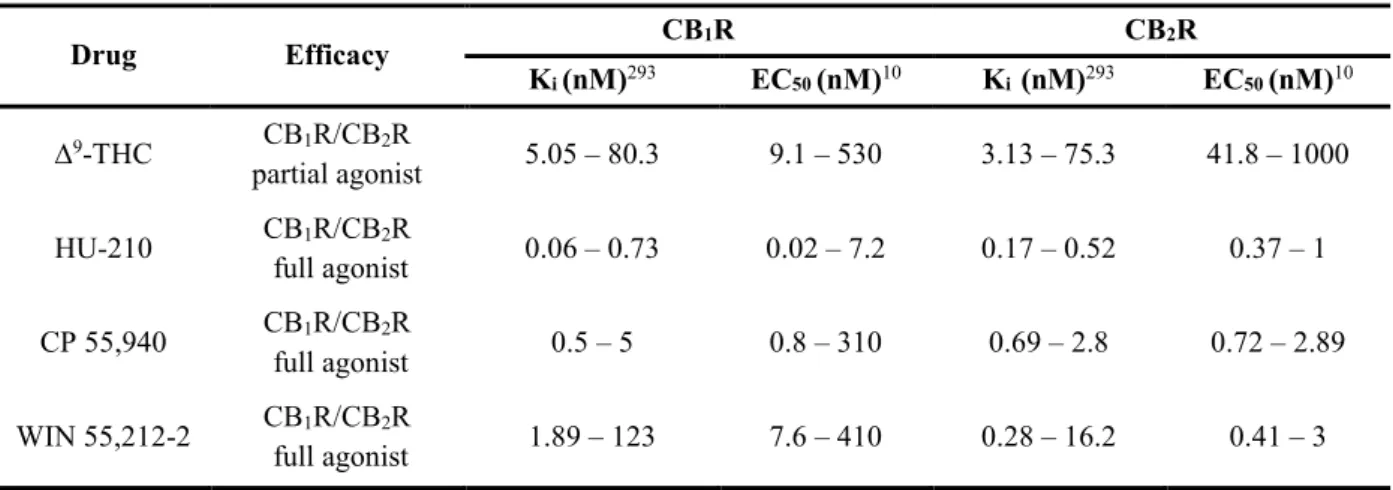
Table 1.1 – Pharmacological characteristics of the four CBRAs most used in research. ... 29 Table 4.1 – Primary antibodies used for Western Blot. ... 83 Table 4.2 – Secondary antibodies used for Western Blot. ... 83
xiv
List of Abbreviations
2-AG – 2-Arachidonoyl-glycerol
5-HT – Serotonin (5-hydroxytryptamine) 5-HT1AR – Serotonin type 1A receptor 5-HT2AR – Serotonin type 2A receptor AA – Arachidonic acid
ABHD – Alpha/beta-hydrolase domain-containing AC – Adenylyl cyclase
aCSF – Artificial cerebral spinal fluid ACTH – Adrenocorticotropic hormone
AEA – Anandamide (N-arachidonoyl-ethanolamine)
AHA1 – Activator of 90 kDa heat shock protein ATPase homolog 1
AMPAR – α-amino-3-hydroxy-5-methyl-4-isoxazolepropionic acid (AMPA) receptor APAP – Active place avoidance paradigm
APS – Ammonium persulfate
Arc – Activity-regulated cytoskeleton-associated protein ATP – Adenosine triphosphate
BDNF - Brain derived neurotrophic factor BrdU – 5-bromo-2'-deoxyuridine
BSA – Bovine serum albumin Ca2+ – Calcium (2+) ion CaCL2 – Calcium chloride
cAMP – 3',5'-cyclic adenosine monophosphate CB1R – Cannabinoid receptor type 1
CB2R – Cannabinoid receptor type 2 CBRA – Cannabinoid receptor agonist CMS – Chronic mild stress
xv
CORT – Corticosterone COX-2 – Ciclooxigenase-2 CPu – Caudate-putamen
CRF – Corticotropin releasing factor CSF – Cerebral spinal fluid
CUD – Cannabinoid use disorder CZ – Central zone of the OFT D1R – Dopamine receptor type 1 D2R – Dopamine receptor type 2 DA – Dopamine
DAG – Diacylglycerol DAT – Dopamine transporter DG – Dentate gyrus
DGLα – DAG lipase α DMSO – Dimethyl sulfoxide
DOPAC – L-3,4,-dihydroxyphenylacetic acid dRN – Dorsal raphe nuclei
DSI/E – Depolarization induced suppression of inhibition/excitation eCB – Endocannabinoid
eCB-LTD – Endocannabinoid mediated long-term depression eCB-STD – Endocannabinoid mediated short-term depression ECS – Endocannabinoid system
EDTA – Ethylenediamine tetraacetic acid EMT – Endocannabinoid membrane transporter EPM – Elevated plus maze
ER – Endoplasmatic reticulum
ERK – Extracellular signal-regulated kinase FAAH – Fatty acid amide hydrolase
xvi
FABP – Fatty acid binding protein
FST/mFST – Forced swim test/modified forced swim test GABA – γ-aminobutyric acid
GABAAR - GABA A receptor GABABR – GABA B receptor GAD – Glutamate decarboxylase
GAPDH – Glyceraldehyde-3-phosphate dehydrogenase GAT-1 – GABA transporter 1
GIRK – G-protein coupled inwardly rectifying K+ channels GluA1 – AMPAR subunit 1
GluA2 – AMPAR subunit 2 GluN2A – NMDAR subunit 2A GluN2B – NMDAR subunit 2B GPCR – G-protein coupled receptor HBT – Holeboard test
HPA axis – Hypothalamic-pituitary-adrenal axis HSP90 – 90 kDa heat-shock protein
i.p. – Intraperitoneal injection
Iba1 – Ionized calcium-binding adapter molecule 1 IL-10 – Interleukin-10
iNOS – Inducible nitric oxide enzyme IQ – Intelligence quotient
IQR – Interquartile range
IZ – Intermediate zone of the OFT JNK – c-Jun N-terminal kinase K+ – Potassium ion
KCl – Potassium chloride
xvii
LC – Locus coeruleus LDBT – Light-dark box test LFP – Local field potential LHb – Lateral habenula LOX – Lipoxygenase
MAGL – Monoacylglycerol lipase
MAPK – Mitogen activated protein kinase MBT – Marble burying test
mGluR – Metabotropic glutamate receptor MgSO4 – Magnesium sulfate
MOR – Mu (µ) opioid receptor mPFC – Medial prefrontal cortex
MSI/E – Metabotropic induced suppression of inhibition/excitation
MTORC1 – Mammalian/mechanistic target of rapamycin complex 1 pathway MWM – Morris water maze
NA – Noradrenaline
Na3VO4 – Sodium orthovanadate
NAAA – N-acylethanolamine hydrolyzing acid amidase NAc – Nucleus accumbens
NaCl – Sodium chloride NaF – Sodium fluoride
NaH2PO4 – Sodium dihydrogen phosphate NAPE – N-acylphosphatidylethanolamine
NMDAR – N-methyl-D-aspartate (NMDA) receptor NO – Nitric oxide
NOPRT – Novel object place recognition test NORT – Novel object recognition test NP40 – Nonidet® P40 substitute
xviii
NSFT – Novelty suppressed feeding test O2 – Molecular oxygen
OCD – Obsessive-compulsive disorder OFC – Orbitofrontal cortex
OFT – Open field test
pCREB – phosphorylated cAMP response element-binding protein PFC – Prefrontal cortex
PFPT – Palatable food preference test PI – Phosphatidylinositol
PI3K – Phosphatidylinositol-3-kinase PKA/PKB/PKC – Protein kinase A/B/C PLC – Phospholipase C
PND – Postnatal day PP – Perforant path
PSA-NCAM – Polysialylated-neural cell adhesion molecule PSD95 – Post-synaptic density protein 95
PTX – Pertussis toxin
PVDF – Polyvinylidene difluoride
PVN – Paraventricular nucleus of the hypothalamus PZ – Peripheral zone of the OFT
Q1 – First quartile Q3 – Third quartile
r – Pearson correlation coefficient
RIPA – Radio immunoprecipitation assay RT – Room temperature
SC/SCs – Synthetic cannabinoid/s SDS – Sodium dodecyl sulfate
xix
SEM – Standard error of mean SERT – Serotonin transporter SIT – Social interaction test
SMSNT – Social motivation and social novelty task SN – Substantia nigra
SPT – Sucrose preference test SSI – Slow self-Inhibition
SSRI – Selective serotonin reuptake inhibitor TBS-T – Tris buffered saline with Tween®20 TCA – Tricyclic antidepressant
TEMED – N,N,N',N'-tetramethylethane-1,2-diamine TH – Tyrosine hydroxylase
THC – Δ9-tetrahydrocannabinol
t-LTD – Spike-timing dependent long-term depression TNFα – Tumor necrosis factor α
TRPV 1 – Transient receptor potential cation channel subfamily V member 1 VAMP2 – Vesicle-associated membrane protein 2
VEGF – Vascular endothelial growth factor VEH – Vehicle solution
VGCC – Voltage-gated calcium channel vGluT1 –Vesicular glutamate transporter 1 vSub – Ventral subiculum
VTA – Ventral Tegmental Area WT – Wild type
1
Resumo
Os canabinóides, agonistas dos receptores do sistema endocanabinóide (SEC), são as drogas ilegais mais consumidas no mundo, sendo os adolescentes um dos grupos etários em que o consumo destas substâncias é mais prevalente.
A adolescência representa um período crítico do neurodesenvolvimento, no qual o sistema nervoso central é extensamente reorganizado – sendo, também, um período de vulnerabilidade aumentada aos efeitos de influências externas, como por exemplo drogas de abuso. Criticamente, grande parte destas alterações neuronais são mediadas e/ou moduladas pelo SEC sendo, portanto, expectável que o uso de drogas que interagem com esse sistema cause alterações profundas, e possivelmente persistentes, no funcionamento do sistema nervoso. Congruentemente, dados obtidos quer com humanos, quer com roedores, sugerem que a exposição crónica adolescente a canabinóides tem efeitos deletérios quer ao nível da função neurocognitiva, quer ao nível do funcionamento afectivo.
Assim, estudos epidemiológicos em populações humanas têm demonstrado que indivíduos adultos, que enquanto adolescentes foram consumidores crónicos de canabinóides, apresentam um risco aumentado de serem diagnosticados com perturbações de ansiedade e/ou perturbações depressivas. Mais ainda, devido a um conjunto ainda não totalmente estudado de factores, este aumento de risco é mais marcado na população do sexo feminino.
Em linha com estas observações, estudos experimentais com roedores têm consistentemente demonstrado que a exposição crónica adolescente a canabinóides induz um conjunto profundo e diversificado de alterações a nível molecular, morfológico, estrutural, funcional e comportamental. Em relação ao último, estudos comportamentais têm repetidamente demonstrado que animais adultos expostos a canabinóides durante a adolescência apresentam défices não só em tarefas de função cognitiva, como em testes de função afectiva – apresentando alterações comportamentais que sugerem um efeito prodepressivo desta exposição, que, replicando os dados humanos, é mais marcado em fêmeas.
Uma limitação da literatura até agora é, no entanto, o uso de um conjunto limitado de canabinóides. De facto, praticamente todos os estudos usaram apenas uma de três substâncias, o que levanta a possibilidade de existirem outros canabinóides cujos efeitos diferem dos até agora observados, o que – a ser verdade – representaria um problema importante na literatura.
2
O presente trabalho pretende averiguar essa possibilidade, ao estudar o HU-210, um potente agonista total e não-selectivo dos receptores canabinóides 1 (CB1R) e 2 (CB2R). Apesar de este fármaco ser amplamente usado em investigação sobre o SEC, e já ter sido encontrado em substitutos sintéticos de canábis, não existe – à data – nenhum relato publicado acerca do impacto que a exposição crónica adolescente a HU-210 possa ter no funcionamento afectivo. Assim, o presente trabalho consiste num conjunto de quatro experiências desenhadas para caracterizar esses efeitos.
Na primeira experiência ratos, Wistar fêmea com 35 dias de idade (PND 35) foram administrados HU-210 diariamente, durante 15 dias, num plano de doses ascendentes (PND 35-39: 25μg/kg; PND 42-46: 50μg/kg; PND 49-53: 100μg/kg, ou solução veiculo equivalente). Após o fim da administração, os animais foram deixados durante 27 dias – de modo a que efeitos residuais, ou resultantes de abstinência, pudessem ser minimizados e permitindo que os animais atingissem a idade adulta – ao fim dos quais foram testados numa bateria de testes comportamentais. Especificamente, para medir alterações ao nível do comportamento ansioso, os animais foram testados no Elevated Plus Maze (EPM; PND 80), Open Field
Test (OFT; PND 80 e 81) e Marble Burying Test (MBT; PND 91). Para determinar os efeitos do tratamento
no comportamento social foi utilizado o Social Interaction Test (SIT; PND 82). Finalmente, para avaliar os efeitos da exposição a HU-210 nas dimensões de stress-coping e de resposta à recompensa, do comportamento depressivo, os animais foram testados no Modified Forced Swim Test (mFST; PND 85) e no Sucrose Preference Test (SPT; PND 88-91), respectivamente. Adicionalmente, o peso dos animais foi registado ao longo da duração da experiência.
Análise dos resultados obtidos revelou que, tal como descrito previamente para outros canabinóides, a exposição a HU-210 não induziu alterações persistentes ao nível do comportamento ansioso, em nenhum dos três testes. Contrariamente ao anteriormente descrito, não foi observada qualquer alteração no SIT, indicando a ausência de efeitos persistentes. No que diz respeito ao comportamento depressivo, foi registado um decréscimo ligeiro no comportamento de trepar, no mFST – sugerindo a possibilidade de alterações de stress-coping – sem qualquer diferença nos outros comportamentos. No SPT não foram encontradas diferenças quer na quantidade de sacarose consumida, quer na preferência relativa por sacarose, indicando que a resposta à recompensa não está alterada. Finalmente, em linha com estudos anteriores, a exposição a HU-210 induziu decréscimos marcados no ganho de peso, que persistiram durante
3
15 dias após o fim da administração.
Dado que os resultados da experiência 1 não foram os esperados, e que várias limitações foram identificadas no protocolo, a experiência 2 foi desenhada para – de novo – testar os efeitos a longo-termo da exposição crónica adolescente a HU-210. Assim, ratos Sprague-Dawley fêmea receberam duas injecções diárias de HU-210 durante 11 dias, seguindo um padrão de doses ascendentes (PND 35-37: 25μg/kg; PND 38-41: 50μg/kg; PND 42-45: 100μg/kg ou veículo equivalente). Após o término da administração, os animais foram deixados em repouso durante 30 dias, ao fim dos quais lhes foi aplicada a mesma bateria de testes comportamentais usada na experiência anterior. Adicionalmente, de modo a determinar os efeitos da exposição adolescente nos níveis de proteína CB1R, amostras de tecido do hipocampo, estriado e córtex pré-frontal, foram recolhidas após o fim da bateria comportamental (PND 88), tendo os níveis de CB1R sido avaliados através de western blotting.
Tal como na experiência 1, não foram encontradas quaisquer alterações no comportamento ansioso, ou no comportamento social, e foi observado um decréscimo marcado no ganho de peso que – mais uma vez – persistiu durante 15 dias pós-última administração. No entanto, em contraste quer com a experiência anterior, quer com a literatura, no mFST também não foram observadas alterações. Semelhantemente, no SPT, o desempenho foi igual entre grupos. Em linha com a ausência de alterações comportamentais, não se detectaram alterações nos níveis de proteína CB1R em nenhuma das três regiões estudadas.
Dada a discordância entre os resultados obtidos na experiência 2 e o descrito na literatura, a experiência 3 consistiu em avaliar se a administração adolescente de HU-210 tinha de facto algum efeito mensurável a curto-prazo que poderia ter desaparecido, durante o interregno de 30 dias entre a última administração e o início dos testes. Para isso uma nova série de ratos Sprague-Dawley fêmea foi manipulada como descrito na experiência 2, e testada no OFT e mFST, nos dois dias após a última administração de HU-210 (PND 46-47). Adicionalmente, amostras de tecido para western blot foram recolhidas no dia após o fim dos testes comportamentais (PND 48).
Surpreendentemente, apesar de nenhuma alteração ter sido encontrada no OFT, no mFST os animais mostraram um padrão comportamental marcado e sugestivo de um efeito antidepressivo do tratamento, com decréscimos no tempo passado em imobilidade e aumentos no tempo passado a trepar. Mais ainda, análise do western blot revelou um decréscimo de cerca de 50% nos níveis de proteína CB1R, na região
4
hipocampal, sem alterações nas restantes.
Uma vez que os resultados na experiência 3, no que respeita aos parâmetros do mFST, foram inteiramente inesperados, e as alterações moleculares encontradas foram incongruentes com um efeito antidepressivo, a experiência 4 foi desenhada para mais uma vez avaliar os efeitos imediatos da exposição adolescente a HU-210, recorrendo a outros dois testes, frequentemente usados para testar ansiedade e depressão, o EPM e o SPT, respectivamente. Assim, uma nova série de ratos Sprague-Dawley fêmea foi manipulada como descrito nas experiências 2 e 3, e testada no EPM (PND 46) e SPT (PND 46-49) nos dias que se seguiram à última administração de HU-210.
Em linha com o encontrado na experiência 3, não foram observadas alterações no comportamento ansioso no EPM. No entanto, em contraste marcado, no SPT, o grupo tratado com HU-210 consumiu significativamente menos sacarose que os controlos e mostrou, igualmente, um decréscimo acentuado na preferência pela mesma – alterações sugestivas de um efeito prodepressivo.
No geral os resultados obtidos ao longo das quatro experiências sugerem que, apesar de o HU-210 ser capaz de induzir alterações marcadas no funcionamento afectivo, estas alterações desaparecem após algum tempo. Mais ainda, o facto deste fármaco não induzir efeitos a longo termo sugere a possibilidade de que diferenças nas características farmacológicas dos vários canabinóides possam ter influência importante e imprevisível, nos resultados observados na literatura. Especificamente, é possível que a ausência de efeitos duradouros após exposição crónica adolescente a HU-210, derive de diferenças farmacodinâmicas deste canabinóide quer ao nível da sua interacção com o SEC, quer ao nível de possíveis interacções com outros sistemas de neurotransmissão/neuromodulação. Assim, a principal conclusão derivada deste trabalho prende-se com a noção de que, ao usar apenas um conjunto limitado de agonistas dos receptores canabinóides, se está a incorrer dois riscos: por um lado, o risco de ignorar a totalidade dos possíveis efeitos da modulação do SEC, e por outro, se formarem conclusões extrapoladas a partir de dados obtidos com vários fármacos diferentes, cuja farmacologia e efeitos podem não ser totalmente comparáveis. Ambos estes riscos têm fortes implicações para a interpretabilidade e utilidade da investigação feita usando canabinóides e acerca do potencial benéfico e/ou deletério da manipulação farmacológica do SEC.
5
Abstract
Cannabinoids, drugs acting as agonists at the cannabinoid receptors comprising the endocannabinoid system (ECS), are the most widely used illegal drug class in the world, with adolescents being one of the age group where the use of such drugs is most prevalent. Adolescence represents a critical neurodevelopmental period, during which the central nervous system undergoes extensive reorganization, with this development being heavily mediated by the ECS. Thus, it is expectable that the adolescent use of drugs targeting that system will lead to profound, and possibly permanent, alterations in nervous system functioning. Accordingly, both human and rodent studies suggests that chronic adolescent exposure to cannabinoids induces deleterious effects at both the cognitive and affective functioning levels:
Epidemiological studies of human populations have shown adults, who were chronic cannabinoid users as adolescents, to be at an increased risk of being diagnosed with both anxiety and/or depressive disorders, with risk being even greater for females. Similarly, rodent experimental studies have consistently demonstrated that chronic adolescent cannabinoid exposure leads to lasting deficits not just in tasks of cognitive function, but in tests of affective functioning, as well – with behavioral alterations suggesting a prodepressant-like effect of cannabinoid treatment which, as in humans, is more marked in females.
One limitation of the literature is, however, the overreliance on a limited set of cannabinoids, raising the possibility that other, yet unstudied cannabinoids, may have differing effects from those reported thus far – a significant problem for the field, if true. As such the present work aimed, through four experiments, to test that possibility, by characterizing the affective impact of chronic adolescent exposure to HU-210 – a potent non-selective full-agonist at both cannabinoid receptors 1 (CB1R) and 2 (CB2R) – that, despite being widely used in ECS research, has yet to be studied in this regard.
In the first experiment, female Wistar rats aged 35 days (PND 35) were administered HU-210 daily, for a 15-day, in an escalating dosing schedule (PND 35-39: 25μg/kg; PND 42-46: 50μg/kg; PND 49-53: 100μg/kg, or equivalent vehicle solution). Following a 27-day washout period animals were put through a battery of behavioral tests: to assess anxiety-like behavior the Elevated Plus Maze (EPM; PND 80), Open Field Test (OFT; PND 80-81) and Marble Burying Test (MBT; PND 91) were used; to assess social behavior, the Social Interaction Test (SIT; PND 82) was employed; to assess the stress-coping and reward
6
functioning dimensions of depressive-like behavior the Modified Forced Swim (mFST; PND 85) and the Sucrose Preference Tests (SPT; PND 88-91) were used, respectively.
Data showed that, as previously described for other cannabinoids, adolescent exposure to HU-210 did not lead to persistent alterations at the level of anxiety-like behavior. However, contrarily to what had been previously described, in the SIT no alteration was observed, suggesting no lasting treatment-induced impairments. With regards to depressive-like behavior, a slight decrease in climbing behavior was observed in the mFST – suggesting the possibility of altered stress-coping – but no changes were found in any of the other behaviors scored. Moreover, no changes were found in the SPT, pointing towards reward functioning being intact.
Experiment 2 was designed to again test the long-term effects of chronic adolescent HU-210 exposure, controlling for confounds that may have biased the results of experiment 1. As such, female Sprague Dawley rats, received twice-daily intraperitoneal injections of HU-210 for a period 11-days, following an escalating dosing schedule (PND 35-37: 25μg/kg; PND 38-41: 50μg/kg; PND 42-45: 100μg/kg or equivalent vehicle solution). After a 30-day washout period, animals were tested using the same behavioral testing battery used in the previous experiment. Additionally, so as to determine the effects of exposure on CB1R protein levels, through western blotting, tissue samples were collected from the hippocampus, striatum and prefrontal cortex (PND 88).
As was the case in experiment 1, no changes were found in anxiety-like or social behaviors. However, in contrast with both the previous experiment and the literature, no changes were observed in any of the mFST parameters, nor in the SPT. Furthermore, in line with the absence of behavioral alterations, CB1R protein levels were found to be unaltered in all the three brain regions studied.
Given the discrepancy between the results obtained in experiment 2 and those described in the literature, experiment 3 was performed so as to determine whether adolescent HU-210 administration did, in fact, have any measurable short-term effect, that might be normalized during washout. To that end a new set of female Sprague-Dawley rats, was manipulated as described in experiment 2, and tested in both the OFT and the mFST, in the two days following the last drug injection (PND 46-47). Additionally, tissue samples for western blotting were collected from the same brain regions (PND 48).
7
Surprisingly, in the mFST animals presented a behavioral pattern suggestive of an antidepressant-like effect of treatment – with decreased in the time spent in immobility and increased time spent climbing. Critically, this effect occurred in the absence of any alterations in the OFT. Moreover, a decrease of approximately 50% in hippocampal CB1R protein levels was observed, with no changes in the remaining regions studied.
Since the results of experiment 3 were unexpected, and the molecular alterations observed were incongruous with an antidepressant-like effect, experiment 4 was performed to complement them, by using two other tests: the EPM and the SPT. As such, a new set of female Sprague-Dawley rats was manipulated as described in experiments 2 and 3, and tested in the EPM (PND 46) and SPT (PND 46-49) on the days following the last drug administration.
In line with the previous experiment, no alterations were observed in anxiety-like behavior. However, contrastingly, the HU-210-treated group presented markedly decreased sucrose intake and relative sucrose preference, in the SPT – suggesting a prodepressant treatment effect.
In general, the results obtained across the four experiments suggest that, despite HU-210 being able to alter affective functioning, these alterations are normalized after sufficient washout time. Moreover, the fact that this drug did not induce long-term effects suggests the possibility that differences in the pharmacological properties of cannabinoids – either in terms of ECS or non-ECS interactions – may influence results observed in the literature, in important and unpredictable ways. As such, the primary conclusion derived from the present work pertains to the notion that, by relying on a limited set of cannabinoid receptor agonists, one may be at risk of ignoring the totality of the possible effects of ECS modulation and of forming possibly erroneous conclusions, extrapolated from data obtained with different drugs, whose pharmacology and effects may not be totally comparable. Both of these risks have strong implications for the interpretability and usefulness of cannabinoid research, and into the beneficial and/or deleterious potential of pharmacological manipulation of the ECS.
8
Chapter 1 – Introduction
1 – Cannabinoid Use and Abuse Worldwide
Cannabis sativa (along with its many derivative preparations, such as hashish) is the most widely
consumed illegal drug in the world, having been used at least once in the last 12 months by an estimated 2.7-4.9% of the global population (183±55 million people)1, and is used daily by an estimated 1% of the European population2. Indeed, the use of this substance is so prevalent that it is only surpassed by the, more widely available, legal psychoactive substances, such as alcohol, tobacco, and caffeine1. Critically, a large segment of cannabis consumers are adolescents: the 2015 European School Survey Project on Alcohol and Other Drugs, found that, in a sample of 96046 students aged 15-16, from 35 European nations, 16% reported having used cannabis at least once in their lives, 7% reported using it in the last 30 days, and 3% reported having first used cannabis at ≤ 13 years of age3. These figures, which represent increases from previous years1,3, are likely to grow in the future, given the increasing support for the decriminalization/legalization of cannabis across the American and European continents4, and the simultaneous decrease in the perceived harmfulness of cannabis use amongst teenagers5.
Concomitantly with the increase in cannabis use in recent years, there have been reports, starting in 2008, of the use of synthetic cannabinoids (SCs)6, as legal alternatives to cannabis. Indeed, between 2008 and 2016 more than 240 different new cannabinoid compounds, spanning multiple chemical classes, were identified in commercially available products1. These products – sold in highly branded packaging, under the guise of being incense or potpourri, and “not for human consumption”, in an attempt to skirt drug laws6 – are often attractive to adolescents due to their perceived legality, as well as their lower cost and greater ease of access7,8, relative to cannabis. However, despite this, there is a dearth of global epidemiological data regarding, the prevalence and patterns of SC adolescent use: a recent study found that 3.5% of American high school seniors reported having used SCs in the past year9, whereas studies done in Spain, Sweden and Germany have estimated adolescent use of SCs to have a prevalence of 0.8%, 3.2% and 6%, respectively6.
Despite being sold as legal alternatives to it, SCs possess characteristics that make them markedly different from and, indeed, likely more dangerous than cannabis. Unlike Δ9-tetrahydrocannabinol (THC),
9
the main psychoactive compound in cannabis – which is responsible for the majority of both its pleasant (e.g., euphoria, increased appetite, heightened sense perception, relaxation and pain reduction) and unpleasant effects (e.g., short-term memory deficits, xerostomia, increased anxiety and motor impairment), by acting as a partial agonist at the human cannabinoid receptor type 1 (CB1R)10 – the overwhelming majority of SCs are high potency, high affinity, full agonists at this receptor. Furthermore, given the well-established biphasic dose-effect relationship that characterizes THC – whereby at low doses users report mainly pleasant effects, whereas unpleasant effects become more prevalent at higher doses11 – these pharmacological differences are likely to underlie the more severe effects and adverse psychological reactions reported by SC users, such as extreme anxiety, hallucinatory phenomena, as well as psychotic and suicidal episodes12. Moreover, there is increasing evidence that SCs may also be more physically toxic than cannabis, with reports detailing cases of hyperemesis, hyperthermia, cardiovascular problems, acute kidney injury, seizures, and loss of consciousness, following SC use, which have in some cases led to fatalities12. Importantly, at least a few of these physical symptoms (e.g., seizures13) have been directly tied to activity of SCs at the CB1R.
Given both the increases in access to, and use of, both cannabis (especially higher potency strains/preparations14–16) and SCs, it is unsurprising that concomitant increases in the prevalence of, and search of treatment for, cannabinoid use disorders (CUD) have been reported. Importantly, not only do the majority of treatment entrants report having begun cannabis use during adolescence17,18, but there has also been a steady decrease in the age at which individuals first seek treatment for these disorders1. Moreover, despite increasing access to treatment19, it is estimated that only 15-37% of individuals treated for CUD will maintain abstinence20, with one of the main reasons for this being the manifestations of cannabinoid withdrawal syndrome21. This well characterized syndrome presents mostly in heavy chronic users, and consists of muscle weakness, restlessness, sweating, dysphoria, insomnia, anxiety and craving22. Furthermore, as is the case with their acute effects, this syndrome seems to be much more pronounced in consumers of SCs23,24, often requiring hospitalization25, and is reported to begin as early as 15 minutes after the last use24.
Given this picture of widespread cannabinoid use and abuse, it is quite interesting that there is also a parallel, and increasingly higher, interest in the medicinal use of cannabinoid-based therapeutics26. Indeed,
10
cannabinoids and other drugs targeting the endogenous cannabinoid system (endocannabinoid system; ECS) are being investigated as possible therapeutics for numerous conditions such as chronic and neuropathic pain27,28, chemotherapy induced nausea and vomiting29,30, obesity31–33, AIDS and cancer induced cachexia/anorexia34–36, glaucoma37, cancer38,39, epilepsy40, neurodegenerative diseases such as Huntington’s disease and multiple sclerosis41–45 and neuropsychiatric diseases such as post-traumatic stress disorder46–48, schizophrenia49,50, anxiety disorders51,52 and depressive disorders53. However, despite being a highly promising field for experimental therapeutics, development and introduction of new drugs targeting the ECS has been hindered by two main concerns: the psychoactive “on target” side-effects of many of the experimental substances tested54, and the concerns regarding the consequences of long-term use of these drugs, especially in more vulnerable populations such as children and adolescents55,56.
As such, a better understanding of the consequences of, and mechanisms behind, the consequences of prolonged treatment with cannabinoids – whether recreational or therapeutic – is key to facilitate the development of strategies to mitigate or revert any lasting effect that this type of exposure may entail, and, thus, both unlock the full therapeutic potential of cannabinoid-based therapeutics, as well as lift the increasing burden imposed on national health systems57.
2 – The Endocannabinoid System
Cannabinoids exert their actions through interactions with the ECS. This highly conserved neuromodulatory system58 is known to be expressed at very early stages of embryonic development, being involved in the specification and development of neuronal tissue59. Moreover, the ECS has been found to be critically involved in numerous relevant physiological processes, such as neurogenesis60, the shaping of neuronal connectivity61, neuroplasticity and the regulation of synaptic activity (see section 2.2), thus explaining the its involvement in processes such as memory and learning62,63, pain perception64,65, stress responses66–68, motor control69,70, homeostatic regulation71–73, reproductive functioning71, reward processing68,74 and, critically for the present work, affective functioning75,76.
To more deeply understand how the ECS is involved in this last process (section 2.3) and how the chronic use of cannabinoids during adolescence may impact affective functioning (section 3), the ECS must first be described.
11
2.1 – Endocannabinoid System Structure
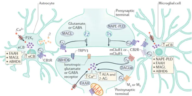
The extensive and diverse processes in which the ECS is known to be involved, derive from the widespread distribution of its constituting elements. Thus, the ECS is comprised of endogenous ligands (endocannabinoids, eCBs), the enzymes responsible for the synthesis and degradation of those ligands, protein transporters, and cannabinoid receptors (fig. 1.1).
Fig 1.1 – Schematic representation of the endocannabinoid system. The ECS is comprised of receptors, eCBs, the enzymes responsible for the
synthesis and inactivation of these ligands, and of the EMT (not pictured). The CB1R is primarily located on the presynaptic membrane, being
activated by AEA and 2-AG released from the postsynaptic neuron. Synthesis occurs in an “on demand” fashion, in response to a number of stimuli, such as the activation of metabotropic glutamate receptors, 2-AG being primarily synthetized postsynaptically, by the DAGLα pathway. AEA synthesis also occurs at the presynaptic neuron, by activation of the NAPE-PLD pathway, and can be released to activate postsynaptic TRPV1R. Fittingly, the primary degradative enzyme for 2-AG, MAGL, is predominantly located in the presynaptic neuron, whereas FAAH – primarily responsible for AEA inactivation – is located postsynaptically. In addition, a number of other enzymes are known to have a role in
2-AG inactivation, such as ABHD6. Moreover, the ECS is also expressed in both astrocytical and microglial cells. In the former, CB1R couples to a
different G-protein than in neurons (Gq and Gi/o, respectively), and is thought to mediate the release of gliotransmitters, In the former, CB2R and
possibly CB1R are involved in immune responses mediated by these cells. Figure taken and adapted from Lutz et al.66
2.1.1 – Endocannabinoid Synthesis and Release
There are at least seven recognized eCBs77, of which the most studied are N-arachidonoyl-ethanolamine (anandamide, AEA; fig. 1.2a)78 and 2-Arachidonoyl-glycerol (2-AG; fig. 1.2b)79. Unlike most neurotransmitters and neuromodulators, which are synthetized and stored in vesicles for posterior use, eCBs are primarily synthetized in the postsynaptic neuron, in an “on demand” fashiona, in response to depolarization-induced calcium (Ca2+) increases and/or the activation of G
q/11 coupled receptors77.
a It should be noted that, in recent years, evidence has emerged that there may non-on demand production of eCBS, whereby these
12
Anandamide, which acts as a partial agonist at both the CB1R and CB2R10, is synthetized by several pathways81, the most well characterized of which is the canonical pathway – in which membrane-lipid derived phosphatidylethanolamine is transacylated by an acyltransferase, to form N-acylphosphatidylethanolamine (NAPE), which is then hydrolyzed to AEA by the Ca2+-sensitive phospholipase D (NAPE-PLD)81.
2-AG, which, unlike AEA, is a full agonist at both the CB1R and CB2R82, is synthetized by several different pathways, the most widely studied of which is the PLCβ-DAGα pathway. This pathways seems to be triggered by Gq/11 activation, leading to phosphatidylinositol (PI) being hydrolyzed by phospholipase C β (PLCβ) to form diacylglycerol (DAG), which is then hydrolyzed by DAG lipase α (DGLα) to form 2-AG81,83. Interestingly there is evidence to support the idea that different 2-AG synthesis pathways (some which are PLCβ-independent), started by different stimuli, have distinct physiological roles84,85. Once synthetized, these endogenous compounds diffuse across the cellular membrane and into the synaptic cleft where they activate cannabinoid receptors.
Fig 1.2 - Molecular structures of anandamide and 2-arachidonoyl-glycerol. AEA (a) and 2-AG (b) are the primary eCBs present in the brain, being responsible for the majority of ECS actions. While AEA acts as a partial agonist at both the CB1R and CB2R, 2-AG is a full agonist at both receptors.
2.1.2 – Cannabinoid Receptors
All known cannabinoid receptors belong to the A-class of the G-protein coupled receptor (GPCR) superfamily86, with two – the CB
1R and CB2R – having been amply characterized. In addition, there are several other previously orphan GPCRs (e.g., GPR18, GPR55, and GPR119), whose belonging to the ECS is still disputed – with the most prominent of these being the GPR5586. Moreover, in recent years the transient receptor potential cation channel subfamily V member 1 (TRPV1), has also been recognized to be an important element in the ECS77.
13
2.1.2.1 – Cannabinoid Receptor Type 1 (CB1R)
The CB1R is one of the most abundant receptors in the human central nervous system87 and is also found in peripheral nervous system87, as well as in non-nervous tissue such as the liver and adipose tissue88. In the brain it is found in high levels in the inner layers of the hippocampus and the olfactory bulb, in the striatum, and in the molecular layer of the cerebellum, with intermediate levels having been found in the frontal, parietal and cingulate cortexes, the amygdala, the hypothalamus, and in some brainstem nuclei77,87. At the cellular level, this receptor is predominantly located on the presynaptic terminals of both γ-aminobutyric acid (GABA) releasing neurons (GABAergic neurons), and, to a lesser extent, in glutamatergic neurons77, where it modulates presynaptic activity. In addition, it also expressed in the postsynaptic membrane – where it has been shown to work as an auto-receptor89,90 – as well as in astrocytes91,92, microglia93 and oligodendrocytes94.
The CB1R is typically coupled to Gi/o protein, whereby its activation results in the inhibition of adenylyl cyclase (AC) and, therefore, in a decrease of 3',5'-cyclic adenosine monophosphate (cAMP) accumulation95. However, it has been shown that this effect is dependent on the specific isoform of AC being expressed, with the opposite effect (i.e., stimulation of AC and cAMP accumulation) occurring in cells expressing AC isoforms 2, 4 and 7, likely through the action of the dissociated Gβγ heterodimer96. Moreover, several studies have found that, in some conditions, CB1Rs are capable of signaling through other G-proteins: CB1R activation has been demonstrated to lead to increases in AC activity, through Gs protein, in cells where Gi/o activation is limited – such as in cells previously treated with pertussis toxin (PTX)97, or in which other G
i/o coupled receptors, such as the dopamine (DA) receptor type 2 (D2R), are simultaneously activated98. Furthermore, some reports have found that CB
1R are capable of signaling through Gq protein, leading to intracellular Ca2+ increases99, most notably doing so in hippocampal astrocytes92.
Gi/o protein activation is also the key component in CB1R-mediated modulation of ion channels. Indeed, CB1R activation is known to both increase potassium (K+) conductance, via activation of A-type and G-protein coupled inwardly rectifying K+ channels (GIRK)88,100, and to decrease Ca2+ conductance, via inhibition of L-, N- and P/Q-type voltage gated Ca2+ channels (VGCC) – through G
βγ mediated interactions88,100.
14
Furthermore, in addition to modulating the AC-cAMP pathway and ion channel activity, CB1R activation also leads to the stimulation of several mitogen activated protein kinase (MAPK) family kinases: depending on the cell type, CB1R activation has been demonstrated to lead to stimulation of extracellular signal-regulated kinase 1/2 (ERK; alternatively named p42/44 MAPK)88, through G
i/o protein activation, phosphatidylinositol-3-kinase (PI3K) activity via protein kinase B (PKB, also known as Akt kinase)101, inhibition of AC and protein kinase A (PKA)102, vascular endothelial growth factor (VEGF) receptor transactivation103, Src tyrosine kinase FYN activation104, and activation of Raf MAP kinase through the synthesis of the lipid second messenger ceramide105. Finally, CB
1R activation, has also been shown to lead to increased activity of both p38 MAPK and c-Jun N-terminal kinase (JNK), in a cell-type dependent manner88,100.
2.1.2.2 – Cannabinoid Receptor Type 2 (CB2R)
The CB2R was, for a long time, thought to be a peripheral cannabinoid receptor, with no significant expression in the nervous system106. Indeed, this receptor is found in high levels in peripheral and immune tissues, such as the spleen, bone, as well as in the gastrointestinal and reproductive systems106. However, in the last 15 years, reports have increasingly – but not without significant controversy106,107 – found CB
2R expression in the nervous system, albeit in much smaller numbers, in comparison with CB1R. The highest levels of CB2R have been reported in pyramidal neurons of layers III and V of the orbital, visual, auditory, motor and piriform cortexes, in pyramidal neurons of the CA2 and CA3 regions of the hippocampus, in the striatum, amygdala, and in Purkinje and granular cells of the cerebellum, and more moderate expression levels are found in several brainstem nuclei, such as the substantia nigra pars reticulata and periaqueductal gray108,109. At the synaptic level, this receptor, unlike the CB
1R, is found primarily in the postsynaptic membrane110, and has been demonstrated to act as an auto-receptor via 2-AG signaling, in the CA2 and CA3 regions of the hippocampus111. Moreover, CB
2R is also expressed in both microglia and astrocytes, in an activation state-dependent manner106,112,113 .
CB2R activation is tied to many of the same intracellular cascades as CB1R activation: by coupling to Gi/o protein, CB2R activation leads to inhibition of the cAMP-PKA pathway86,95, and to both increased K+ conductance and decreased Ca2+ conductance, through G
βγ interactions with GIRK114 and VGCCs115, respectively. Additionally, CB2R activation leads to stimulation of the Raf-MAPK cascade, leading to
15
increased ERK1/2 activity, in four possible ways: through Gi/o-dependent activation of PLC116, through decreased PKA activity117, through stimulation of the PI3K/PKB pathway94, and through the synthesis of ceramide118. Furthermore, CB
2R mediated activation of both the p38 MAPK119,120 and JNK120 pathways has also been reported. Finally, CB2R activation has been shown to lead to an increase in intracellular Ca2+ concentration, through PLC mediated Ca2+ release from IP
3 controlled calcium stores121.
However, two important distinctions have to be made between these two receptors: first, unlike CB1R, CB2R has not yet been shown to be capable of coupling to G proteins other than Gi/o, and, secondly, the inhibitory effect that CB2R activation has over AC is strongly modulated by both expression levels and cell environment, such that in some cells CB2R activation leads to little or no inhibition of AC activity, whereas in others it fully inhibits it117.
2.1.3 – Endocannabinoid Uptake
Once eCBs have activated the cannabinoid receptors, they are removed from the synaptic cleft so as to be intracellularly degraded81. However, the mechanism through which eCB uptake occurs is highly debated, and not yet fully elucidated122,123. Indeed, there is a long standing idea of an eCB membrane transporter (EMT)124, which posits the existence of a (yet to be characterized) membrane transporter which would transport eCBs (especially AEA) to the intracellular space122,123. This transporter is purported to be saturable, to work in a time and temperature-dependent manner, and to be capable of being selectively inhibited123. Accordingly, drugs have been developed that greatly decrease the rate at which AEA is removed from the extracellular space125. Moreover, experiments have demonstrated AEA uptake to be ATP- and ion gradient-independent, thus excluding the possibility of EMT working like other transporters (such as the DA transporter)125–127. However, several aspects of this hypothesis have been questioned, giving rise to alternative models:
Based on the findings that the original drugs used to inhibit the putative EMT have shown to also inhibit the activity of the AEA degradative enzyme fatty acid amide hydrolase (FAAH)128, that selective inhibition of FAAH greatly reduces AEA uptake129, and that inhibition of EMT does not, on a short time scale (<40s), inhibit AEA uptake130,131, a passive diffusion model was proposed130. In this model it is suggested that it is FAAH – by creating a concentration gradient – that drives AEA uptake, such that AEA passively diffuses across the cellular membrane, and is hydrolyzed intracellularly130,131. However, this model has been put
16
into question, due to the fact that AEA uptake still occurs (albeit in a reduced manner) in cells derived from FAAH-/- knockout mice132,133, and that newer, more selective, inhibitors of EMT, that do not inhibit FAHH, have shown to still be effective in blocking AEA uptake132.
Another proposed model for how AEA is uptaken proposes that, instead of entering the cell through the EMT or passive diffusion, AEA might instead do so via endocytosis134–136. Specifically, it is suggested that AEA binds to proteins in cholesterol rich domains of the cell membrane (lipid rafts), undergoes rapid endocytosis, and is then transported in vesicles, to be hydrolyzed by FAAH134–136. This model is supported by the fact that depletion of cholesterol reduces AEA uptake135, whereas increased cholesterol augments it137, that exogenously administered AEA and 2-AG were shown to congregate towards lipid rafts138, and that AEA has been shown to be accumulated intracellularly in adiposomes139. Despite the fact that this model has yet to be fully refuted, it cannot explain why FAAH inhibitors also reduce AEA efflux140, given it only posits a mechanism for uptake.
A third model posits that AEA is indeed uptaken via passive diffusion, but that, once inside the cell, AEA binds to carrier proteins which take it to be enzymatically degraded141,142. Of these carrier proteins the most widely studied are fatty acid binding proteins 5 and 7 (FABP5/7), as well as albumin and heat-shock protein 70, which have already been demonstrated to transport eCBs intracellularly143,144. Supporting this model, there are reports that FABP overexpression increased both AEA uptake and hydrolysis, whereas FABP inhibition had the opposite effects143, and that FABP5-/- knockout mice had a 50% increase in whole-brain AEA levels145. However, contrary to this model, there is conflicting data regarding the levels of FABP expression in adult neurons146, and there is yet no concluding data that excludes the possibility of the existence of an EMT. Additionally, much like is the case with the endocytosis model134– 136, this model cannot explain the inhibitory effects of AEA uptake inhibitors on AEA efflux140.
Finally, a more recent model, combines the previous model with the idea of the EMT, by suggesting that AEA does indeed bind to a membrane located transporter, which facilitates its diffusion to the cytoplasm123. Once in the intracellular space, AEA is suggested to bind to carrier proteins, which then take it to be degraded by FAAH123. By suggesting the existence of an EMT, this model allows for the inhibitory effects of AEA uptake inhibitors on AEA efflux.