Marta Machado Pereira dos Santos
Characterization of TRIB2-mediated
resistance to anti-cancer drugs
Mestrado em Oncobiologia:
Mecanismos Moleculares do Cancro
Trabalho efetuado sob a orientação de:
Professor Doutor Wolfgang Link
Professor Doutor Richard Hill
UNIVERSIDADE DO ALGARVE
Departamento de Ciências Biomédicas e Medicina
I
Characterization of TRIB2-mediated
resistance to anti-cancer drugs
Declaração de autoria de trabalho
Declaro ser a autora deste trabalho, que é original e inédito. Autores e trabalhos consultados estão devidamente citados no texto e constam da listagem de referências incluída.
II
Copyright em nome do estudante da UAlg, Marta Machado Pereira dos Santos
A Universidade do Algarve reserva para si o direito, em conformidade com o disposto no Código do Direito de Autor e dos Direitos Conexos, de arquivar, reproduzir e publicar a obra, independentemente do meio utilizado, bem como de a divulgar através de repositórios científicos e de admitir a sua cópia e distribuição para fins meramente educacionais ou de investigação e não comerciais, conquanto seja dado o devido crédito ao autor e editor respetivos.
III
Acknowledgements
I could not start without thanking my supervisors Dr. Wolfgang Link, for the opportunity to work in the lab in a theme that I liked, for the patience and guiding, and to Dr. Richard Hill for all the knowledge, for this challenging year, for all the patient (a lot needed!), but mainly for showing me that “A person who never made a mistake never tried anything new”, because they are not the easiest things that make us grow.
I want to thank to all that contributed to make this thesis possible, especially Dra. Patrícia Madureira for her help and hard work with all in vivo study, I couldn’t have done without her help and knowledge, is always a privilege work with you. To Dr. José Luís and their pathology Unit of Algarve Hospital Center-Faro for the technical support in the of the ex-vivo samples, to Prof. Kiss-Toth and his team for sharing their studies conducted in Sheffield, to Murat Isbilen Ali O. Gure, and to Dr Selma Ugurel (Julius-Maximilians- Universität Würzburg, Germany) that provided to our group normal and melanoma tissue samples.
Of course I need to thank my lab colleagues Laura that were there before me and gave a big help with everything in the lab and Neuton, Eduarda, Inês and Gisela for all the support, all the craziness and friendship because working with happiness is a privilege! I will miss those times.
Thank you to my last supervisor Iris Silva, she made me the scientist I am today, stronger, always trying to see the bigger picture, you will always be an example for me. Thank you for never leave alone and for all the advices, you are more than a supervisor, you are a friend for life.
To my colleagues from the Restaurant Pontapé thank you for all the help and support in these days, you are a real family to me. To all my friends a huge thank you, since you did exactly what I need in the right time, always distracting me from all the stress and keeping me motivated and sorry for my bad mood. A special thank you for Daniel Pestana, João Moreira, Bernardo Baião and Bruna Ramos for never let me be alone, for never give up on me, for all the companionship, I could never do this without you. Thank you Ricardo Vieira for all the jokes and the words, for being always present in my life, for being one of my best friends. Carina Maranga, I will never forget every word, all days, all the motivation. I would gave up without you, no doubt. Thank you for although Km far away, you were always by my side doing this hard road. Thank you for being the best friend I could ever have.
“Walking with a friend in the dark is better than walking alone in the light.”
Finally, thank in special to my mother and all my family, to all the support and dedication to help me, and to teach me always to fight for the better things. I want to dedicate my thesis to my father, that died last year. I would gave everything to have you in this moment with me, you will always be my star and idol. Thank you for leaving me so many word, so much of you, because I am stronger and I handle the hard roads because you always told that I am stronger than I think.
“One moment of pain is worth a lifetime of glory.”
IV
Abstract
Cancer results from the accumulation of multiple mutations and is characterized by deregulated cell mechanisms including cell growth, tissue invasion, angiogenesis and metastasis, being responsible for a large number of worldwide deaths. Malignant melanoma is the most aggressive type of human skin cancer, being just 5% of all skin cancer cases but accounting for over 80% of all skin deaths. This is primarily the result of melanoma being highly resistant to conventional chemotherapy. The PI3K/AKT signaling pathway is involved in many cell processes like survival, proliferation, growth and is one of the most mutated pathways in all human cancers, particularly in melanomas. FOXO3a, a transcriptional factor, is a crucial component of the PI3K/AKT pathway regulating the transcription of genes that promote apoptosis and cell cycle arrest. Following its activation, AKT negatively regulates FOXO3a, via phosphorylation, leading to FOXO3a export from the nucleus into the cytoplasm where it is ubiquitinated and is degraded. Consequently, if AKT is constitutively active, this enables a cancer cell to avoid apoptosis and to keep proliferating. The TRIB2 protein was discovered to be a FOXO3a repressor and is over expressed in many cancers although how TRIB2 mediates this effect is unknown. This project evaluates TRIB2 drug resistance to a dual PI3K/mTOR inhibitor BEZ235 in a clinically representative in vivo BEZ235 treatment model to confirm if our in vitro data (published and unpublished) correlates in a 3D in vivo model and demonstrates that AKT and TRIB2 interact in both in vitro cell models and in ex vivo clinical samples.
V
Resumo
O cancro é a nível mundial, uma das principais causas de morte. É o resultado da acumulação de várias mutações ao longo do tempo, e caracteriza-se pela desregulação dos mecanismos celulares. Há várias características que uma célula normal necessita para que se tornar cancerígena, sendo designadas como as “hallmarks of cancer” e neste momento são conhecidas dez: resistência à morte celular (apoptose), serem capazes de proliferar, serem capazes de escapar à vigilância e destruição por parte do sistema imune, serem imortais através da sua contínua replicação, serem genomicamente instáveis, serem capazes de induzirem a angiogénese (produção de novos vasos sanguíneos), desregularem os mecanismos de produção de energia celular, terem a capacidade de invadir outros tecidos, e por fim serem capazes de metastizar.
Melanoma maligno, é um tipo de cancro da pele que embora não sendo o mais comum, contemplando apenas 5% dos casos totais de cancro da pele, é o tipo mais agressivo, visto que é responsável por 80% das mortes por cancro da pele e a idade média de aparecimento é aos 50 anos, tendo maior incidência em indivíduos caucasianos. Este tipo de cancro é causado pela transformação maligna das células produtoras de melanina, os melanócitos que se encontram situados na epiderme. É também caracterizado por ser dos cancros no ser humano com pior prognóstico, e este facto deve-se à sua rápida metastização e invasão de outros tecidos e ao facto de atualmente não existirem drogas eficazes para o seu tratamento, havendo muitos casos verificados de resistência a quimioterapêuticos convencionais por parte destes pacientes. Este tipo de cancro é inicialmente designado por melanoma in situ sendo que nesta primeira fase, o seu desenvolvimento ocorre horizontalmente, dentro da epiderme. Ao longo do tempo, o seu crescimento é em profundidade, penetrando a derme e neste caso passa a ser designado por melanoma invasivo. Caso a doença seja detetada até esta fase, o prognóstico do paciente será bom uma vez que através de intervenção cirúrgica a fim de remover o tumor confere cerca de 95% de taxa de sobrevivência de 5 anos aos pacientes.
VI A via de sinalização PI3K/AKT está envolvida em múltiplos processos celulares, desde mecanismos relacionados com sobrevivência, crescimento e proliferação celular, e é uma das vias metabólicas mais mutadas em cancros humanos. PI3Ks são enzimas que desencadeiam a cascata de ativação de várias proteínas. A desregulação da via PI3K/AKT está associada a um elevado número de pacientes com melanoma, pelo que é necessário que se encontrem novas drogas de forma a melhorar o prognóstico dos pacientes, através da compreensão desta via de sinalização e dos seus componentes.
Sendo o AKT uma quinase, identificada como estando constantemente ativa nesta via, e estando descrito que esta ativação por parte do PIP3 (um dos substratos das PI3Ks), vai fazer com que o fator de transcrição FOXO3a, responsável pela transcrição de genes que promovem a paragem do ciclo celular (para que sejam remendados erros através dos checkpoints), e a apoptose (necessária para que as células cancerígenas morram) seja exportado do núcleo (onde está ativo), para o citoplasma onde através da fosforilação por parte do AKT, vai ser ubiquitinado e marcado para destruição proteossomal, impedindo assim que os genes com características anticancerígenas sejam transcritos.
Por ser uma das vias mais mutadas, e por não haver terapias eficazes, a via PI3K/AKT está a ser alvo de vários estudos e há várias drogas a ser testadas em ensaios clínicos, sendo BEZ235 principal foco no meu projeto de investigação, tendo já sido descrito por vários autores como tendo um potencial terapêutico anticancerígeno, visto que é um inibidor das PI3Ks.
Atualmente, sabe-se que a grande maioria dos pacientes com melanoma, adquire ao longo do tempo resistência aos tratamentos, pelo que se tem tentado compreender os mecanismos envolvidos nessa resistência, de forma a saber como reverter este problema.
VII É também conhecido na literatura que o TRIB2 é um gene cuja sobrexpressão está associada a vários tipos de cancro, entre os quais melanomas, estando este gene também associado a mecanismos de resistência a quimioterapêuticos, bem como está descrito o seu envolvimento na regulação negativa do FOXO3a. Não é ainda evidente de que forma é feita esta regulação, pelo que é crucial que haja mais investigação acerca destes mecanismos, uma vez que sabendo de que forma FOXO3a é inativado através do TRIB2, será possível encontrar uma forma de inverter esta situação. A principal hipótese colocada pelo nosso grupo de investigação com base em estudos feitos anteriormente, é a de que embora os inibidores das PI3Ks estejam a atuar, e que estas sejam de facto inibidas, se o TRIB2 estiver constantemente a ativar o AKT hipoteticamente através da fosforilação deste, o inibidor não estará a sortir o efeito desejado, pelo facto de o AKT estar constantemente ativo, visto que consequentemente vai inativar o FOXO3a, impedindo a transcrição dos genes com propriedades anticancerígenas, como por exemplo BIM, FasLG, p27, entre outros.
Neste projeto de investigação, é feita a avaliação do inibidor das PI3Ks BEZ235, de forma a compreender se a inativação do FOXO3a tem ou não relação com a interação entre o AKT e o TRIB2. Para isso fizeram-se vários estudos para não só verificar se há uma co-localização do TRIB2 e do AKT, através de CO-IP e de microscopia de fluorescência, bem como para compreender se realmente se verifica esta resistência a drogas por parte das células tumorais dos melanoma, e se a sobreexpressão de TRIB2 está em concordância um aumento do de AKT no seu estado ativo, ou seja fosforilado (pAKT/PO4-AKT) através de estudos in vitro,
VIII Atualmente está aprovado apenas um biomarcador para melanoma, LDH. Assim, o ideal seria que o TRIB2 fosse igualmente aprovado como biomarcador para a doença, para que desta forma fosse possível que através de um “screening” a pacientes com melanoma, verificar se há sobreexpressão de TRIB2. Verificando ser o caso, evitar-se-ia a administração de inibidores das PI3Ks tal como BEZ235, uma vez que esta droga não terá um efeito benéfico para o paciente e este estará apenas a sofrer efeitos citotóxicos desnecessários.
Devem ser feitos mais estudos de forma a elucidar os mecanismos que levam a que haja esta resistência, para que se descubram alternativas terapêuticas eficazes de forma a melhorar o prognóstico destes pacientes, já que este é dos menos promissores para a sobrevivência destes doentes.
IX
TABLE OF CONTENTS
Pages Acknowledgements III Abstract IV Resumo V Figures List X Tables List XI Appendix List XIAbbreviations List XII
CHAPTER 1 ... 1
1. Introduction ... 2
1.1. Cancer ... 2
1.1.1. Epidemiology ... 4
1.1.2. Cancer treatment methodologies ... 4
1.1.3. Novel cancer therapeutics ... 4
1.2. Melanoma ... 5
1.2.1. Epidemiology ... 6
1.2.2. Stages of disease/diagnosis. ... 7
1.2.3. Current treatment options for melanoma. Conventional therapy. 11 1.2.4. Novel therapeutic approaches useful against melanoma? ... 12
1.3. PI3K/AKT pathway ... 13
1.3.1. The role of the PI3K network in Melanoma ... 15
1.3.2. Forkhead transcription factors ... 16
1.4. Tribble2 homolog. ... 18
1.5. Hypothesis ... 20
X
2. Materials and Methods ... 22
2.1. In vitro studies ... 22
2.1.1. Cell culture ... 22
2.1.2. Transfection ... 23
2.1.3. Protein fragment complementation assays (PCA) ... 23
2.1.4. Co-Imunnoprecipitation (Co-IP) ... 23
2.1.5. Immunofluorescence ... 24
2.2. In vivo studies ... 26
2.2.1. BEZ235 treatment of Xenografted mice. ... 26
2.2.2. Western Blotting ... 27
2.2.3. RNA Extraction/cDNA Synthesis... 30
2.3. Ex Vivo: Patient Tissue Samples ... 32
CHAPTER 3 ... 33
3. Results ... 34
3.1. Increased TRIB2 protein expression confers significant resistance to PI3K/mTORC1 inhibitors in vivo... 34
3.2. In vitro cell characteristics are retained in our in vivo model. ... 39
3.3. The TRIB2 and AKT proteins interact both in vitro and in ex vivo clinical samples. ... 45 CHAPTER 4 ... 50 4. Conclusion ... 51 4.1. Future directions ... 54 CHAPTER 5 ... 55 5. References ... 56 CHAPTER 6 ... 60 6. Appendix ... 61
XI
FIGURES LIST
Pages
Figure 1.1.1 Genetic alterations in cancer 2
Figure 1.1.2. Hallmarks of cancer, 2011 3
Figure 1.2.2.1. ABCDE system for melanoma diagnosis 7
Figure 1.3.1.Regulators, effectors, and somatic alterations in the PI3K– AKT pathway in melanoma
14
Figure 1.3.1.1. AKT activation through PDK1 and mTORC2 phosphorylation
15
Figure 1.3.2.1. A) FOXO active in the nucleus, transcribing genes. B) FOXO regulation through AKT leads to their exportation
17
Figure 1.4.1. TRIB2 domains: N-terminal, kinase domain and C-terminal two binding motifs, one for MEK1 and one for COP1)
18
Figure3.1.1. Representative immunoblot analysis of TRIB2 status in our 293T isogenic cell lines
35
Figure 3.1.2. TRIB2 conferred resistance to PI3K inhibitors in vivo. 37 Figure 3.1.3.TRIB2 expression 293T tumour bearing mice survival 38 Figure 3.2.1: Representative immunoblot analysis from in vivo
treated/mock treated isogenic 293T-Empty/TRIB2 tumors
39
Figure 3.2.2. Spleen morphology 42
Figure 3.2.3. Spleen length measures 43
Figure 3.2.4. Spleen weight measures 44
Figure 3.3.1. Representative yellow fluorescent protein (YFP) complementation microscopy analysis assay
45
Figure 3.3.2. Fluorescent intensity of YFP positive cells 47
Figure 3.3.3. Co-immunoprecipitation of TRIB2 or total AKT1 48
Figure 3.3.4. Total protein levels in each indicated cell line for co-IP targets
48
Figure 3.3.5. Representative immunofluorescent images of stage IV metastatic melanoma
XII
Figure 4.1. AKT/TRIB2 interaction and FOXO3a inactivation. 52
TABLES LIST
Pages
1.2.2.1: TNM classification
9
1.2.2.2: Melanoma patients staging
10
2.4.1.1:Cell lines for Immunofluorescence
24
2.2.1.1. Xenografted mice groups.
27
2.2.2.1: Primary antibodies
28
2.2.2.2: Secondary antibodies
29
2.3.1:Melanoma and Normal tissue samples
32
APPENDIX LIST
Pages
Figure A. 10: Mounting solution protocol for immunofluorescence. 61 Figure A. 11: Poster done by me for the Oncobiology Retreat Session and
XIII
ABBREVIATIONS LIST
AJCC American Joint Committee on Cancer
AKT Protein kinase B
ATCC American Type Culture collection
ATF4 Activating transcription factor 4
BRAF B-type Raf kinase
CTLA-4 CTL antigen 4
Co-IP Co-Imunnoprecipitation
DNA Deoxyribonucleic acid
DTIC Dacarbazine
FACS Fluorescent Activated Cell Scanning
FasL Fas ligand
FDA Food and Drug Administration
FOXO Forkhead transcription factor
GPCR G-protein coupled receptor IGF-1 Insulin-like growth factor 1
Il-2 Interleukin-2
ING3 Inhibitor of growth family 3
hPTTG1 Human pituitary tumour-transforming gene HSP90 Heat shock protein 90
KD Kinase Domain
LDH Lactate dehydrogenase levels MAPK Mitogen-activated protein kinases
MAPKK Mitogen-activated protein kinase kinase
MDM2 Mouse double minute 2 homolog
mTOR Mechanistic target of rapamycin
NCOA3 Nuclear receptor coactivator-3
p70S6K Phosphorylation of 40S ribosomal protein S6
PBS Phosphate buffered saline
PDK1 Pyruvate dehydrogenase lipoamide kinase isozyme 1
PH Pleckstrin homology
PI3K Phosphatidylinositol 3 kinase
PIP2 Phosphatidylinositol-4,5- bisphosphate PIP3 Phosphatidylinositol (3,4,5)-trisphosphate
PRAS40 proline-rich AKT substrate of 40 kDa
PTEN Phosphatase with tensin homology
RGS1 Regulator of G-protein signaling 1
RTK Receptor tyrosine kinases
S100B S-100 calcium binding protein B SPP1 Secreted phosphoprotein-1
TRIB2 Tribbles2
TYR Tyrosinase
USA United States of America
Page 1 of 62
CHAPTER 1
Page 2 of 62
1. Introduction
1.1. Cancer
The development of organisms involves mechanisms such as proliferation, division and growth of cells. The problem arises when the organism is already developed and the cells keep proliferating and dividing which in case of wound repair is crucial, but when the cells get mutations in their genome, allowing genes to acquire new functions, driving the cells to new phenotypes that will no more follow their normal process and in the worst case, driving our body to the appearance of cancer.1,2
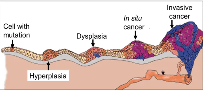
Unbalance between cell proliferation and apoptosis as well as the accumulation of several genetic mutations in DNA are needed for a normal cell to become a cancer cell, passing from one mutated cell that goes out of control, to a state of neoplasia, when the cancer spreads to other parts of the body. (Figure 1.1.1).1,3,4
Figure 1.1.1. Cancer development starts with a single mutated cell and progresses with the
accumulation of more several mutations followed by changes in gene expression. This genetic alterations are represented in different histopathological stages (Hyperplasia, Dysplasia, In situ cancer and finally invasive cancer).4
Page 3 of 62 Long ago, cancer was a concept to describe just one disease, but nowadays incorporates over 100 different diseases, different cancer types wherein each of them consists in a multistage process, consequence of genetic factors like genetic mutations or environmental factors that will have an effect in gene expression (i.e., drugs, diet, UV).5,6
For tumor progression, in order to survive and avoid apoptosis, is crucial that a cancer cell acquire particular characteristics which are named “The hallmarks of
cancer”. In 2000, were defined six leading characteristics2, however nowadays with
the new recent discoveries, are known in total ten (Figure 1.1.2).1
Page 4 of 62
1.1.1. Epidemiology
Cancer is a worldwide urgent problem since it was responsible for 14.1 million new cases and 8.2 million deaths in 2012, making cancer one of the most deadly diseases in the world whether in non-developed or in developed countries. Worldwide most frequent cancers are breast, colorectal and lung cancer. Is currently called “the disease of the future” considering one likely 70% increase of new cancer cases for the following 2 decades, having 22 million cases instead of the 14 million in 2012 due to many factors: the increase of an older population, their lifestyle with associated behavioral changes like a sedentary lifestyle, smoking and a poor diet.7
1.1.2. Cancer treatment methodologies
Cancer treatment is chosen according to the cancer type and stage, because each cancer is unique; each cancer will have a different treatment approach. People could have just one kind of treatment or a combination of more than one, for example surgery (procedure made by a surgeon to remove the tumor from your body) is the first approach in case the tumor can be removed, plus radiation therapy or chemotherapy in order to kill cancer cells by damaging DNA and avoid cells to keep diving.5,8
1.1.3. Novel cancer therapeutics
Optionally, we have also new therapies to address this problem, like immunotherapy that is used with the aim of stimulating the immune system to fight cancer, through the activation of immunologic cells (i.e. using dendritic cells vaccine).9
To avoid tumor progression and induce their regression, targeted therapy is another alternative approach which uses drugs to block the action of mutated proteins in signaling pathways that are deregulated and that are allowing the tumor cells to survive and keep growing.8,10
Page 5 of 62
1.2. Melanoma
Skin cancer is classified as non-melanoma (basal or squamous cell) or melanoma (only 5% of all skin cancer cases), according to the cell type that gave rise to cancerous cells. Melanoma arises from the transformation of the melanocytes, cells that are located in the bottom of epidermal layer of the skin and that are responsible for the production of the pigment melanin. This pigment is involved in skin protection mainly against ultraviolet sun rays exposure, which is the major cause of melanocytes transformation. This exposure triggers mutations that will lead to an abnormal cell multiplication and consequently to malignant tumors formation.11
Furthermore mutations in CDKN2A and CDK4 genes (involved in cell division control) have been shown to be associated with familial malignant melanoma (10-15% of the cases). However, the most part of the melanoma cases appear sporadically, being named sporadic melanoma. Having blue or green eyes and fair skin, if the person gets easily sunburns, freckles and moles which (could develop to melanoma) are risk factors as well.12,13
Melanomas can arise from any part of the skin, appearing commonly in the neck and face, but preferably on the legs in women and on the chest and back in men.11
Page 6 of 62
1.2.1. Epidemiology
On average, each year 132,000 melanoma are diagnosed cases worldwide. Caucasian populations have a higher risk to develop melanoma skin cancer comparatively with dark-skinned populations, due to a lower melanin pigmentation protection, for these factor Australia population is the one with highest incidence of melanoma since is predominantly Caucasian.14,15 The age more susceptible to the
appearance of melanoma is the late fifties and are diagnosed more cases in men than in women (1.3/1).16
Melanoma although not the most common skin cancer type, contemplating 5% of all skin cancer cases, is the most dangerous one, being responsible for 80% of all skin cancer deaths, making it one of the most aggressive of all human cancers. The American Cancer Society (AJCC) predicts that in 2015, there will be diagnosed around 73,870 new melanoma cases in the US, showing an increase of 200% since 1973, wherein 31,200 in women and 42,670 in males, estimating in total 9,940 deaths. 5,7,8
Statistical studies show that New Zealand/Australia has the highest melanoma mortality rate in the world with 3.5 deaths per 100 000 habitants, followed by the United States of America (USA) with 1.7/100 000 and the third is Europe with 1.5 deaths/100 000.7,17
In Portugal, it was registered in 2007 by the National Cancer Registry, that per 10.000 habitants we have 6 cases of malignant melanoma, corresponding to an increase of 15% since 2001 (with 5.2 cases per 10.000 habitants). Annually, appear about 700-800 new cases being the incidence higher in women than in men (1/1.14) and the average upper the 50.15,18
Page 7 of 62
1.2.2. Stages of disease/diagnosis.
In order to know how far the cancer is spread, thereby giving a more appropriate and accurate treatment, melanoma patients need to have a good diagnosis, to consequently have a better prognosis. For that, is used the staging system from the AJCC.11
Prior to melanoma staging, ABCDE nomenclature (Figure 1.2.2.1) is performed. This method is used for an early detection and is limited, once the asymmetry, border, color, diameter and evolution parameters are self-skin checked. If there are abnormal changes, next step is to go see an dermatologist.11
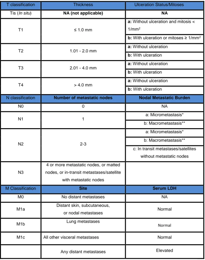
Page 8 of 62 Melanoma staging is usually performed by the American Joint Commission on Cancer (AJCC) TNM system, that evaluates the tumor thickness (T), how many lymph nodes are affected with cancerous cells (N) and finally if there are any metastases, to know how far the cancer spread or not to other organs (M). In combination with these factors, are measure as well the tumor ulceration, mitotic rate and lactate dehydrogenase levels (LDH). (all described in table 1.2.2.1)19,20
Nowadays, only LDH is established by the AJCC as a biomarker for melanoma prognostic classification system, over more than 60 years ago and is used because of their high specificity and sensitivity to detect this disease. A good biomarker must obey to some criteria, such as to be measured in an accessible body fluid like blood, must be minimally invasive to the patient and to have a low cost.20
Page 9 of 62
T classification Thickness Ulceration Status/Mitoses
Tis (In situ) NA (not applicable) NA
T1 ≤ 1.0 mm
a: Without ulceration and mitosis <
1/mm2
b: With ulceration or mitoses ≥ 1/mm2
T2 1.01 - 2.0 mm a: Without ulceration b: With ulceration T3 2.01 - 4.0 mm a: Without ulceration b: With ulceration T4 > 4.0 mm a: Without ulceration b: With ulceration
N classification Number of metastatic nodes Nodal Metastatic Burden
N0 0 NA N1 1 a: Micrometastasis* b: Macrometastasis** N2 2-3 a: Micrometastasis* b: Macrometastasis** c: In transit metastases/satellites without metastatic nodes
N3
4 or more metastatic nodes, or matted nodes, or in-transit metastases/satellite
with metastatic nodes
M Classification Site Serum LDH
M0 No distant metastases NA
M1a Distant skin, subcutaneous,
or nodal metastases Normal
M1b Lung metastases Normal
M1c All other visceral metastases Normal
Any distant metastases Elevated
Table 1.2.2.1: TNM classification according to the 7th edition of the Melanoma staging system
Page 10 of 62 To determine the melanoma patient stage, is made the combination of the TNM parameters in order decide the most appropriate treatment according to the disease stage (table 1.2.2.2).20
Stage Melanoma
Characteristics
Classification
5 year survival rate
T N M
0
Carcinoma in situ (in epidermis and has not
spread to dermis)
Tis N0 M0 99 – 100 %
I
A/B Lesions up to 2 mm but no
nodal or distant metastases
T1a N0 M0 A – 95% B –88 - 92 % T1b N0 M0 T2a N0 M0 II A/B/C
Lesions greater than 2 mm, no positive nodes or distant
metastases T2b N0 M0 A – 77 - 79 % B – 61 - 70% C – 43 - 45% T3a N0 M0 T3b N0 M0 T4a N0 M0 T4b N0 M0 III
A/B/C Lesions of any size with
positive lymph nodes
Tx N1 M0 A – 57 – 73 % B – 41 – 57 % C – 20 – 34 % Tx N2 M0 Tx N3 M0
IV Lesions of any size with
distant metastases Tx Nx M1 5 – 22 %
Table 1.2.2.2: Melanoma patients staging based in TNM classification according to the 7th
edition of the Melanoma staging system by the American Joint Commission on Cancer (AJCC) plus the measurement of lactate dehydrogenase levels (LDH), tumor ulceration and mitotic rate. (Tx-Primary tumor cannot be assessed; NX- Regional lymph nodes cannot be assessed; MX-Presence of distant metastasis cannot be assessed.)
Page 11 of 62
1.2.3. Current treatment options for melanoma. Conventional therapy.
Early melanoma detection makes possible that through surgery (surgical excision of the tumor and the nearby tissue), most part of primary melanomas (about 80%) could be treated and curable. For stage III melanoma patients usually have for option one year treatment with adjuvant immunotherapy with interferon-α. In advanced stages, with the cancer spreading to other organs, making hard to define the best treatment, the prognosis is a real challenge. In these cases, around 45% of patients with metastatic melanoma receive immunotherapy or chemotherapy. For the patients with the worst prognosis, melanoma patients in stage IV, surgery is not a viable option, wherein the suggested treatment is systemic drug therapy. 16,22–24
Conventional chemotherapy uses drugs with aggressive cytotoxic effects, mostly alkylating agents, to avoid cell replication. Food and Drug Administration (FDA) is the entity that approves these compounds, such as Dacarbazine (DTIC) a drug approved in 1975, Fotemustine (Muphoran), Temozolomide (Temodal). The problem with these treatments is the fact of just 10% triggers an effective response and do not have an overall survival increase. 17,25FDA recently approved the BRAF
inhibitor vemurafenib and the MEK inhibitor trametinib for melanoma patients that harbor BRAF mutations.26,27
Regarding immunotherapy, in 1998 FDA have approved Interleukin-2 (IL-2), the first immunotherapy for advanced melanoma, however has the same issue of DTIC because even with high-doses of de drug the response is not significant. Nowadays, is used in combination with DTIC (since 2011). 28
Since 2009, new systemic cancer treatments have been emerging wherein monoclonal human antibody against the CTL antigen 4 (CTLA-4) commercially known as Ipilimumab, was one of the most important discoveries in the research for immunotherapeutic drug crucial in immune system stimulation to respond against tumor antigens. Recently, was found that Ipilimumab in combination with GM-CSF instead Ipilimumab administrated alone is more effective to treat metastatic melanoma patients since there is an overall survival increase and lower toxicity to the patients.29–33
Page 12 of 62
1.2.4. Novel therapeutic approaches useful against melanoma
Presently, with the aim to provide more quality of life to melanoma patients, it is important to search novel therapeutic approaches. Currently, several compounds are being tested in clinical trials, including NPV-BEZ235 (BEZ235).34,35.
For this project, BEZ235 (Novartis) was our particular research focus. BEZ235 is a selective PI3K and mTOR dual inhibitor, classified as an imidazoquinoline and tested already in many clinical trials that have showed anti-cancerous properties, such as the induction of apoptose, suppression of cell proliferation, induction of G1 cell cycle arrest and inhibiting the function of many proteins such as AKT, S6, S6K. This drug is given orally to the patients, having side effects like nausea, headache diarrhea, with a stable response demonstrated in clinical trials.36–40
Page 13 of 62
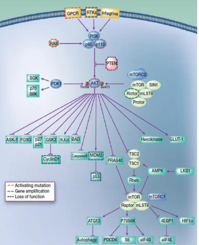
1.3. PI3K/AKT pathway
PI3K/AKT signaling pathway (showed in Figure 1.3.1) has been a crucial target for research not only because it is involved in many regulatory cell mechanisms (differentiation, migration, growth, proliferation and survival)41,42 , but
also for being one of the most mutated pathways in human cancers.43
Phosphoinositide 3-kinase (PI3K) is a lipid kinase. It is a heterodimer since it is constituted of two subunits: catalytic (p100) and regulatory (p85). 40,44 PI3K is
one of the most important component in this pathway, having a central role in the regulation of angiogenesis (formation of new blood vessels), apoptosis (programmed cell death), cellular metabolism, and Deoxyribonucleic acid (DNA) repair.45,46
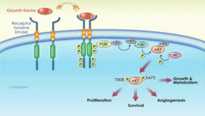
PI3K activation could be made through many signals, such as the interaction between transmembrane tyrosine kinase-linked receptors (RTKs), G-protein coupled receptor (GPCR), or simply by cell-to-cell interaction.47 This activation will
trigger the phosphorylation of PI3K substrate phosphatidylinositol-4,5- bisphosphate (PIP2, present in the cellular membrane) into phosphatidylinositol- 3,4,5- triphosphate (PIP3), a fast action that takes just seconds.42,45,47 After this
conversion, PIP3 will recruit to the cell membrane proteins that contain a pleckstrin
homology (PH) domain, like the protein kinase B (PKB or more known as AKT). AKT named as the first downstream mediator of PI3K signaling pathway, belongs to serine-threonine kinase family, and in mammals exists in three isoforms (AKT1/ AKT2/AKT3) encoded by three different genes, been expressed in variable levels, according to the tissue in question.48 In order to achieve total activity, AKT must
have both their conserved residues (threonine 308 and serine 473) phosphorylated, being threonine 308 residue targeted by the kinase phosphoinositide-dependent kinase 1 (PDK1) and serine 473 residue by Mechanistic Target of Rapamycin Complex 2 (mTORC2). At this moment, p-AKT is ready to phosphorylate other downstream substrates that are involved in cell regulation. One negative regulator of this pathway is PTEN (tumour suppressor), that will through their phosphatase function, dephosphorylate PIP3 into PIP2 avoiding that AKT becomes activated.40,45
Page 14 of 62
Figure 1.3.1: Regulators, effectors, and somatic alterations in the PI3K–AKT pathway in
Page 15 of 62
1.3.1. The role of the PI3K network in Melanoma
PI3K/AKT signaling pathway in our particular field of research, is deregulated in a high proportion of melanomas, so by understanding how the molecules of this pathway intact and the mechanisms involved, will make possible the discovery of molecules in order to avoid melanoma progression. 43,44,46
In Melanoma cells, AKT3 isoform has a higher expression than AKT1 and AKT2, since their activation is identified in about 60% of sporadic Melanomas.26,49
AKT promotes a perfect environment to these cancerous cells, since it allows these cells to avoid apoptosis, to keep growing, proliferating, to resist to hypoxia (low levels of oxygen in the cells/tissues) and increasing cell metabolism, due to the fact of AKT is constitutively active. When AKT is activated it translocates from cell membrane to the cytoplasm (figure 1.3.1.1) and then to different cell places to target substrates.47,50,51
Page 16 of 62
1.3.2. Forkhead transcription factors
As mentioned previously, many effector proteins and substrates are phosphorylated by pAKT (active form of AKT) and Forkhead box O3a (FOXO3a) is one of them. FOXO3a is a member of the family FOXO which is a subgroup of Forkhead transcription factor family.52 FOXO3a is a transcriptional factor located
and active in the nucleus, responsible for the regulation of the expression of genes that are involved in many mechanisms such as Cyclin-dependent kinase inhibitor
p27 (p27), a cell cycle inhibitor; Fas ligand (FasL), a ligand responsible for
Fas-dependent cell-death; Bcl-2 Like protein 11 (Bcl-2L11, also known as BIM), a Bcl-2 family member (which possess conserved Bcl-2 homology [BH] domains 34) responsible for pro-apoptotic signaling.53
FOXO3a is negatively regulated by AKT phosphorylation, leading FOXO3a to translocate from the nucleus to cytoplasm, becoming inactive and consequently represses transcription anti-apoptotic genes. In the cytoplasm FOXO3a will be polyubiquitinated and marked for proteosomal destruction (Figure 1.3.2.1).53
A
Page 17 of 62
Figure 1.3.2.1: A) FOXO active in the nucleus, transcribing genes involved in cell cycle arrest and
apoptosis ( p27, FasL and Bim); B) FOXO regulation through AKT leads to their exportation from the nucleous to the cytoplasm.
Page 18 of 62
1.4. Tribble2 homolog.
Being LDH the only biomarker approved by FDA for melanoma, there is a need to find new melanoma biomarkers and actually many are been tested in clinical trials, such as S-100 calcium binding protein B (S100B); tyrosinase (TYR); human pituitary tumour-transforming gene 1(hPTTG1); RGS1 (Regulator of G-protein signaling 1); heat shock G-protein 90 (HSP90); Secreted phosphoG-protein-1 (SPP1); Inhibitor of growth family 3 (ING3); Nuclear receptor coactivator-3 (NCOA3). 54–60
In addition to the discovery of these biomarkers, it was discovered Tribbles2 (TRIB2) correlates with melanoma. TRIB2 is the mammalian homolog of the drosophila gene tribbles2 (was described firstly in this specie). In drosophila, this protein is involved in cell cycle control during morphogenesis and oogenesis, being involved as well as in regulation of cell proliferation, metabolism, motility and cancer development.52
TRIB2 is a kinase-like protein or a pseudokinase as it belongs to the TRIB pseudokinases family, composed for three members (TRIB1/TRIB2/TRIB3) whose catalytic residuesare not conserved, conditioning their catalytic function. Structurally, is divided in three distinct regions: N-terminal region, a serine-threonine kinase-like domain (KD, that has evident homology with serine-serine-threonine kinases) and a C-terminal domain, a binding site for E3 ubiquitin ligases (Figure 1.4.1.).52
Figure 1.4.1: TRIB2 domains: N-terminal, kinase domain and C-terminal (two binding motifs,
Page 19 of 62 The three TRIB members are all related with human cancers, having roles in signaling pathways such as MAPK and PI3K/AKT, interacting with many transcription factors and signaling molecules: MAPKK, ATF4, p65, COP1, CtIP, and AKT, being implicated in regulation of gene expression, apoptosis, cell activation and mitosis.52,61,62
TRIB2 is known to be overexpressed in human malignant melanoma and it is related with FOXO3a export from the nucleus to the cytoplasm, leading to the inhibition of FOXO3a tumor suppressor function. Although it is not known exactly how this mechanism works, it is believed that TRIB2 is a good research target to avoid melanoma cells growth and increase patients overall survival since is implicated in the maintenance of melanoma cells properties. 22,52
TRIB2 is is involved in cell division and has been reported to be up-regulated in a subset of acute myeloid leukemia’s and members of tribbles family have been also reported to interact and modulate the activity of signal transduction pathways, including the PI3K/AKT and the MAPK, our pathways of interest. It is important to say that TRIB2 has been implicated in the negative regulation of FOXO3a and this
knowledge is important to understand the mechanism(s) of action of how TRIB2
mediates PI3K inhibitor resistance and the role of FOXO3a in this response. Thus, if we knew how this pathway works and their mechanisms are related to tumor progression and melanomas appearance, we could design strategies to treat cancer in earlier stages through target therapy.35,61–63
Page 20 of 62
1.5. Hypothesis
It is known that PI3K/AKT is one of the most mutated pathways in human cancer, is also known that FOXO mediates the action of many anticancer drugs and that TRIB2 negatively regulates FOXO. Our research group has demonstrated that TRIB2 is not only a biomarker for melanoma diagnosis, but is also involved in this cancer type progression being our biggest aim to understand how is TRIB2 allowing melanoma patients to acquire resistance to chemotherapeutic drugs, in particular to PI3K inhibitors, like BEZ235. Through in vivo models with an oral administration to be a representative model to the patients in the clinic, and ex vivo samples from melanoma patients to see if there is a drug resistance caused by TRIB2 and knowing that AKT negatively regulates FOXO, to understand this mechanism of resistance, was hypothesized if does this happen because TRIB2 is in AKT network and if they interact. Since preliminary results from our group indicate that TRIB2 confers chemotherapeutic resistance, the central aim is to elucidate the mechanism(s) of this resistance and in particular how the TRIB2 protein responds following PI3K inhibition.
Page 21 of 62
CHAPTER 2
Page 22 of 62
2. Materials and Methods
2.1. In vitro studies
2.1.1. Cell culture
For this project were used isogenic cell lines, SK-MEL-28 derived from a range of melanoma cell lines settled from skin primary tumor samples from a 51-year-old male patient (unknown ethnicity), acquired from American Type Culture collection (ATCC) and SK-MEL-28 TRIB2 shRNA; G-361 also acquired from ATCC was established from a malignant melanoma of a 31 year old male Caucasian. U2OS cell line (Human Bone Osteosarcoma Epithelial Cells) derived from a fifteen-year-old human female suffering from osteosarcoma; 293T shrna TRIB2 cell line originated from human renal cancer cells. All cell lines were defrosted from -80ºC and cultured within 35mm plates in 3ml DMEM (Dulbecco's Modified Eagle Medium Sigma, USA) medium with 10% heat inactivated FCS (Sigma) supplemented with Pen/Strep (Gibco) to be used for BEZ235 (10nM) drug treatment whereby were added 3ul of the drug to get a 1:1000 ratio. After the drug addition, all the cell cultures were kept overnight in a humidified incubator in 5% CO2 at 37ºC.First of all, the medium was aspirated and to detach the cells from the culture plates was used 1ml of Trypsin-EDTA 1x solution (SAFC Biosciences, UK). After trypsinization, the trypsin with cells was transferred to a falcon to centrifuge at 11rpm during 5 minutes.
The pellet was washed with 800ul of Phosphate buffered saline (PBS) 1x and shifted to an eppendorf for protein extraction.
Page 23 of 62
2.1.2. Transfection
We used JetPrime transfection Protocol to transfect our cell lines with a dilution of 2 μg of our DNA into 200 μl jetPRIME® buffer, both posteriorly mixed by vortexing. After, was added 4 μl of jetPRIME®, vortexed for 10 seconds and then spun down briefly. 10 minutes room temperature incubation and thereafter it was added 200 μl transfection mix per plate. Lastly, the plates were slowly shaken and placed into the humidified incubator in 5% CO2 at 37ºC for 24h.
2.1.3. Protein fragment complementation assays (PCA)
5 μg of our each plasmid construct (ZIPV1, ZIPV2, AKT1, AKT2, TRIB2, TRIB3 or JIP1) were transfected (JetPRIME following the manufacturers protocol) into 293 cells. 24 hours post transfection, cells were stained for YFP imaging and visualized on a Leica TCS SP5 II confocal microscope. Duplicate studies were conducted and fluorescent signal intensity was measured 24 hours post dual construct transfection using a BD FACS Calibur scanner. The leucine zipper constructs ZIPV1 and ZIPV2 serve as positive control for these studies.
2.1.4. Co-Imunnoprecipitation (Co-IP)
After protein sample quantification using the Bradford assay followed by Nanodrop measure, were added PBS up to 500ul depending of the protein value to be loaded, plus 5ul of primary antibody and stayed overnight in the rotator at 4ºC. For each sample were needed 20ul of protein A/G-agarose beads (Sigma) which were first washed 2x with PBS. 1300 spin for 2min and only keep the pellet to be mixed with 20ul of PBS and the resuspended. Spin 1300 rpm for 2min and waste the PBS. Were added 20ul of beads to each sample, then resuspended and placed 1h on the rotator. Another 1300 rpm spin for 1min and after that were made one wash with 200ul of PBS. Spin 1300rpm 1min. Once again the pellet was kept and were added 40ul of laemmli, boiled for 5min at 98ºC and 20ul of each sample were used to be loaded in SDS-PAGE gel.
Page 24 of 62
2.1.5. Immunofluorescence
In order to have samples to see in the Microscope, we need to prepare them previously after cell growing in the desired conditions (Table 2.4.1.1). First of all we put each lamella with respective cell line condition in a six well plate with PBS solution (10x).
Table 2.4.1.1: Cell lines for Immunofluorescence.
Then, we transferred the lamellas with the cells side face up to a new six well plate wash 3x with PBS (1x) and after that see in the Microscope if there is a good cell confluence. If the cells look fine, we could proceed with the dual staining protocol. We cover a western blot glass with parafilm paper and put four equal drops (more or less 200ul) of 1:30 block solution (900ul PBS (1x) + 30ul block solution (stock))and after that lay down the lamellas with the cells side face down to be in contact with the solution, one per drop. Incubate in the Humified Chamber for 1h, 37ºC. (Cell culture incubater). We took the lamellas from the Humified Chamber and put them with the cells face up in the parafilm paper, cleaned the block solution from the glass with parafilm and prepared 1:500 1ºAB for 1ul p-AKT (Santa Cruz) and 1ul TRIB2 (Santa Cruz) in PBS (1x) and put four drops of that mix in the glass with parafilm. Then, put the lamellas face down in contact with the 1ºAB and incubate in the Humified Chamber for 1h, 37ºC.
Cell lines for Immunofluorescence
293T Skmel-28
293T EMPTY NT Skmel-28 EMPTY NT
293T EMPTY BEZ Skmel-28 EMPTY BEZ
239T TRIB2 NT Skmel-28 TRIB2 NT
Page 25 of 62 Took lamellas from the Humified Chamber and put them with the cells face up in the six plate wells and wash them 3x with PBS (1x). Clean the 1ºAB from the glass with parafilm. We prepared 1:500 2ºAB for 1ul α-mouse (against TRIB2) (1ul) and 1ul α-rabbit (against p-AKT) in PBS (1x) and put four drops of that mix in the glass with parafilm. Then, put the lamellas face down in contact with the 2ºAB. Incubate in the Humified Chamber for 1h, 37ºC. Took lamellas from the Humified Chamber and put them with the cells face up in the six plate wells and wash them 3x with PBS (1x). Clean the 2ºAB from the glass with parafilm. For each condition/lamella, one lamina where was placed one drop of ClearMount™ Mounting Solution, INVITROGENTM. Then we put the cells face down and incubate
in a 60ºC chamber for 30 minutes. After the 30 minutes, we've saved them there in the dark, very important to not leave them in the light! The Microscope used for image visualization were Axio Imagen Z2.
Page 26 of 62
2.2. In vivo studies
2.2.1. BEZ235 treatment of Xenografted mice.
This work was performed with 21 animals (table 2.2.1.1), which were divided into two initial groups to be injected with different isogenic cell lines, in order to form tumors: 14 mice injected with 2x10⁶ 293T-empty cells into the right flank of
matched NOD/SCID mice and at the same time, the other 7 were injected with 2x10⁶ 293T-TRIB2 cells into the right flank of matched NOD/SCID mice. These
animals were monitored daily for tumour development, and once tumors were visible, they were measured prior to in vivo treatment. The 14 mice containing 293T-empty tumors were then separated into two groups of 7 mice prior to the BEZ235 in vivo study commencing, having after one group with endogenous levels of TRIB2 and no drug treatment, 293T Empty Vehicle (n=7); other group with endogenous levels of TRIB2 and BEZ235 drug treatment, 293T Empty BEZ235 (n=7) and finally the last group with overexpression of TRIB2 plus BEZ235 drug treatment, 293T TRIB2 BEZ235 (n=7). Drug aliquots were prepared for one or two days (since the compound is stable for only two days), being the average mice weight 25g, and the concentration to be administrated to the mice 30mg/Kg, the calculation is 10,5mg of BEZ235 dissolved in 2,1ml of 20% NMP/80% PEG 300 for one day. This compound must be protected from the light and be stored at 4ºC, the NMP is added to BEZ235 and must stir for 30 min at 40ºC (protected from light), PEG300 as well must stir but for 60 min at 40ºC (protected from light) and then keep at RT to be used in the drug administration.
The animals had a daily tumor measurement and daily drug treatment with BEZ235: 30mg/kg, orally administrated (syringes provided by Prof. Inês Araújo) but with the mice anaesthetized/sedated with isoflurane (Cell line injection to the mice, tumour measures, and drug administration was made by Dra. Patrícia Madureira).
Page 27 of 62
Table 2.2.1.1. Xenografted mice groups.
In the end of the study, the animals were sacrificed after been exposed with CO2 (by Dr. Richard Hill), and the organs (spleen and tumors) were surgically removed (by Dra. Patrícia Madureira) for protein and RNA extraction.
2.2.2. Western Blotting
We’ve collected the following organs for protein extraction and quantification: the liver, the spleen and the tumors. The organs homogenization was made in a manual potter homogenizer (Sigma) with 200ul-500ul (depending on the organ size) of RIPA Buffer (Tris-HCL ph 7.4, NaCl, 10% Nonidet P-40, 10% sodium deoxycholate, 100 mM EDTA, PIC, 200 mM NA-F, 100mM Na3VO4 and protease inhibitors cocktail) for the tumors and livers or 300ul for the spleens. Then, the homogeneized samples incubated for 30 minutes on ice. The lysed samples were spun at 13000 rpm and the supernatant transferred to a new eppendorf for posterior protein quantification. Extracted proteins were stored at -80ºC until use. For protein quantification to determine their concentrations we used the BCA assay (Thermus Scientific Protein Assay Kit). Then, to be quantified we used 2ul of that mix to be read in the NanoDrop 2000/2000c (ThermoScientific) UV-Vis Spectrophotometer.
Xenografted mice (n = 21)
Groups Number of animals Number of injected cells 293T Empty Vehicle n=7 2x10⁶
293T Empty BEZ235 n=7 2x10⁶
Page 28 of 62 Our extracted protein samples were diluted into 2x lammeli loading buffer (containing β-mercaptoethanol) and heat for 5 minutes at 95ºC to be loaded into 10% SDS-PAGE gels with the NZYBlue Protein marker. Once the proteins were separated, the gel was transferred to a nitrocellulose membrane (Amersham Hybond ECL) Nitrocellulose Membrane through TRANS-BLOT® SEMI-DRY TRANFER CELL (BioRad) system for 1h10min. The membranes were blocked with skimmed milk powder (exception when we used p-MDM2 Antibody, in which case BSA was used) and placed on a shaker for 1 hour to prevent the binding of non-specific antibodies and probed with many primary antibodies (table 2.2.2.1) overnight at 4ºC.
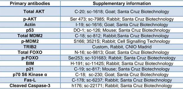
Table 2.2.2.1: Primary antibodies.
Primary antibodies Supplementary information
Total AKT C-20; sc-1618; Goat; Santa Cruz Biotechnology
p-AKT Ser 473; sc-7985; Rabbit; Santa Cruz Biotechnology
Actin I-19; sc-1616; Goat; Santa Cruz Biotechnology
p53 DO-1; sc-126; Mouse; Santa Cruz Biotechnology
Total MDM2 C-18; sc-812; Rabbit;Santa Cruz Biotechnology
p-MDM2 S166; 3521S; Rabbit; Cell Signalling Technology
TRIB2 Custom, Rabbit, CNIO Madrid
Total FOXO N-16; sc-9813; Goat; Santa Cruz Biotechnology
p-FOXO Ser253; sc-101683; Rabbit; Santa Cruz Biotechnology
BIM H-191; sc-11425; Rabbit; Santa Cruz Biotechnology
p21 C-19; sc-817; Mouse; Santa Cruz Biotechnology
p70 S6 Kinase α C-18; sc-230; Goat; Santa Cruz Biotechnology
Fas-L C-178; sc-6237; Rabbit; Santa Cruz Biotechnology
Page 29 of 62 After incubation, were washed for 10 minutes with TBS 0.1% tween20 (3x)
and then, probed it with matched secondary antibodies for 1 hour at room temperature in the shaker.
Table 2.2.2.1: Secondary antibodies.
Membranes were once again washed for 10 minutes with TBS 0.1% tween20
(3x) and perform an image visualization, the membranes were placed in a ECL solution which is a mix of a Buffer 1 (5ml 1M Tris pH 8.5 100nM, 0.5ml luminol 2.5nM, 222.3ul p-coumaric-acid 360uM, 45.5ml H2O) and Buffer 2 (4ml 1M Tris pH
8.5 100nM, 46ml H2O, 31ul 30% H2O2) and then achieved through a Molecular
Imager® ChemiDoc™ XRS System (BioRad).
Secondary
Antibodies Secondary Antibodies
Anti-mouse IgG-HRP; sc-2314; Donkey; Santa Cruz Biotechnology
Anti-rabbit IgG-HRP; sc-2004; Goat; Santa Cruz Biotechnology
Page 30 of 62
2.2.3. RNA Extraction/cDNA Synthesis
RNA extraction and isolation was made from the tissue samples of the animals and to each were added 500ul of TRI Reagent® (Sigma). After 5 minutes of room temperature incubation, was added 100ul of chloroform for each sample and we’ve mixed them manually up and down and then centrifuged at 12000xg for 15 min at 4ºC, resulting in 3 visually and distinct phases: in the bottom of the Eppendorf the organic phase which contains protein, the interphase with the DNA and finally our phase of interest in the top, containing RNA, the aqueous phase. We just collected the aqueous phase, without taking any interphase to avoid contamination with DNA, transferred to a fresh Eppendorf adding 250ul of isopropanol to each sample, incubated for 10 minutes at room temperature, and centrifuged at 12000xg for 10 minutes at 4ºC, obtaining a white pellet of RNA. The supernatant was discarded and the pellet washed with 500ul of 75% ethanol. Next step was a centrifugation at 7500xg for 5 minutes at 4ºC, thereon the ethanol was discarded and the pellets air dry around 5 minutes. Finally we ressuspended the RNA pellets in RNase-free water (DEPC-treated water) aiming to be used for cDNA synthesis. To obtain cDNA we used NZY First-strand cDNA Synthesis Kit (NZYTech, Portugal), wherein the protocol is described the following steps: on ice, add for each sample 10ul NZYRT 2x MasterMix, 2ul EnzymeMix, 6ul of DEPC-treated water and 2ul of RNA. After slightly mixed, were used the BioRad C1000 Therm machine with the Programe BioRad manager where the samples incubated for 10 minutes at 25ºC and then at 50ºC for 30 minutes. It was made the inactivation of the synthesis reaction through samples heating at 85ºC for 5 minutes and then placed on ice to add 1ul of NZY RNAse H (E.coli) for the last step at 37ºC for 20 minutes.
Page 31 of 62
2.2.4. Statistical studies with two-tailed ANOVA analysis
ANOVA analysis could be either one-way/one-tailed if we want to evaluate just one categorical factor, or could be made a two-way/two tailed analysis if we want to evaluate two categorical factors.
Statistical analysis were done using two-tailed ANOVA analysis, once the goal were to see if there was a significant difference in the weight and length of the spleens among the different animals groups (293T Empty Vehicle, 293T Empty BEZ235, 293T TRIB2 BEZ235) and if that could be related to the drug treatment. For that, graphs were made based on the average of the values obtained with the spleens measures. Statistical analysis results were treated using unpaired two-tailed student t-test (Graph pad PRISM). The P value generated for each comparison is used to show if the results are or not significant. The symbols used in order to see if there is or not a significant difference, were based in the following values: * P≤0.05; ** P≤0.01; *** P≤0.001; **** P≤ 0.0001. When P≥0,05 there is no significant difference, where N/S is indicated and in this case P value is not significant. For example, if the value is P ≤ 0,01, the result is more statically significant comparatively with a P≤0.05.
Page 32 of 62
2.3. Ex Vivo: Patient Tissue Samples
Dr Selma Ugurel (Julius-Maximilians- Universität Würzburg, Germany) provided to our group normal and melanoma tissue samples (represented in the table 2.3.1) being cryo-preserved until processing. These samples were sectioned for Immunohistochemistry (at Faro Hospital) and part of the tissue was used for protein and RNA analysis using TRI-Reagent (Sigma). This tissue samples were surgically excised prior to first-line treatment from patients with stage IV metastatic lesions.
Page 33 of 62
CHAPTER 3
Page 34 of 62
3. Results
3.1. Increased TRIB2 protein expression confers significant resistance to PI3K/mTORC1 inhibitors in vivo.
Previous data from our group (published and unpublished) indicated that TRIB2 is a negative regulator of the forkhead transcription factor FOXO3a.52 As a
critical cellular protein that mediates the transcription of several pro-apoptotic and cell cycle arrest genes, we hypothesized that TRIB2 could confer resistance to many standard front line chemotherapeutics as well as a number of novel PI3K inhibitors, including BEZ235. A large in vitro screen using isogenic cell lines generated within our group confirmed this hypothesis. Critically, it was unknown if high TRIB2 expression could confer resistance to the PI3K/mTORC1 inhibitor BEZ235 in vivo. This is a crucial as patient tumors do not present as a flat monolayer and rather, are large, tumour masses that contain leaky blood vasculature, hypoxic regions and are in general, highly inaccessible for direct exposure to these therapeutics.
To address this question, we first grew low passage isogenic 293T cells with stable TRIB2 over expression. Using an immunoblotting approach, we confirmed that our cell lines had matched TRIB2 protein expression (Figure 3.1.1). We collected and ran 100 μg of total cell lysate (RIPA lysis buffer) on a 10% SDS-PAGE gel.
This immunoblot analysis also confirmed that our shRNA constructs against TRIB2 were effective (although opted against using these in our in vivo study) and rather, for our in vivo studies, compared the endogenous 293-GFP and 293T-TRIB2 cell lines.
Page 35 of 62
Figure 3.1.1: Representative immunoblot analysis of TRIB2 status in our 293T isogenic cell lines.
Stable scramble shRNA sequences sc.) in the lanes indicated with no knock down of target. Our two shRNA constructs (#1 and #2) knock TRIB2 down over 90%. β-Actin is used as a loading control for immunoblot analysis.
Having confirmed the status of our 293T cell lines, we questioned if 1), TRIB2 status affected the rate of in vivo tumour growth and 2). If TRIB2 status correlated with in vivo tumour response following BEZ235 treatment. To address question 1, we injected 2x10⁶ 293T-TRIB2 cells into the right flank of 7 age and sex matched NOD/SCID mice. At the same time we injected 2x10⁶ 293T-empty cells into the
right flank of 14 matched NOD/SCID mice. These mice were monitored daily for tumour development. Once tumors were noted they were measured prior to in vivo treatment. The 14 mice containing 293T-empty tumors were separated into two groups of 7 mice prior to the BEZ235 in vivo study commencing.
Page 36 of 62 Palpable tumors were present in each mouse (100% tumour take) and at the time the study as initiated, there was no difference between any of the test groups (Figure 3.1.2, time post-treatment day 1). When the average tumour volumes were calculated at day 1 and the averages compared, we note a P value of 0.915 indicating that there is no significant difference at all between the in vivo tumour growth rates prior to BEZ235 treatment. Strikingly, we note that following the daily oral administration of BEZ235, 293T-empty cells start to show reduced tumour burden as early as day 5 post-treatment.
In contrast, 293T-TRIB2 tumour bearing mice treated with an identical dose of BEZ235 (30mg/kg) do not display any delay or change in tumour growth. The 293T-TRIB2 growth plot was very similar to the profile noted for the 293T-empty-vehicle only treatment group.
At the completion of the in vivo study, we noted that there was a highly significant difference between the treated 293T-empty BEZ235 treated group versus the matched 293T-TRIB2 BEZ235 treated group (P value = 0.0299) and the 293T-Empty BEZ235 treated group versus the 293T-empty vehicle treatment only group (P value = 0.0062) (Figure 3.1.2).
Page 37 of 62
Figure 3.1.2. TRIB2 conferred resistance to PI3K inhibitors in vivo. TRIB2 overexpressing 293T
tumors (n = 7) show little to no response to daily administration of BEZ235 compared to isogenic lines with endogenous (low, n =7) TRIB2 expression. (Tumors measures done by Dra. Patrícia Madureira)
In contrast, the 293T-TRIB2 BEZ235 treated group versus the 293T-empty vehicle only treatment, had no statistically significant difference (P value = 0.6459). From this study, we can conclude that BEZ235 treatment drives significant tumour reduction only in 293T tumors with endogenous (low) TRIB2 expression. When TRIB2 protein level is significantly increased, there is little to no effect following daily BEZ235 administration.
Based on this data, we questioned if the significantly reduced tumour burden correlated with an equivalent difference in survival. From this study, Kaplan-Meier survival plots were generated and each treatment group was evaluated (Figure 3.1.3).
Page 38 of 62
Figure 3.1.3. Elevated TRIB2 expression significantly reduced the survival of 293T tumour bearing
mice. Kaplan-Meier analysis of isogenic TRIB2 cell lines grow subcutaneously in NOD/Scid mice treated with vehicle (n = 7), or BEZ235 (n = 7). The presence of TRIB2 significantly reduced survival (log rank P = 0.033) when treated daily with BEZ235.
As we would have predicted based on our in vivo tumour measurement data, we note that there is a highly significant difference in overall survival. The 293T-empty treated mice compared to 293T-TRIB2 BEZ235 treated mice show a highly significant survival difference (P value = 0.0339) and when the 293T-empty BEZ235 group is compared to the 293T-empty vehicle only group, this difference is even more pronounced (P = 0.0083). In contrast, when the 293T-TRIB2 BEZ235 group is compared to the 293T-empty vehicle only group, there is no statistically significant difference (P = 0.6374). Based on these findings, we conclude that the over expression of TRIB2 significantly reduces the effectiveness of BEZ235 following daily oral administration. This was not related to the delivery agent at all (as empty vehicle only treated tumors grew at the same rate as the 293T-TRIB2 BEZ235 treated tumors) and was not a result of the oral treatment procedure itself as every mouse underwent the same procedure (including volume of drug/vehicle directly administered).