*Correspondence: C. H. R. Serra. Departamento de Farmácia, Faculdade de Ciências Farmacêuticas, Universidade de São Paulo. Av. Prof. Lineu Prestes, n.580, 05508-000 – São Paulo – SP, Brasil. E-mail: [email protected]; [email protected]
A
vol. 49, n. 4, oct./dec., 2013
Equilibrium solubility
versus
intrinsic dissolution: characterization
of lamivudine, stavudine and zidovudine for BCS classification
André Bersani Dezani, Thaisa Marinho Pereira, Arthur Massabki Caffaro,
Juliana Mazza Reis, Cristina Helena dos Reis Serra
*Department of Pharmacy, University of São Paulo, São Paulo, Brazil
Solubility and dissolution rate of drugs are of major importance in pre-formulation studies of pharmaceutical dosage forms. The solubility improvement allows the drugs to be potential biowaiver candidates and may
be a good way to develop more dose-eficient formulations. Solubility behaviour of lamivudine, stavudine
and zidovudine in individual solvents (under pH range of 1.2 to 7.5) was studied by equilibrium solubility
and intrinsic dissolution methods. In solubility study by equilibrium method (shake-lask technique),
known amounts of drug were added in each media until to reach saturation and the mixture was subjected to agitation of 150 rpm for 72 hours at 37 °C. In intrinsic dissolution test, known amount of each drug was compressed in the matrix of Wood’s apparatus and subjected to dissolution in each media with agitation of 50 rpm at 37 °C. In solubility by equilibrium method, lamivudine and zidovudine can be considered as highly soluble drugs. Although stavudine present high solubility in pH 4.5, 6.8, 7.5 and water, the solubility determination in pH 1.2 was not possible due stability problems. Regarding to intrinsic dissolution, lamivudine and stavudine present high speed of dissolution. Considering a boundary value presented by Yu and colleagues (2004), all drugs studied present high solubility characteristics in intrinsic dissolution method. Based on the obtained results, intrinsic dissolution seems to be superior for solubility studies as
an alternative method for biopharmaceutical classiication purposes.
Uniterms: Drugs/solubility. Drugs/dissolution rate. Intrinsic dissolution/method/biopharmaceutical
classiication. Biopharmaceutical Classiication System.
A solubilidade e a taxa de dissolução de fármacos são de grande importância em estudos de pré-formulação de formas farmacêuticas. A melhora na solubilidade permite que os fármacos sejam candidatos
potenciais à bioisenção, podendo ser uma boa maneira para desenvolver formulações dose-eicientes. O
comportamento de solubilidade da lamivudina, estavudina e zidovudina em solventes individuais (sob faixa de pH 1,2 a 7,5) foi estudado pelos métodos de solubilidade em equilíbrio e dissolução intrínseca. No estudo de solubilidade pelo método do equilíbrio (método de agitação de frascos), conhecidas quantidades do fármaco foram adicionadas em cada meio até atingirem a saturação e a mistura foi submetida à agitação de 150 rpm por 72 horas a 37 °C. No ensaio de dissolução intrínseca, conhecida quantidade de cada fármaco foi comprimida na matriz do aparato de Wood e submetida à dissolução em cada meio com agitação de 50 rpm a 37 °C. No método de solubilidade em equilíbrio, a lamivudina e a zidovudina podem ser consideradas como fármacos altamente solúveis. Embora a estavudina apresente alta solubilidade nos meios pH 4,5, 6,8, 7,5 e água, a determinação da solubilidade em pH 1,2 não foi possível devido a problemas de estabilidade. Com relação à dissolução intrínseca, a lamivudina e a estavudina apresentaram alta velocidade de dissolução. Considerando o valor limite apresentado por Yu e colaboradores (2004), todos os fármacos apresentaram características de alta solubilidade no método de dissolução intrínseca. Com base nos resultados obtidos, a dissolução intrínseca parece ser superior para os
estudos de solubilidade como um método alternativo para o propósito de classiicação biofarmacêutica.
Unitermos: Fármacos/solubilidade. Fármacos/taxa de dissolução. Dissolução intrínseca/método/
INTRODUCTION
Biopharmaceutical Classification System (BCS)
is a scientiic tool proposed by Amidon and colleagues
(1995) and it is based on aqueous solubility and intestinal permeability characteristics of drug substances. Thereby,
the drugs can be classiied into four classes: class I (high
solubility and high permeability), class II (low solubility and high permeability), class III (high solubility and low permeability), and class IV (low solubility and low permeability) (Amidon et al., 1995; Lennernäs, Abrahamsson, 2005; Löbenberg, Amidon, 2000; United States, 2000).
The BCS is used to develop new pharmaceutical formulations, it assists in biowaiver processes for class I drugs as a regulatory tool and it predicts the in vivo behavior of a compound (Amidon et al., 1995; EMA, 2001; United States, 2000).
In 2000, a guide published by FDA (Food and Drug Administration), based on BCS, provides waiver for bioavailability and bioequivalence studies in vivo for immediate release dosage forms (class I drugs) (United States, 2000). Thus, it is possible to reduce time, costs and exposure of healthy volunteers to develop new medicinal products (Chen, Yu, 2009; Cook, Addicks, Wu, 2008; Lennernäs, 2007; Lennernäs, Abrahamsson, 2005; Lindenberg, Kopp, Dressman, 2004; Lobenberg, Amidon, 2000; United States, 2000).
For a biowaiver process, FDA guidance (2000) recommends solubility and permeability tests in order to classify drugs according to BCS (United States, 2000).
For permeability studies, several methods can be used to determine intestinal permeability of drugs such as: in situ intestinal perfusion in animals or humans (in vivo perfusion), in vitro permeation studies using artiicial membranes or cell culture monolayers and ex vivo method using isolated intestinal segment (Dezani et al., 2013; Reis et al., 2013 United States, 2000).
The methods most recommended for solubility
studies are: equilibrium method (shake-lask technique)
and acid or base titration methods (employment must be justified). In the equilibrium solubility method, the evaluation of drug is achieved with the addition of an amount of substance in an aqueous solvent until it reaches the saturation of the media, followed by stirring for a prolonged period until the attainment of equilibrium (Avdeef, 2003; EMA, 2008; United States, 2000).
Solubility studies must be conducted under physiological pH 1.0 – 7.5 according to FDA guidance, while for EMA (European Medicines Agency) the pH range should be between 1.2 and 6.8, with test temperature
of 37.0 ± 1 °C (EMA, 2008; United States, 2000). The solubility test allows to obtain the dose:solubility ratio, which corresponds to the ratio between the highest dose available in a pharmaceutical product and the solubility of the drug. Thus, according to FDA guidance, highly soluble drugs exhibit dose:solubility ratio less than 250 mL in a pH range of 1.0 to 7.5 at 37 °C (Lindenberg, Kopp, Dressman, 2004; United States, 2000).
Other method for solubility studies, the intrinsic
dissolution, has been used to characterize solid drugs (Amidon, Higuchi, Ho, 1982; Yu, Amidon, 1999; Yu et al., 2004; Zakeri-Milani et al., 2009). This property has been studied in order to elucidate the relationship between the dissolution rate and the crystalline form. In addition, it is possible to determinate a dissolution profile of a drug and also study the surfactants and pH effects on the dissolution of poorly soluble drugs (Chan, Grant, 1989; Dahlan, McDonald, Sunderland, 1987; Jinno et al., 2000; Wadke, Reier, 1972; Yu et al., 2004).
The intrinsic dissolution rate (IDR) is generally
deined as the dissolution rate of a drug under constant
surface area, stirring speed, pH and ionic strength of the dissolution media. The effective IDR is described as the rate of mass transferred from the solid surface to the liquid phase (Yu et al., 2004; Zakeri-Milani et al., 2009).
For the application of IDR and equilibrium methods for solubility studies, antiretroviral drugs were used in this study.
Lamivudine (3TC), stavudine (d4T) and zidovudine (AZT) are some examples of nucleoside reverse
transcriptase inhibitors for HIV (Human Immunodeiciency
Virus) treatment (Aymard et al., 2000; Balint, 2001; Checa et al., 2005). Solubility data related to these antiretroviral drugs are scarce in the literature.
Based on these conditions, the present study aims to evaluate the solubility behavior of 3TC, d4T and AZT using equilibrium solubility and intrinsic dissolution, and compare the results between these methods for BCS application, since steps such as disintegration and dissolution of a solid dosage form are crucial for drug release and subsequent absorption and bioavailability.
MATERIAL AND METHODS
Buffers
Four buffer solutions were prepared from different proportions of hydrochloric acid, monobasic potassium phosphate (0.2 M) and sodium hydroxide (solution 0.2 M), as follows: pH 1.2 (HCl), pH 4.5 (KH2PO4), pH 6.8 (KH2PO4 and NaOH), pH 7.5 (KH2PO4 and NaOH).
Milli-Q puriication system (Millipore, MA, USA) and
used for components dissolution. The pH values were
adjusted using HCl 0.1 M and NaOH 0.2 M solutions.
All dissolution media were prepared according to USP XXXIII, British Pharmacopoeia and Portuguese Pharmacopeia IX (British Pharmacopoeia, 2009; Farmacopéia Portuguesa IX, 2008; United States Pharmacopeia, 2010).
Equilibrium solubility method (shake-flask)
Solubility studies of antiretroviral drugs were determined by equilibrating excess amount of each drug in
buffer solutions of pH 1.2, 4.5, 6.8, 7.5 and puriied water.
Assays were performed in plastic flasks with a capacity of 50 mL. In each flask were added 10 mL of media and the amount of drug separately. The amount was
suficient to saturate each media, which was characterized
by depositing of substance not solubilized.
Incubator shaker was used to keep samples at 37 °C during the test with agitation of 150 rpm for 72 hours (until achieve the equilibrium condition) (Marín, Margarit,
Salcedo, 2002; Okumu, Dimaso, Löbenberg, 2009; United
States, 2000; Won et al., 2005). After this period, samples were immediately filtered (0.45 µm) and diluted in a
volumetric lask with the corresponding media.
For quantiication processes of antiretroviral drugs, a
UV-Vis spectrophotometer (Varian) was used at maximum absorbance wavelength for each media and the solubility values were calculated using calibration curves determined for each substance.
With solubility results, the dose:solubility ratio was calculated, which is obtained by dividing the highest commercially available dose of the drug (in milligrams) by the solubility (in milligrams per milliliter) obtained in the study. The values of the dose:solubility ratio are then compared with the criteria established by the FDA guidance to verify if the drug is highly soluble or not (Lindenberg, Kopp, Dressman, 2004; United States, 2000).
Intrinsic dissolution method
Two types of apparatus are described for intrinsic
dissolution tests: a ixed-disk system and a rotating-disk
system (Wood’s apparatus) (British Pharmacopoeia, 2009; European Pharmacopoeia, 2008; United States Pharmacopeia, 2010; Viegas et al., 2001).
Dissolution studies were conducted using the Wood’s apparatus, which was originally developed by John Wood and enables the calculation of the dissolution rate per square centimeter (Wood, Syarto, Letterman, 1965; Yu et al., 2004; Zakeri-Milani et al., 2009).
During the intrinsic dissolution method development, the antiretroviral drugs were subjected to different
compression force until deine the optimal compression
force. Previous intrinsic dissolution tests indicate that there were no changes in the dissolution profiles, indicating absence of a polymorphic transition. The compression force used was sufficient to form the drug disk, which remained stable until its complete dissolution in the media. According to Yu and colleagues (2004), the intrinsic dissolution method presents robustness and it is not influenced by compression force, dissolution volume, distance of the drug disk from the bottom of the dissolution vessel, and drug disk rotation speed (Yu et al., 2004).
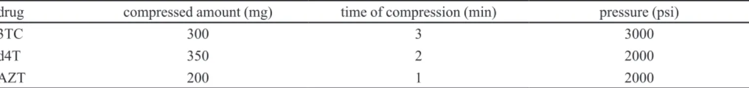
An amount of drug was compressed to make a non-disintegrating disk and the exposed surface area was 0.5 cm². Table I summarizes the conditions used to obtain disks for intrinsic dissolution studies for each drug.
For each assay, three vessels were previously filled with 900 mL of buffer solution at a temperature of 37.0 ± 0.5 ºC with rotation of 50 rpm. Samples were withdrawn at 2, 4, 6, 8, 10, 12, 14, 16, 18, 20, 25, 30, 35, 40, 45, 50, 55, 60, 70 and 80 min with replacement of fresh media. To highly soluble drugs, it was necessary to withdraw a known aliquot from the dissolution media and make a volumetric dilution before determining absorbance values by UV-spectroscopy (United States Pharmacopeia, 2010; Yu et al., 2004). It is important to consider that the solution containing a compound with low solubility and high molar absorptivity may be diluted, as solution containing a compound with high solubility and low molar absorptivity may not be necessarily diluted. Thus, the dilution mainly depends on molar absorptivity of the drugs (Alves et al., 2010; Filippin, Souza, 2006; Pereira et al., 2011; Ruela, Araújo, Pereira, 2009).
TABLE I - Conditions adopted for each antiretroviral drug to obtain disks for intrinsic dissolution study
drug compressed amount (mg) time of compression (min) pressure (psi)
3TC 300 3 3000
d4T 350 2 2000
The cumulative quantity of dissolved drug was corrected for each sample interval according to volume collected from the each vessel. For IDR values, a graph was constructed through accumulated amount of the drug dissolved versus time and a linear regression was performed. Thus, the dissolution rate was obtained (Issa, Ferraz, 2011).
Absorbance values were determined in triplicate at the maximum absorbance wavelength. The amount dissolved per surface unit of the disk was plotted against the time for each dissolution vessel. The slope of the linear regression (R2≥0,99, p<0,005) was considered as IDR, which can be calculated by equation below:
where j is the disk intrinsic dissolution rate, V is the volume of the dissolution media, c is the drug concentration, A is the disk area, and t is the time (Yu et al., 2004).
RESULTS AND DISCUSSION
Equilibrium solubility method (shake-flask)
In order to set a reliable condition for BCS
classiication of compounds and considering that small
intestine is the major site for drug absorption, this study has considered the pH range of 1.2 to 7.5 for equilibrium solubility and intrinsic dissolution studies.
Based on solubility results, it is possible calculate the dose:solubility ratio, which represents a volume of media necessary to solubilize the highest dose of the administered drug. An API (active pharmaceutical ingredient) is considered highly soluble when the dose:solubility ratio is less than 250 mL (Lindenberg, Kopp, Dressman, 2004; United States, 2000) in a pH range of 1.0 to 7.5 (United
States, 2000) or pH 1.2 to 6.8 (EMA, 2008; WHO, 2005)
at 37 °C (Strauch et al., 2011).
However, differences may exist in relation to the dosages of the drug, depending on the country and adopted protocols, which can determine variability in
the biopharmaceutical classiication of a compound. For
example, due to the higher dose available for a medicinal product, the same active substance may be considered as high solubility drug in a country and low solubility drug in another country (Lindenberg, Kopp, Dressman, 2004;
United States, 2012; WHO, 2005).
During equilibrium solubility test of d4T, the buffer solution pH 1.2 showed, at the end of the test, changes in smell and color, suggesting a possible degradation reaction due the test conditions (150 rpm 37 °C for 72 hours), thus
making it impossible to obtain results of solubility in this media.
Studies conducted by Silva and colleagues (2008) have reported that the main degradation product of d4T is thymine. This product is mainly formed after hydrolytic and oxidative processes (Silva et al., 2008).
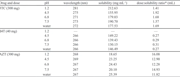
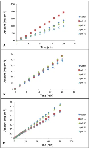
Table II shows solubility results (in mg mL-1) and dose:solubility ratio (in mL) for antiretroviral drugs and the Figure 1 represents average solubility of 3TC, d4T and AZT, respectively.
The recommended dose for 3TC is 300 mg, for d4T is 40 mg and for AZT is 300 mg. These substances
are clearly classiied as high solubility drugs at pH 1.2,
4.5, 6.8, 7.5 and water (except d4T, where solubility in pH 1.2 was not possible), since they satisfy the criteria of
dose:solubility ratio < 250 mL, as demonstrated in Table II (United States, 2000; United States, 2012; WHO, 2006).
The results of solubility indicate that 3TC and AZT present high solubility characteristics based on FDA criteria. By the other hand, d4T presents high solubility in water, pH 4.5, pH 6.8, pH 7.5, but the solubility determination of d4T was not possible at pH 1.2 due to stability problems in this media. Thus, the solubility is not a limiting factor for bioavailability of 3TC and AZT (United States, 2000).
The stability problems can be identified through differences in the smell and color of the solution prepared. In the case of d4T, a difference in the smell and color of the solution was noticed but for some drugs these differences may not be evident. To ensure the integrity of the drugs solubilized in the different media studied, the antiretroviral drugs were subjected to a stability assay and
the quantiication was performed using a chromatographic
method (HPLC system Merck-Hitachi LaChrom®, USA). The stability assay was performed for each drug in
each media with different pH values. Briely, the solutions
were subjected to the conditions of the solubility assay considering the time and the temperature. The total time of the stability assay was 72 hours at 37 °C. Analyses of the solutions were performed at 12, 24, 36, 48, 72 hours and the results were compared to standard solutions of the drugs freshly prepared. The chromatograms of the drugs were equivalent to standard solution of the drugs for 3TC, AZT and d4T, except for pH 1.2, where the chromatograms
indicate signiicant differences for stavudine.
Solubility data from literature about 3TC, d4T and AZT are not easily found. The few existing reports show
differences that can be justiied by: technique or type of
TABLE II - Solubility at 37 °C and dose:solubility ratio of 3TC, d4T and AZT for each media used obtained through equilibrium method
Drug and dose pH wavelength (nm) solubility (mg mL-1) dose:solubility ratio* (mL)
3TC (300 mg) 1.2 281 212.63 1.41
4.5 275 155.95 1.92
6.8 271 179.03 1.68
7.5 273 190.70 1.57
water 272 177.53 1.69
d4T (40 mg) 1.2 — — —
4.5 266 149.22 0.27
6.8 266 139.43 0.29
7.5 266 130.15 0.31
water 266 146.49 0.27
AZT (300 mg) 1.2 268 18.65 16.08
4.5 269 23.25 12.90
6.8 267 24.43 12.28
7.5 267 20.10 14.93
water 267 25.39 11.82
* Dose:solubility ratio ≤ 250 indicates high solubility related to dose (United States, 2000)
FIGURE 1 - Average solubility of 3TC (A), d4T (B) and AZT
(C) in water, pH 1.2 (except d4T), pH 4.5, pH 6.8 and pH 7.5.
(at 25 °C). The results presented in this study are different from Merck Index, but test conditions such as temperature and method can justify theses differences. The temperature
is a crucial factor, since that this parameter can inluence the
solubility, i.e., if temperature increases, the drug solubility is higher (Merck Index, 2001). It is important to note that this study followed FDA recommendations.
Singh and colleagues determined the equilibrium solubility of AZT in water and in phosphate buffer pH 6.8
employing the technique of shake-lask under stirring for
48 hours at temperatures of 37 °C and 25 °C. The results indicated that AZT had a solubility of 30.6 mg mL-1 at 37 °C and 19.5 mg mL-1 at 25 °C in water. In phosphate buffer pH 6.8, the solubility was 24.4 mg mL-1 at 37 °C and 20.3 mg mL-1 at 25 °C. AZT results of this study are in accordance to results presented by Singh and colleagues (2010) (Singh et al., 2010).
In another study, Prakash and colleagues (2008) examined the solubility of antiretroviral drugs, 3TC, d4T
and AZT, in different media (puriied water, 0.01 M HCl
solution, acetate buffer pH 4.5 and phosphate buffer pH 6.8). The results obtained by these authors are presented in Table III.
As explained above, the solubility determination of 3TC, d4T and AZT in the present study was performed according to FDA guidance (United States, 2000) in an incubator with orbital shaking platform. This equipment
enables the heating of lasks and adequate control of the
temperature (maintained at 37.0 ± 0.5 °C). Moreover, the orbital shaking platform promotes speed control (150 rpm). Thus, the conditions adopted in this study remained constant and standardized. The stirring time used in this work was 72 hours, which is important period to reach the saturation of the media.
Furthermore, in studies conducted by Prakash and colleagues (2008), the heating of media was located not ensuring proper distribution of temperature, and stirring was promoted using a magnetic stirrer. It should be noted also that the total test time for the cited study was 5 hours. Another factor is the dissolution media composition (Prakash et al., 2008).
Intrinsic dissolution method
Intrinsic dissolution has been recently studied and discussed for solubility determination of a compound as an alternative method to equilibrium solubility. Values of intrinsic dissolution rate can be used to determine the solubility class of a drug (Issa, Ferraz, 2011; Yu et al., 2004; Zakeri-Milani et al., 2009).
The standardization of intrinsic dissolution test
should be made carefully because this method is inluenced
by internal and external factors (properties of solid state, surface area, hydrodynamic condition, composition of dissolution media, vibration, rotation speed, presence of dissolved gases in dissolution media, sample sites) (Issa, Ferraz, 2011; Souza, Freitas, Storpirtis, 2007). Thus, all these conditions were controlled and standardized to minimize any interference.
Figure 2 shows a typical plot of amount of drug versus time for 3TC, d4T and AZT at different values of
pH and water. The insigniicant discrepancies between
three runs using three disks in three dissolution vessels indicate an excellent reproducibility (RSD less than 10%). Linearity was also excellent, which can be demonstrated
by a correlation coefficient higher than 0.99 for all solutions used.
From the dissolution proiles were obtained intrinsic
dissolution rates (IDR). The IDR speed of the sample is determined from the slope of the line (United States Pharmacopeia, 2010).
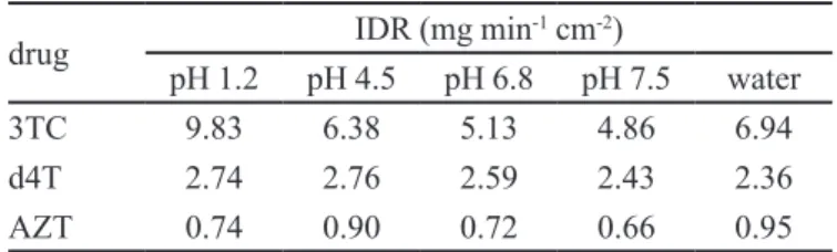
The determined IDR for each drug are shown in Table IV. The presence of a sink condition in the dissolution media during the experiment is upheld by
comparison of the inal concentration of drugs and their
solubility in dissolution media.
IDR results showed in Table IV demonstrate a good
TABLE III - Solubility values obtained from literature (Prakash et al., 2008)
Solubility (mg mL-1)
water HCl 0.01 M pH 4.5 (acetate buffer) pH 6.8 (phosphate buffer)
3TC 140.01 276.08 230.50 92.76
d4T 75.36 78.19 101.23 76.13
AZT 28.90 27.36 21.36 20.10
FIGURE 2 - Amount versus time proile for 3TC (A), d4T (B)
TABLE IV - Intrinsic dissolution rate (IDR) of 3TC, d4T and AZT.
drug IDR (mg min
-1 cm-2)
pH 1.2 pH 4.5 pH 6.8 pH 7.5 water
3TC 9.83 6.38 5.13 4.86 6.94
d4T 2.74 2.76 2.59 2.43 2.36
AZT 0.74 0.90 0.72 0.66 0.95
qualitative correlation with the equilibrium solubility. IDR is a rate phenomenon instead of an equilibrium phenomenon. In addition, IDR values correlate more closely with in vivo drug dissolution dynamics than equilibrium solubility because dose recommended is taken into account in this method while the intrinsic dissolution does not consider this effect (Issa, Ferraz, 2011).
Thus, when the drug dose is extremely high or extremely low (for exemple 1.0 mg mL-1 and recommended dose of 0.25 mg or 4.0 mg mL-1 and recommended dose of 1000 mg) a discrepancy between the equilibrium solubility and the IDR may occur. Further, when the drug dose is extremely high, the in vivo absorption may be limited by solubility (Singh et al., 2010).
At least 10 points were used for the construction
of intrinsic dissolution proiles of drugs. Due to the high
rate of dissolution, short intervals were imposed to reach sampling points and obtain meaningful results. 3TC and d4T presented higher dissolution speed compared to AZT, which takes a longer time to reach 10% of dissolution (United States Pharmacopeia, 2010).
As regard to IDR, it is noteworthy that there are no reports in the literature about the intrinsic characteristics
of antiretroviral drugs dissolution but it possible to ind
limits to characterize high soluble drugs.
According to Yu and colleagues (2004), compounds which have IDR greater than 0.1 mg min-1 cm-2 can be classified as highly soluble substances. Based on this
information, all antiretroviral drugs studied can be classiied
as highly soluble compounds in all dissolution media due to IDR values higher than 0.1 mg min-1 cm-2 (Yu et al., 2004).
Another study developed by Zakeri-Milani and colleagues (2009) suggests that drugs with IDR value higher than 1 mg min-1 cm-2 can be considered as highly soluble substances, while compounds with low solubility show an IDR value lower than 1 mg min-1 cm-2 (Zakeri-Milani et al., 2009).
Equilibrium solubility versus intrinsic dissolution
In recent years, solubility and permeability studies
have received focus from scientiic community due to BCS
and, consequently FDA guidance for biowaiver purposes. Thereby, comparison between equilibrium solubility and intrinsic dissolution methods has been grown, since that solubility is a crucial factor for the absorption (and bioavailability) of drugs.
Equilibrium solubility presents some factors that can difficult the achievement of results, such as:
temperature, iltration processes, substance characteristics
(as crystalline structure or salts formation) (Baka, Comer, Takács-Novak, 2008; Issa, Ferraz, 2011; Yu et al., 2004).
However, this does not allow obtain a solubility proile
of drugs or identify their amorphous because it is an equilibrium condition that must not be interfered during the test (sampling is not possible), otherwise result in the impairment of reliable data (Issa, Ferraz, 2011).
Although equilibrium solubility method adopts physiological conditions as temperature and solution pH, other aspects must be observed. The temperature is a factor that interferes a lot in this test and the loss of temperature can cause the appearance of crystals and loss of equilibrium condition, compromising results. The intrinsic dissolution test, the same temperature and solutions are used but it is a safer method because the loss of temperature causes no
major inluences compared to equilibrium test because the
dissolution media is not saturated.
In addition, the test time is too long (until 72 hours) in equilibrium method and the amount of drug used for this study is very high compared to intrinsic dissolution, which requires a less amount of drug (which is suitable for the early stages of development of new compounds). A less amount of drug in intrinsic solution allows realize any interference in the results due to factors such as any change in crystalline structure or salts formation characterized by deviation from linearity (Issa, Ferraz, 2011; Yu et al., 2002; Yu et al., 2004; Zakeri-Milani et al., 2009).
The conditions of amount of drug and dissolution media volume deviate from the in vivo condition because the administered drug does not remain so long in the dissolution step in the stomach and the substance does not saturate the dissolution media in vivo. In intrinsic dissolution, the volume of dissolution media is major than equilibrium method and the dissolution of drug is constant and linear.
CONCLUSION
The BCS was developed based on two properties: solubility/dissolution rate and intestinal permeability of drugs. In this study, 3TC, d4T and AZT were assessed by two methods for solubility study: equilibrium solubility and intrinsic dissolution. Based on results, 3TC and AZT present high solubility characteristics in equilibrium
solubility, which was conirmed by intrinsic dissolution
(high speed of dissolution). The solubility determination of d4T in pH 1.2 was not possible due to stability problems in equilibrium solubility method. Based on FDA criteria, a drug must be soluble in a pH range of 1.0 – 7.5 at 37 °C and the d4T does not match this criteria. By the other hand, d4T did not present any inconvenience in intrinsic dissolution method and d4T presents high speed of dissolution as 3TC and AZT.
According to discussed in this study, IDR has demonstrate adequate method for solubility studies instead equilibrium solubility method, since these method does not consider dose effect of drug. Thus, the behavior of drugs can be predicted through IDR because this method is closest to in vivo conditions.
Yu and colleagues (2004) consider 0.1 mg min-1 cm-2 as a boundary value, while Zakeri-Milani and colleagues (2009) consider 1.0 mg min-1 cm-2 for classiication of a drug according to their intrinsic solubility. Then, based on Yu and colleagues, 3TC, d4T and AZT may be considered
as highly soluble drugs. On the other hand, when the
boundary value proposed by Zakeri-Milani and colleagues is considered, 3TC and d4T may be considered as highly soluble drugs, while AZT may be considered a compound of low solubility.
According to FDA, a criterion which is based on Biopharmaceutical Classification System, AZT is classified as highly soluble drug (Soares et al., 2013), since the equilibrium method is employed for solubility studies. As intrinsic dissolution is not yet used for regulatory purposes, this study aimed to discuss the feasibility of this technique and once appropriately standardized, may be accept as an alternative methodology for equilibrium solubility assay of drugs. Moreover, the intrinsic dissolution is a rate phenomenon, different from the equilibrium solubility (Yu et al., 2004; Zakeri-Milani et al., 2009).
Due to great diversity of drugs with different
physicochemical characteristics, a limit value is dificult
to be purpose and it depends on data and more consistent studies, as discussed by Yu and colleagues (2004). The establishment of a boundary value for intrinsic dissolution is still considered a challenge, which is being explored
by researchers and the results of these studies are being published in the literature.
In this study, we emphasize that conditions adopted to obtain intrinsic dissolution results is in according to conditions described in the study published by Yu and colleagues (2004), where variables were evaluated, determining the robustness of the method.
In summary, the data obtained from the intrinsic
dissolution test conirm the results obtained in equilibrium
solubility when a boundary value of 0.1 mg min-1 cm-2 is
considered and the drugs can be classiied as highly soluble
substances, as proposed by Yu and colleagues (2004) (Yu et al., 2004). Based on the boundary value (1 mg min-1 cm-2) proposed by Zakeri-Milani and colleagues (2009), 3TC
and d4T can be classiied as highly soluble drugs, while
AZT would be considered as low solubility drug (Zakeri-Milani et al., 2009).
ACKNOWLEDGMENTS
The authors wish to thank the Conselho Nacional de Desenvolvimento Científico e Tecnológico (CNPq, Brazil), Fundação de Amparo à Pesquisa do Estado de São Paulo (FAPESP, Brazil) and Coordenação de Aperfeiçoamento de Pessoal de Nível Superior (CAPES,
Brazil) for inancial support and Fundação para o Remédio
Popular (FURP, Brazil) and Cristália Produtos Químicos Farmacêuticos for donation of chemicals substances (lamivudine, stavudine and zidovudine).
REFERENCES
ALVES, L.D.S.; ROLIM, L.A.; FONTES, D.A.F.; ROLIM-NETO, P.J. Desenvolvimento de método analítico para quantiicação do efavirenz por espectrofotometria no
UV-VIS. Quím. Nova, v.33, n.9, p.1967-1972, 2010.
AMIDON, G.L.; HIGUCHI, W.I.; HO, N.F. Theoretical and
experimental studies of transport of micelle-solubilized
solutes. J. Pharm. Sci., v.71, p.77-84, 1982.
AMIDON, G.L.; LENNERNÄS, H.; SHAH, V.P.; CRISON, J.R. A theoretical basis for a biopharmaceutic drug classiication:
the correlation of in vitro drug product dissolution and in
vivo bioavailability. Pharm. Res., v.12, n.3, p.413-420,
1995.
AVDEEF, A. Absorption and drug development: solubility,
AYMARD, G.; LEGRAND, M.; TRICHEREAU, N.; DIQUET, B. Determination of twelve antiretroviral agents in human plasma sample using reversed-phase high-performance
liquid chromatography. J. Chromatogr. B Biomed. Sci.
Appl., v.744, n.2, p.227-240, 2000.
BALINT, G.A. Antiretroviral therapeutic possibilities for human immunodeficiency virus/acquired immunodeficiency
syndrome. Pharmacol. Ther., v.89, n.1, p.17-27, 2001.
BAKA, E.; COMER, J.E.; TAKÁCS-NOVÁK, K. Study of
equilibrium solubility measurement by saturation
shake-lask method using hydroclorothiazide as model compound.
J. Pharm. Biomed. Anal., v.46, n.2, p.335-341, 2008.
BRITISH Pharmacopoeia. London: Her Majesty’s Stationary
Ofice, 2009. A768 p.
CHAN, H.-K.; GRANT, D.J.W. Inluence of compaction on the intrinsic dissolution rate of modiied acetaminophen
and adipic acid crystals. Int. J. Pharm., v.57, n.2,
p.117-124, 1989.
CHECA, A.; SOTO, V.G.; HERNÁNDEZ-CASSOU, S.;
SAURINA, J. Fast determination of pKa values of reverse
transcriptase inhibitor drugs for AIDS treatment by using
pH-gradient low-injection analysis and multivariate curve
resolution. Anal. Chim. Acta, v.554, n.1-2, p.177-183, 2005.
CHEN, M.-L.; YU, L. The use of drug metabolism for prediction
of intestinal permeability. Mol. Pharm., v.6, n.1, p.74-81,
2009.
COOK, J.; ADDICKS, W.; WU, Y.H. Application of the biopharmaceutical classiication system in clinical drug
development: an industrial view. AAPS J., v.10, n.2,
p.306-310, 2008.
DAHLAN, R.; MCDONALD, C.; SUNDERLAND,
V. B . S o l u b i l i t i e s a n d i n t r i n s i c d i s s o l u t i o n r a t e s
of sulphamethoxazole and trimethoprim. J. Pharm.
Pharmacol., v.39, n.4, p.246-251, 1987.
DEZANI, A.B.; PEREIRA, T.M.; CAFFARO, A.M.; REIS,
J.M.; SERRA, C.H.R. Determination of lamivudine and zidovudine permeability using a different ex vivo method
in Franz cells. J. Pharmacol. Toxicol. Methods, v.67,
p.194-202, 2013.
EUROPEAN MEDICINES AGENCY. Evaluation of
medicines for human use. Committee for proprietary
medicinal products. Note for guidance on the investigation
of bioavailability and bioequivalence. London, 2001.
(CPMP/EWP/QWP/1401/98). Available at: <http://www.
ema.europa.eu/docs/en_GB/document_library/Scientiic_
guideline/2009/09/WC500003519.pdf>. Accessed on: 20 Aug 2012.
EUROPEAN MEDICINES AGENCY. Pre-Authorization
Evaluation of Medicines for Human Use. Committee for
Medicinal Products for Human Use. DRAFT: Guideline
on the investigations of bioequivalence. London, 2008.
(CPMP/EWP/QWP/1401/98). Available at: <http://www.
emea.europa.eu/docs/en_GB/document_library/Scientiic_
guideline/2009/09/WC500003011.pdf>. Accessed on: 16 Aug 2012.
EUROPEAN Pharmacopoeia. 6.ed. France: European
directorate for the quality of medicines. Strasbourg: Council of Europe, 2008. p.309-211
FARMACOPÉIA Portuguesa IX. Lisboa: Infarmed/Ministério da
Saúde e Instituto Nacional da Farmácia e do Medicamento, 2008. 3827 p.
FILIPPIN, F.B.; SOUZA, L.C. Eficiência terapêutica das
formulações lipídicas de anfotericina B. Rev. Bras. Ciênc.
Farm., v.42, n.2, p.167-194, 2006.
ISSA, M.G.; FERRAZ, H.G. Intrinsic dissolution as a tool for evaluating drug solubility in accordance with the
biopharmaceutics classiication system. Dissolut. Technol., v.18, n.3, p.6-13, 2011.
JINNO, J.; OH, D.-M.; CRISON, J.R.; AMIDON, G.L.
Dissolution of ionizable water-insoluble drugs: the
combined effect of pH and surfactant. J. Pharm. Sci., v.89,
n.2, p.268-274, 2000.
LENNERNÄS, H. Animal data: the contributions of the Ussing
chamber and perfusion systems to predicting human oral
drug delivery in vivo. Adv. Drug Deliv. Rev., v.59, n.11,
p.1103-1120, 2007.
LENNERNÄS, H.; ABRAHAMSSON, B. The use of biopharmaceutic classiication of drugs in drug discovery
and development: current status and future extension. J.
LINDENBERNG, M.; KOPP, S.; DRESSMAN, J.B. Classiication of orally administered drugs on the World Health Organization model list of essential medicines according to the biopharmaceutics classiication system.
Eur. J. Pharm. Biopharm., v.58, n.2, p.265-278,2004.
LÖBENBERG, R.; AMIDON, G.L. Modern bioavailability, bioequivalence and biopharmaceutics classiication system.
New scientific approaches to international regulatory
standards. Eur. J. Pharm. Biopharm., v.50, n.1, p.3-12, 2000.
MARÍN, M.T.; MARGARIT, M.V.; SALCEDO, G.E.
Characterization and solubility study of solid dispersions
of lunarizine and polyvinylpyrrolidone. Farmaco, v.57, n.9, p.723-727, 2002.
MERCK index: an encyclopedia of chemicals, drugs and biologicals. 13.ed. Whitehouse Station: Merck, 2001. p.959-1802.
OKUMU, A.; DIMASO, M.; LÖBENBERG, R. Computer
simulations using Gastroplus™ to justify a biowaiver
for etoricoxib solid oral drug products. Eur. J. Pharm.
Biopharm., v.72, n.1, p.91-98, 2009.
PEREIRA, A.V.; GARABELI, A.A.; SCHUNEMANN, G.D.;
BORCK, P.C. Determinação da constante de dissociação
(Ka) do captopril e da nimesulida – Experimentos de
química analítica para o curso de farmácia. Quím. Nova,
v.34, n.9, p.1656-1660, 2011.
PRAKASH, K.; RAJU, P.N.; KUMARI, K.S.; NARASU, M.L. Solubility and dissolution rate determination of different antiretroviral drugs in different pH media using UV visible
spectrophotometer. E-J. Chem., v.5, n.S2, p.1159-1164,
2008.
REIS, J.M.; DEZANI, A.B.; PEREIRA, T.M.; AVDEEF, A.; SERRA, C.H.R. Lamivudine permeability study: A comparison between PAMPA, ex vivo and in situ
single-pass intestinal perfusion (SPIP) in rat jejunum. Eur. J.
Pharm. Sci., v.48, p.781-789, 2013.
RUELA, A.L.M.; ARAÚJO, M.B.; PEREIRA, G.R.
Desenvolvimento de um teste de dissolução para comprimidos de nimesulida em meio que assegure
condições sink. Lat. Am. J. Pharm., v.28, n.5, p.661-667,
2009.
SILVA, G.R.; CONDESSA, F.A.; PIANETTI, G.A.; NUNAN, E.A.; CAMPOS, L.M.M. Desenvolvimento e validação de método por cromatograia líquida de alta eiciência para
determinação simultânea das impurezas timina e timidina
na matéria-prima estavudina. Quím. Nova, v.31, n.7,
p.1686-1690, 2008.
SINGH, S.; DOBHAL, A.K.; JAIN, A.; PANDIT, J.K. CHAKRABORTY, S. Formulation and evaluation of solid
lipid nanoparticles of a water soluble drug: zidovudine. Chem. Pharm. Bull., v.58, n.5, p.650-655,2010.
SOARES, K.C.C.; REDIGUIERI, C.F.; SOUZA, J.; SERRA, C.H.R.; ABRAHAMSSON, B.; GROOT, D.W.; KOPP, S.; LANGGUTH, P.; POLLI, J.E.; SHAH, V.P.; DRESSMAN,
J. Biowaiver monographs for immediate-release solid oral
dosage forms: zidovudine (azidothymidine). J. Pharm. Sci.,
v.102, n.8, p.2409-2423, 2013.
SOUZA, J.; FREITAS, Z.M.; STORPIRTIS, S. Modelos in vitro para determinação da absorção de fármacos e previsão da
relação dissolução/absorção. Rev. Bras. Ciênc. Farm., v.43,
n.4, p.515-527, 2007.
STRAUCH, S.; JANTRATID, E.; DRESSMAN, J.B.;
JUNGINGER, H.E.; KOPP, S.; MIDHA, K.K.; SHAH,
V.P.; STAVCHANSKY, S.; BARENDS, D.M. Biowaiver monographs for immediate release solid oral dosage forms:
lamivudine. J. Pharm. Sci., v.100, n.6, p.2054-2063,2011.
UNITED STATES. Department of Health & Human Services.
Food and Drug Administration. Drugs @ FDA: FDA
approved drug products. Available at: <http://www.
accessdata.fda.gov/scripts/cder/drugsatfda/>. Accessed on: 14 Mar. 2012.
UNITED STATES. Department of Health & Human Services. Food and Drug Administration. Center for Drug Evaluation
and Research. Guidance for industry: waiver of in vivo
bioavailability and bioequivalence studies for immediate-release solid oral dosage forms based on a biopharmaceutics classification system. Rockville: FDA, 2000. p.1-13.
Available at: <http://www.fda.gov/downloads/Drugs/
GuidanceComplianceRegulatoryInformation/Guidances/ ucm070246.pdf>. Accessed on: 20 July 2012.
VIEGAS, T.X.; CURATELLA, R.U.; WINKLE, L.L.V.; BRINKER, G. Measurement of intrinsic drug dissolution
rates using two types of apparatus. Pharm. Technol., v.25,
p.44-53, 2001.
WADKE, D.A.; REIER, G.E. Use of intrinsic dissolution rates to determine thermodynamic parameters associated with
phase transitions. J. Pharm. Sci., v.61, n.6, p.868-871, 1972.
WORLD HEALTH ORGANIZATION. Proposal to waive in vivo biequivalence requirements for the WHO model list of essential medicines immediate release, solid oral dosage
forms. Geneva: WHO, 2005. Available at: <http://www.
who.int/medicines/services/expertcommittees/pharmprep/ QAS04_109Rev1_Waive_invivo_bioequiv.pdf>. Accessed on: 10 May 2012.
WORLD HEALTH ORGANIZATION. Who expert committee on specifications for pharmaceutical preparations. Lamivudine: inal text for addiction to The International
Pharmacopeia. Geneva: WHO, 2006. Available at: <http://
www.who.int/medicines/publications/pharmacopoeia/ QAS_124rev2_Lamivudine.monoFINAL.pdf>. Accessed on: 3 July 2012.
WON, D.-H.; KIM, M.-S.; LEE, S.; PARK, J.-S.; HWANG, S.-J.
Improved physicochemical characteristics of felodipine solid dispersion particles by supercritical anti-solvent
precipitation process. Int. J. Pharm., v.301, n.1-2,
p.199-208, 2005.
WOOD, J.; SYARTO, J.; LETTERMAN, H. Improved holder
for intrinsic dissolution rate studies. J. Pharm. Sci., v.54,
n.7, p.1068, 1965.
YU, L.X.; AMIDON, G.L. Analytical solutions to mass transfer. In: AMIDON, G.L.; LEE, P.I.; TOPP, E.M. (Eds.).
Transport processes in pharmaceutical systems. New York: Marcel Dekker, 1999. p.23-54.
YU, L.X.; AMIDON, G.L.; POLLI, J.E.; ZHAO, H.; MEHTA, M.U.; CONNER, D.P.; SHAH, V.P.; LESKO, L.J.; CHEN,
M.L.; LEE, V.H.L.; HUSSAIN, A.S. Biopharmaceutics classification system: the scientific basis for biowaiver
extensions. Pharm. Res., v.19, n.7, p.921-925, 2002.
YU, L.X.; CARLIN, A.S.; AMIDON, G.L.; HUSSAIN, A.S.
Feasibility studies of utilizing disk intrinsic dissolution
rate to classify drugs. Int. J. Pharm., v.270, n.1-2,
p.221-227, 2004.
ZAKERI-MILANI, P.; BARZEGAR-JALALI, M.; AZIMI,
M.; VALIZADEH, H. Biopharmaceutical classiication of
drugs using intrinsic dissolution rate (IDR) and rat intestinal
permeability. Eur. J. Pharm. Biopharm., v.73, n.1,
p.102-106, 2009.
Received for publication on 26th January 2013