Universidade do Algarve
Faculdade de Ciências do Mar e do Ambiente
m 7
Cloning of the Bone Gla Protein gene from the teleost fish Sparus aurata (gilthead seabream).
Molecular organization, developmental appearance and evolutionary implications
Faculdade de Ciências do Mar e do Ambiente
Cloning of the Bone Gla Protein gene from the teleost fish Sparus auraía (gilthead seabream).
Molecular organization, developmental appearance and evolutionary implications
Dissertation presenteei at the University of Algarve, Portugal, to obtain the degree of Doctor in Biological Sciences, Area of Molecular Biology.
.V £ i.'.' A".1 "j . z . z C-
CÍCé oZj Íoé^í J 2 50^7, s^.fl 2.
No já longínquo ano de 1980? frequentava eu o T ano de escolaridade, uma professora de ciências, hoje anónima, porque esqueci o seu nome, cativou-me. Ao revelar-me a magia e o mistério das ciências, e da Biologia em particular, desvendou- me o meu destino. A Biologia, numa ou noutra vertente, nunca mais me iria deixar e a essa senhora, à professora dos bichos-da-scda, devo um sincero e tardio obrigado.
Numa universidade pequena, como era a Universidade do Algarve em 1987, quando iniciei a licenciatura em Biologia Marinha e Pescas, encontrei um conjunto dc professores empenhados em fazer evoluir uma universidade periférica, em condições muito adversas. Àqueles que, talvez sem o saberem, mais me deram, em particular aos professores Karim Erzini, Margarida Castro, Fátima Rosado. Dclminda Moura. Lucília Coelho e Helena Galvão, o meu agradecimento.
Num dia, que não sei já localizar, dirigi-me ao gabinete da professora Leonor Cancela e disse-lhe que gostaria de fazer um doutoramento em Biologia Molecular e de passar algum tempo nos Estados Unidos. Alguns dias passados e eu estava já certo de que encontrara a pessoa que me iria orientar. Ao longo destes anos fizemos juntos esta viagem e, no seu decorrer, a professora Leonor ensinou-me, não só a conduzir o barco, mas, acima de tudo, onde ele nos pode levar. E mostrou-me também que não é fácil chegar ao porto, e que não chegamos como partimos. À professora Leonor, por tudo o que me ensinou, e pela amizade que nos une, um enorme e sentido obrigado.
A realização deste trabalho não teria sido possível sem o apoio da Fundação para a Ciência e Tecnologia, através da atribuição da bolsa de doutoramento PRAXIS XXI /B D / 3734/94, e do financiamento dos projecto de investigação BIA 469/94 e BIA
11159/98. nos quais este trabalho se integrou.
Para todos aqueles que com os seus conselhos ou ajuda técnica directamente contribuíram para a realização de alguns pontos deste trabalho, em particular para o Doutores Mare Ohresser, Ester Serrão e Deborah Power, o meu agradecimento.
Aos doutores Pierre Marie e Marie Christine de Vemejoul o meu agradecimento pelo estágio facultado no Hôpital Lariboisière, em Paris, no decorrer do qual foi efectuada uma parte deste trabalho. Pela assistência então prestada, e pela simpatia, agradeço às técnicas Monique e Aischa.
lodos tentei igualmente dar algo. À Carla Viegas, Rute Martins, Paulo Gavaia. Nuno Henriques, João Paulo Reia, Cláudia Ribeiro, Lénia Ferrão, Dina Simes (a "mulher desvairada"), Dr. Vincent Laizé, Sara Mira. António Pombinho e Ricardo Leite um sentido "muito obrigado".
Em termos mais pessoais, o meu primeiro e maior agradecimento vai para aqueles a quem dedico esta tese. Para os meus pais, que não só me educaram e incentivaram para que fizesse o que mais gostava, como me apoiaram, nos sucessos c fracassos, mostrando-me que os terei sempre ali, alegrando-se ou sofrendo, como se deles se tratasse.
Para agradecer aos meus amigos Teresa Modesto e Luís Faísca não bastaria uma página inteira, tanto é o que lhes devo e tanto o carinho que lhes tenho. Não posso ocupar uma página, mas posso dizer-lhes que este obrigado que aqui deixo é muito mais do que isso. Obrigado.
A Ana Barbosa contagiou-me em primeiro lugar com o entusiasmo pelas bactérias, depois pelo plâncton, e, com o passar do tempo, com o entusiasmo por tudo o que nos rodeia. À minha amiga Anita, que me ensinou tanto e de quem tanto gosto, um beijinho de agradecimento.
Estava eu no segundo ano do curso de Biologia Marinha e Pescas e, a propósito de um trabalho sobre radiolários, dirigi-me ao Professor Sadat Muzavor em busca de orientação. Encontrei a orientação, mas encontrei também um amigo a quem muito estimo e a quem agradeço tudo o que me tem dado.
Houve bons e maus momentos nesta viagem. Tempos de acalmia em que o vento soprava a favor e era apenas uma questão de tempo chegar a bom porto. E tempos de tormenta, em que as vagas cavavam abismos de onde parecia não haver engenho nem arte que nos pudessem salvar. Nos bons e nos maus momentos tive quase sempre alguém a meu lado, para me dar razão, mesmo quando a não tinha. À minha amiga Natércia, a minha parceira da "dupla invencível", obrigado por tudo.
E por fim, só porque foi o último a chegar, para o Marco, que trouxe o que faltava e fez de mim uma pessoa melhor, um derradeiro e enorme obrigado.
symbols (e.g., HC1), the abbreviations and acronyms below are used throughout this work. l,25(OH)2D3 aa: AP: bp: BGP: BMP: DNA: ddHjO: dph: dsDNA: EDTA: Gla: GRE: KO: MGP: nt: O/N: OP: ORG: PGR: RNA: rpm: R/T: RXR: S.L. spBGP: Tris-HCI: U: VDR VDRE: vol.: approximately 1,25-dihydroxyvitamin D3 amino acid Alkaline Phosphatase base pair
Bonc Gla Protein
Bone Morphogenetic Protein Deoxyribonucleic acid Bidestilled water Days post-hatching double-stranded DNA Ethylenediaminotetracetate y-carboxyglutamic acid
Glucocorticoid Responsive Element Knock Out. Depleted
Matrix Gla Protein nucleotides
Ovemight Osteopontin
Osteocalcin-Related Gene Polymerase Chain Reaction Ribonucleic Acid
rotations per minute Room Temperature Retinoid X Receptor
Standard Length (length measured from the jaw to the base of the caudal fin)
Sparus aurata BGP
Tris(hydroxymethyl)aminomethane adjusted to the referred pH with HC1 Enzyme units (the amount of restriction enzyme necessary to digest one microgram of DNA under established conditions)
Vitamin D Receptor
Vitamin D Responsive Element volume
RESUMO
A proteína Bone Gla (BGP, osteocalcina) é uma pequena proteína dependente- da.vitamina K que apresenta resíduos de ácido glutâmico y-carboxilados. A presença destes aminoácidos modificados permite à proteína ligar-se a iões Ca2 e interagir com os cristais de hidroxiapatite dos tecidos mineralizados.
Embora a BGP tenha sido isolada pela primeira vez em 1976, só recentemente se demonstrou a sua função de modo convincente, permanecendo o mecanismo de acção ao nível molecular essencialmente desconhecido.
O alinhamento das sequências de aminoácidos de todas as BGPs previamente conhecidas revela uma notável conservação de certas regiões, as quais se acredita serem essenciais para o funcionamento correcto da proteína, ao longo de vários milhões de anos de evolução. O tacto de o gene da BGP ter sido clonado apenas em mamíferos (humano e roedores) e de apenas se encontrar disponível a sequência parcial de aminoácidos da BGP de um vertebrado inferior (um peixe teleósteo) impedia a realização de estudos evolutivos desta proteína, assim como a avaliação do seu papel no aparecimento e evolução do osso.
O presente relatório descreve a clonagem do gene da BGP de um vertebrado inferior, o peixe teleósteo Sparus aurata (Dourada), a sua expressão ao longo do desenvolvimento e correspondente distribuição tecidular d do RN Am. A organização molecular do gene da BGP de Sparus (spBGP) é semelhante à dos genes das BGPs de mamíferos, diferenciando-se apenas por um Intrão II mais longo e pela localização dos sítios de inserção dos três intrões. Tal como nos mamíferos, a expressão do gene da BGP em Sparus está restricta aos tecidos ósseos e o início da sua expressão ao longo do desenvolvimento segue-se ao início da calcificação do esqueleto. Estes resultados foram obtidos independentemente por diferentes técnicas de detecção.
Desenvolveu-se uma cultura celular de células derivadas de osso de Sparus a fim de testar a funcionalidade de uma construção promotor spBGP/vector ppGal, demonstrando-se a capacidade deste promotor em induzir a transcrição deste vector de expressão nestas células.
Com base na sequência do gene de spBGP e em outras sequências parciais de RNAm de peixe e amííbio igualmente obtidas pelo nosso grupo, efectuou-se uma análise filogenética e avançaram-se hipóteses relacionando as BGPs com outra proteína Gla (proteína Matrix Gla), em particular, e com a família das proteínas Gla, em geral. Os nossos dados suportam a hipóteses de que todas as BGPs terão tido a mesma origem, partilhando um ancestral comum com a proteína Matrix Gla. Juntamente com as semelhanças observadas ao nível da distribuição tecidular e do
milhões de anos sem alterações significativas, desempenhando provavelmente o mesmo papel desde a alvorada dos vertebrados.
ABSTRACT
The Bone Gla Protein (BGP, osleocalcin) is a small vilamin K-dependent protein which presenls Ihree y-carboxylated glutamic acid residues. lhe prcsence oí these modified amino acids enables the protein to bind to Ca" ions and to interact with hydroxyapatite ciystals of mineralized tissues.
Although BGP was first isolated in 1976, only recently has its function been convincingly demonstrated, the mechanism of action at the molecular levei remaining essentially unknown.
Amino acid sequence alignment of ali previously known BGPs show a remarkable conservation of certain regions, which are believed to be essential for the correct functioning of the protein, throughout several million years of evolution. The fact that the BGP gene had only been cloned in mammalian systems (human and rodents) and that parlial BGP amino acid sequences were available only írom one lower vertebrate (the teleost bluegill) prevented evolutionary studies ol this protein, as well as the assessment of its role in the appearance and evolution of bone.
This report describes the molecular cloning of the BGP gene from a lower vertebrate. the teleost fish Sparus auraía (gilthead seabream), its developmental expression and corresponding mRNA tissue distribution. The molecular organization of the Sparus BGP (spBGP) gene is similar to that of mammalian BGP genes, the only differences being a longer intron II and the sites of insertion ol the three introns. As in the mammalian models, BGP gene expression in Sparus is restricted to bony tissues and its expression throughout development follows the onset of skeletal calcification. These rcsults were independently obtained by different detection tcchniques.
Different Sparus bone-derived cell strains were obtained and used to test the functionality of an spBGP promoter/pPGal vector construction, proving the ability oí this promoter to drive the transcription of this expression vector in these cells.
Based on the spBGP gene sequence and on other partial fish and amphibian BGP mRNA sequences also obtained by our group, a phylogenetic analysis was performed and evolutionary assumptions were advanced relating BGPs with another Gla protein (Matrix Gla Protein), in particular, and with the Gla family of proteins, in general. Our data support the hypothesis that ali BGPs have a single origin and share a common ancestor with Matrix Gla protein. Together with the similarities observed in
million years with only minor changes, probably playing the same role since the dawn of the vertebrates.
INDEX
AGRADECIMENTOS
ABREVIATIONS AND ACRONYMS ABSTRACT
RESUMO INDEX
FIGURE INDEX TABLEINDEX
CHAPTER I: GENERAL INTRODUCTION 1. Bone Gla Protein
1.1. Discovery and purification from bone
1.2. The BGP Gla residues: location, origin and function 1.3. Biochemical characteristics of Bone Gla protein
1.4. Biosynthesis, tissue distribution and abundance of BGP 1.5. Developmental appearance of BGP
1.6. BGP function
1.7. The Bone Gla Protein gene 1.8. Bone Gla Protein gene regulation
2. Other Vitamin K-Dependent Proteins 3. Sparus aurata: The Gilthead Seabream
3.1. Taxonomical classification of Sparus aurata 3.2. General Sparus aurata biological features
4. Appearance, Structure and Importance of the Skelelon in Vertebrates in General and in Fishes in Particular
4.1. The origin of the skeleton 4.2. Some important notions 4.3. The flsh skeleton l III IV vi 2 2 3 8 10 13 14 20 26 38 43 43 44 46 46 48 49
5. The Bone Tissue: Formation and Major Characteristics ^ 5.1 Mechanisms underlying bone formation and main characteristics 53 5.2. The bone cells
6. General Introduction and Objectives 60
CHAPTER II: IMATERIAL AND IMETHOPS ^ 62 62 1. RNA Extraction
2. Amplification of a Parlial spBGP Clone by RT-PCR
3. Cloning and Sequencing of DNA Fragments Resulting From PCR Amplification 63 4. Amplification and Cloning of the 5'-End of spBGP cDNA by 5' RACE PCR 64 5. Extraction of Genomic DNA
6. Amplification of the spBGP Gene
7. Amplification of the 5'-Flanking Region of the spBGP Gene 8. Genomic Southern Analysis
9. Determination of the Transcription Start Site of the spBGP Gene 10. Northern Blot Analysis
11. RT-PCR Southern Blot Analysis
12. Detection of Cartilaginous and Mineralized Structures in S. auraía 70
13. Inclusion of 5. auraía Bone in Methylmetacrylate 71
14. Histological Detection of Alkaline Phosphatases in Osseous Tissues of 5. auraía 72 15. Flistological Detection of Acid Phosphatases in Osseous Tissues of 5. auraía 72 16. In Situ Hybridization Analysis 73 17. Establishment of Primary Celi Cultures Derived From Different Sparus Tissues 75
75 65 66 66 67 68 69 70
17.2. From scales ^
18. Preservation of .S/rams-Derived Celi Types 76 19. Amplification of the BGP Message From Primary Celi Cullures of S. aurala 77 20. Detection of Alkaline Phosphatase Activity in Vertebra-Derived Primary Celi Cultures of
S. aurala 77 21. Delection of Mineral Deposition in lhe Extracellular Matrix of.S. aurala Bone-Derived Celis 78 22. Cloning the spBGP S^Flanking Region in the pPGal-Basic Expression Vector 79 23. Transienl Transfection of Bone-Derived Primary Cultures of S. aurala 81 24. Cloning of a Parlial Halobalrachus didactylus (Toad fish) BGP cDNA 82 25. Phylogenetic Analysis ^7
CHAPTER III: RESULTS 84_ 1. Molecular Cloning of spBGP cDNA 84
1.1. Cloning of a partial spBGP cDNA containing the 3'-end 84 1.2. Cloning of the 5'- end of spBGP cDNA 85
2. Molecular Cloning and Organization of the spBGP Gene 87 3. Determination of the Start Site of Transcription of the spBGP Gene 90 4. Cloning and Analysis of the 5'-Flanking DNA of the spBGP Gene 91
5. How Many BGP Genes Exist in Sparus aurala? 95 6. Search for More than One spBGP mRNA 96 7. Expression of the spBGP Gene ^00
7.1. Tissue distribution ^ 90 7.2. Developmental expression 191 7.3. Correlation between BGP appearance and bone development in Sparus 104
8. Study of the Functionality of the spBGP Promoter in S. aurala bone-derived cells 110 8.1. Obtention of primary cell cultures derived from S. aurala tissues 110
9.1. Cloning of a parlial Halobatrachus didactylus (load fish) BGP cDNA 116 9.2. Evolutionary analysis of spBGP ^ ^ ^
CHAPTER IV: PISCUSSIQN i^O 1. The Sparus auraía BGP cDNA ^ ^0 2. The Sparus auraía BGP Gene and 5' Flanking DNA 120 3. Sparus auraía Has One Single BGP Gene? 1 2j 4. Expression of lhe spBGP Gene ^ 24 4.1. Tissue distribution: the spBGP gene is only expressed in mineralised tissues 124 4.2. Developmental expression: the onset of spBGP gene expression follows the appearanee
of bony structures 126
5. Sparus auraía Bone-Derived Celis and Assessment of spBGP Gene Promoter activity 130 6. Halobatrachus didactylus BGP Clone Shows High Similarity With its Sparus Counterpart 133 7. Phylogenetic Analysis of spBGP ^ 23
CHAPTER V: GENERAL CONCLUSION AND PERSPECTIVES 140
REFERENCES 142 APPENDIX I
APPENDIX II APPENDIX III
FIGURE INDEX
I-1. Model for Ca2+-induced structure of BGP. 6
1-2. General pathway of BGP biosynthesis and secretion by osteoblasts. 9 1-3. General model of BGP gene regulation. 22 1-4. Drawing of an adult Sparus auraía specimen 43
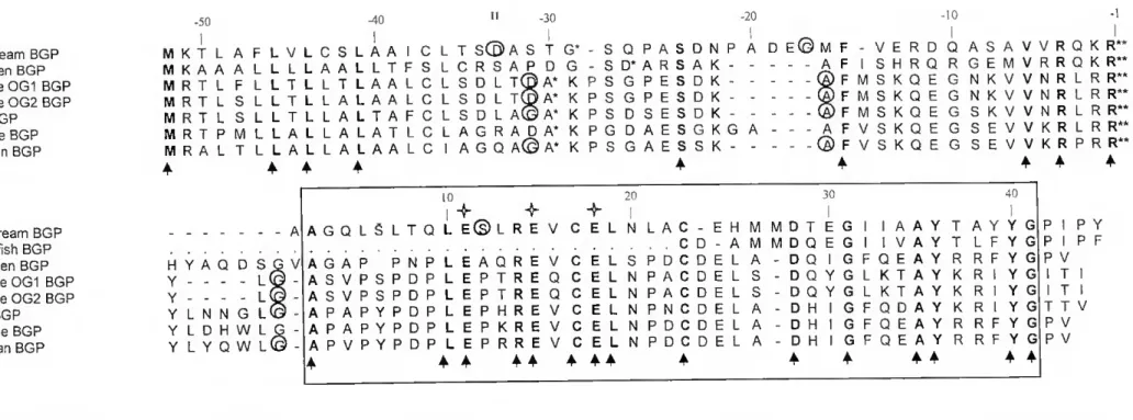
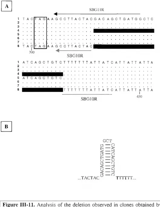
98 99 100 IIL2. Amplification of Sparus mRNA by 5' RACE PCR 85 111.3. Complete nuclcotide sequence of Sparus BGP cDNA 86 111.4. Amino acid sequences of ali known complete pre-proBGPs 88 111.5. Identification of transcription start site of the spBGP gene 90 111.6. Gen Walk primary amplification of Sparus genomic DNA 91 111.7. Gen Walk secondary amplification of Sparus genomic DNA 91 111.8. Sequence oíSparus BGP gene and SMlanking region 93 111.9. Map of the Sparus BGP gene and S^anking region 94 III. 10. Analysis of the Sparus BGP gene locus by Southern hybridisation 96 III. 11. Analysis of the deletion observed in clones obtained by RI -PCR
111.12. RT-PCR amplification oí Sparus RNA lo test the existence of a deletion in the spBGP cDNA sequence
III. 13. Tissue dislribulion of spBGP mRNA by Northern analysis
III 14. Detection by RT-PCR (35 cycles) of BGP from several tissues of Sparus 101 III. 15. Detection of BGP mRNA in Sparus tissues by RT-PCR (20 cycles), coupled with
Southern hybridisation ^1 III. 16. Analysis of developmental expression of spBGP mRNA by Northern blotting 102 IIL17. Detection of BGP mRNA by RT-PCR (35 cycles) in Sparus developmental stages 103 III. 18. Detection of BGP mRNA by RT-PCR (20 cycles) in developmental stages of Sparus 103 III. 19. Alcian blue/alizarin red staining of 15 days Sparus larvae 104 111.20. Alcian blue/alizarin red staining of 20 dph Sparus specimens 105 111.21. Alcian blue/alizarin red staining of 27 dph Sparus larvae _ 106 111.22. Anterior half of an alcian blue/alizarin red-stained Sparus specimen with 35 dph 106 ni.23. Alcian blue/alizarin red staining of 72 dph Sparus juveniles 107 111.24. Alcian blue/alizarin red staining of 110 dph Sparus}uvem\es 108 111.25. Alcian blue/alizarin red staining of 150 dph Sparus]uvem\e (anterior end) 108 111.26. Localization of spBGP mRNA in Sparus tissues by in silu hybridisation 109
111.27. Migration of cells from Sparus vertebrae 110
111.28. Phenotype oí Sparus vertebra-derived cells in active growing 110 111.29. Sparus vertebrae-derived cells kept without passage for 30 days I 1 111.30. Migration of cells from Sparus scales 111 111.31. Differentiation of scale-derived cells 111 111.32. Detection of alkaline phosphatase activity in vertebra-derived cells oí Sparus 1 - 111.33. Von Kossa staining of mineralized structures in Sparus vertebra-derived cells 113 111.34. Staining of mineralized nodules produced by Sparus vertebra-derived cells with alizarin
red S _ • ... 111 111.35. Phenotype of iS/7«mv-derived cells of different origins cultured in mineralising médium 114 111.36. Von Kossa staining of Sparus cells derived from vertebra, jaw and branchial arches
with no signs of mineralization ^5 111.37. Schematic representation of the cloning of the spBGP gene 5' flanking region in the
multiple cloning site (MCS) of the the p(3gal-Basic repórter vector 115 111.38. Transient transfection of bone-derived primary cultures of S. aurata with
spBGPpromoter/pBGal construct ^1 ^
111.39. RT-PCR amplification of a partial Halobatrachus didactylus BGP cDNA 117 111.40. Partial Halobatrachus didactylus BGP cDNA sequence 117 111.41. Strict consensus tree of the 30 Maximum Parcimony Trees generated with the available
data on BGPs, MGPs, Human Coagulation Factor II and Pacific Hagfish Thrombin amino acid sequences . ^ IV. 1. Identification of osteoblasts and osteoclasts in sections oí Sparus by detection of alkaline
TABLEINDEX
LI. Most important ions and molecules that inhibit BGP adsorplion to hydroxyapatite
I.II. Main regulatory factors of BGP gene expression 24 • ^ r\ l.III. Genes regulated by Vitamin D
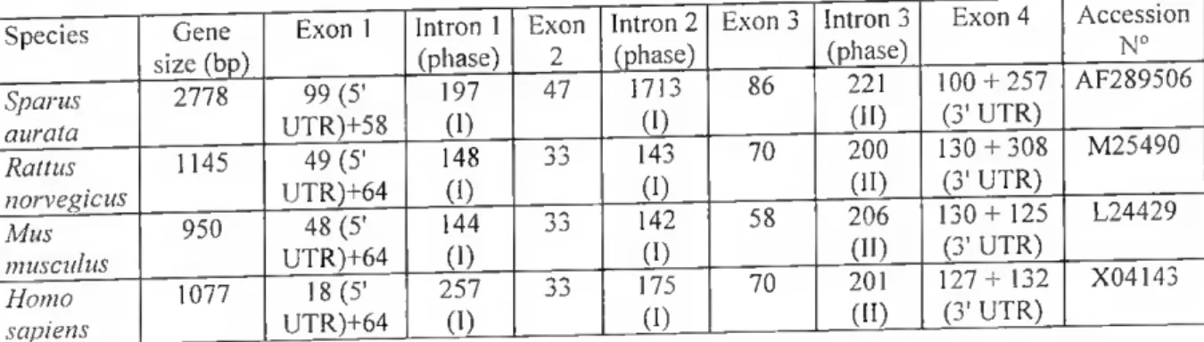
I.IV. Summary of Information on vitamin K-dependent proteins (excepting BGP) 40 ILL Oligonucleotides used for PCR amplificalion of Sparus BGP cDNA and gene 83 IILI. Characteristics of known BGP cDNAs and corresponding proteins 87 IILIL Exon-intron splice junctions and phase of the introns in Sparus aurata BGP gene 89 III.III. Characteristics of known BGP genes structurcs 90
The first chapter of this thesis is meant to give an overview on the current knowledge of Gla proteins, in general, and on Bone Gla protein, in particular. Easily noticeable. lhe vast majority of infonnation arises from studies using maramals. mostly, and avian. more scarcely, as models. Also, the kind of information available is influenced by the "use" this protein has been given throughout most of the 23 years that spanned since its discovery, as an indicator of bone remodelling in clinicai studies. As described, oniy more recently has a shift occurred m the approach chosen to sludy this protein and new infonnation has been obtained conceming such aspects as its ftmction and regulation in mammalian models. In this report we desenhe the first BGP cDNA and gene structure from a lower vertebrate, and give the first results on its tissue distribution in fish and appearance during development. We also report the development of bone-derived primary cell cultures. suitable to further analyse BGP gene expression and regulation in vitro. With this work we introduce a lower vertebrate, the teleost fish Sparus aurala, as a new model for the study of the bone-related proteins.
1. BONE GLA PROTEIN
1.1. DISCOVERY AND PURIF1CATION FROM BONE
Bone Gla protein (BGP, osteocalcin) is a small protein originally isolated from the mineralized phase of bovine bone (420, 421). It is the most abundant non-collagenous protein of bone in ali species analysed (90, 207, 209, 212, 216, 288, 298), with a molecular weight of up to 6500 (chicken; 209). BGP is a very acidic protein, with an isoelectnc point of 4.0 and a net negative charge of 9 at pH 8.0 (414, 434). The name osteocalcin (Gr. osteo, bone + Lat. ca/c, lime salts + in, protein) derives from (i) its Ca2f affimty (209, 214, 422) and (n) its abundance in bone tissue [1-20% of noncollagenous proteins, depending on species, age and site (90, 207, 209, 212, 216, 288, 436)].
INTRODUCTION
Chicken (70, 207, 209) and cow (420) bone were the first tissues from which BGP was isolated. More recently, the protein has been purified and sequenced from bonés of human (415), rnonkey (215), pig, goat, sheep and wallaby (232), cat (486), rat (387, 400), mouse (Hauschka and Gundberg, unpublished observations), rabbit (Hauschka and Tnffitt, unpublished observations), emu (234). toad (64), swordfish (422) and seabream (64), and has been inelusively detected at low leveis in fóssil bonés, possibly protected from degradation through its binding affinity for hydroxyapatite (233, 358).
1.2. THE BGP GLA RESIDUES: LOCATION. ORIGIN AND FUNCTION
Location and origin
One of the most striking features of BGP is the presence of three residues of the vitamin K-dependent aminoacid y-carboxyglutamic acid (Gla) [located at positions 17, 21 and 24 in human, bovine and rat, and at equivalent positions in ali other species], from swordfish to mammals (this study, 117, 261, 371, 527), which are thought to be positioned on the same face of one helix, spaced at intervals of about 5.4 A (214). Gla residues result from a post- translation modification (y-carboxylation) in which specif.c glutamic acid residues are modified in a reaction catalysed by a vitamin K-dependent carboxylase (113, 143, 207, 308, 327, 420, 438, 443, 506), a process that requires the intervention of vitamin K in its reduced form, as a cofactor (385, 517, 544). The enzymatic reaction generates y-carboxyglutamate and vitamin K 2,3,-epoxide, which is then recycled back to the hydroquinone form by a reductase enzyme (173).
Function
Since its discovery in the early 70s, the presence of Gla residues has been implicated in the ability of vitamin K-dependent proteins to bind to divalent cations, hidroxyapatite, and acid phospholipids (301, 370, 501, 505, 541, 545), although the strength of this association is relatively weak (439). Seatchard plot analysis of Ca2+ binding to bovine BGP revealed the presence of three Ca2+ binding sites with an average dissociation constant of 2-3 mM (214,
373. 422). Since there are three Glas in bovine BGP, it seemed resonable to postulate that the three Ca2+ binding sites were provided by the side chain oi Gla. This inlerpretation is supported by the observation that decarboxylation of Gla to Glu abolishes Ca-+ binding to BGP (414) and by the prediction that the organization oí the Gla residues on the BGí structure is similar to the spacing of Ca2+ in the hydroxyapatile crystal (214). Such coordination of Ca2+ by Gla residues in BGP would leave two Ca2' coordination sites unoccupied by protein ligands and therefore free to function in binding interactions with bone mineral (439). Inhibition of the y-carboxylation by the vilamin K antagonist warfarin, by selective proteolysis of lhe Gla domain. or by chelation of the calcium ions results in loss oí binding of Gla-containing proteins to metal ions, hydroxyapatile or membranes (375, 428, 430. 435).
Do Gla residues play a role in BGP secretion?
It is unclear whether Gla residues may play an intracellular role in modulating protein secretion. From the comparatively low Ca2+ affinity of Gla proteins (Kd=0.2-3 mM, far lesser than the affinity for hydroxyapatile) and the paucily of intracellular ionic Ca2+ (<10-6M), it might be expected that functionally important Gla- and Ca2+-dependent conformational changes will occur only as these proteins leave the cell (218). However, carboxylation does appear to play a role in the intracellular fate of vitamin K-dependent proteins (218). In vitro studies performed on cells and tissues (286, 375, 400) demonstrate inhibited secretion of BGP and accumulation of intracellular precursor in the absence of correct y-carboxylation. suggesting that processing occurs intracellularly and that the presence of Gla residues in the protein may be required for processing into the mature secreted peptide. Although the cleavage enzyme [which may be related to the furin/PACE specific endopeptidase known to cleave the propeptide of profactor IX (54)] may prefer the presence of Gla in its substrate, the presence of Gla residues is not absolutely required for production of the mature BGP species, which has been shown to circulate in the non-carboxylated form (for a revision on this subject see 218).
1NTR0DUCTI0N
Calcium binding to Gla shapes the BGP protein
Ca2+ binding to fully carboxylated BGP alters lhe circular dichroism spectrum and the immunochemical properties of the protein (105, 214). lhe apparent a-helical content oí bovine BGP increases from 1% in the absence of Ca2' to 14% in its presence (105), the same phenomenon ocurring for chicken BGP (214). It is interesting to note that the a-helical content of carboxylated BGP in the presence of Ca2+ approaches that of uncarboxylated BGP much more than that of carboxylated BGP in the absence of Ca2 (105). One interpretaiion for this phenomenon is that lhe Gla-conlaining region of BGP can only exist as an a-helix when the Gla residues bind Ca2+. In lhe absence of this metal. Gla residues would destabilize the a- helix. perhaps because of the extra negative charge on the Gla side chains (439).
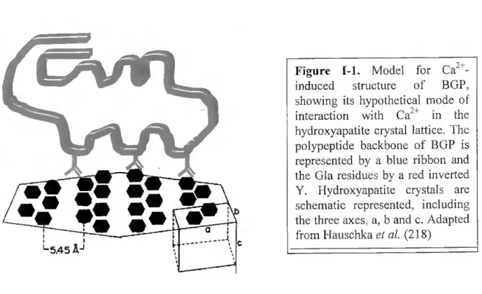
Combined chemical, immunochemical, spectral. and predictive investigations of BGP structure (214) have yielded the model shown in Figure 1-1. The model consists of two antiparalell a-helical domains, the "Gla helix" (residues 16-25 in the chicken protein), and the "Asp-Glu helix" (residues 30-41) connected by a peptide segment containing a p-tum (residues 26-29) and stabilized by the Cys-23-Cys-29 disulfide bond. Olher P-tums occur at positions 5-8 and 12-15, with p-sheel structure in the COOH-terminus from residues 42 to 48. Additional constraints derive from immunochemical studies that indicate contiguity of the NH2- and COOH-termini of BGP (214, 216). From the few two-dimensional NMR spectra data available, it appears that the Ca2+ -induced helical conformation is extremely rigid, with hydrophobic stabilization of the helical domain by the COOH-terminal domain (214, 216). The adjacent carboxyl groups in the Gla residues are aligned in such a way that they project from the helix in a plane, potentially facilitating the protein adsorption to hydroxyapatite (214). Millimolar leveis of Ca2+ cause normal BGP to change from a random-coil to an alpha- helical conformation. Apparently, these milimolar leveis of Ca24 or other specific cations are required to offset electrostatic repulsion if the highly anionic BGP molecule is to achieve its full potential of ~ 40% a-helix (200, 214, 240).
Not only must Gla residues be present, they must also be in helical register to fully achieve the adsorption specificity for hydroxyapatite (414). The binding oí BGP to hydroxyapatite probably involves the bidentate chelation of Ca2 atoms on the crystal
surface by the malonate side chain of Gla, which is supported by the fact that binding to hydroxyapatite completely prevents the thermal decarboxylation of Gla residues to Glu residues (439).
Figure 1-1. Model for Ca2+- induced structure of BGP, showing its hypothetical mode of interaction with Ca2+ in the hydroxyapatite crystal lattice. The polypeptide backbone of BGP is represented by a blue ribbon and the Gla residues by a red inverted Y. Hydroxyapatite crystals are schematic represented, including the three axes, a, b and c. Adapted from Hauschka el ai (218)
The affinity of metal-free BGP for hydroxyapatite is increased fivefold by the addition of 5mM Ca2+ (214, 563). Interestingly, Mg2' is known to induce a somewhat aberrant a- helical conformation in BGP (214) and simultaneously inhibits the binding to hydroxyapatite (219, 560). Magnesium ions compete rather well for the Ca2' binding sites, but Sr2' and Ba2' show little or no competition (200, 209). In contrast, the Gla-rich fragment-1 portion of prothrombin shows increased hydroxyapatite binding in the presence of either Ca" or Mg" (560). Other inhibitors of BGP adsorption to hydroxyapatite are described in Table I-I. Finally, it seems interesting to refer that although BGP is distinguished by its normal content of Gla residues, the human protein may contain only two fully y-carboxylated Glas (415), a fact that may be related to the low concentration of BGP in human bone and plasma (-5% of most other species, including other mammals; 425, 427, 431).
\ l5.45 Âj
1NTRODUCT10N
Table I.I. Most important ions and moléculas that inhibit BGP adsorption to hydroxyapatite, suggested mechanism of action and corresponding references.
Inhibitors of BGP adsorption to
Mode of action References
Magnesium (Mg ) Induces aberranl a-helical conformation in BGP
219,560
Diphosphonates Competes with BGP íor hydroxyapatite
219,561
Dicoumarol Prevents y-earboxylation by inhihitinp vitamin K production
211
Warfarin (TioHifAi)
Prevents y-carboxylation ot Glu residues to Gla in BGP
82,289,375,430, 437, 441
Decarboxylation Reverts Gla residues lo Glu. which are unable to bind
hydroxyapatite
213,219,414, 560
Phenprocoumon CigHiôOs
With a structure very similar to warfarin, inhibits vitamin K epoxide reductase and, indirectly,
the aclivity of vitamin K.
138.226. 277,410,538
BGP in free solution binds between 2 and 3 mol Ca2+ /mol protein with a dissociation constant ranging from 0.8 to 3 mM (200, 209, 214. 422). Binding sites for Ca2t are probably formed by carboxyl groups of Gla resíduas, as well as by oppostng carboxyls of asparUc actd and glutamic acid in the two helical domains of BGP (218), The interaction of Gla wrth Ca
is such that only two of the six to nine likely coordination sites are occupied (62, 214). Thus the sequestered Ca2+ is available for other types of interaction. Candidate ligands for sharing in the interaction of Ca2+ bound to Gla resíduas of BGP include other Ca2+ -binding protems. acidie phospholipid surfaces, and calcium phosphate mineral surfaces, such as hydroxyapatite. Although BGP apparently fails to bind phosphate antons m solution (422. 444) a potential arginine moiety for phosphate interaction is absolutely conserved (Arg-20). The' ATg-20 is positioned optimally to provide for both i) internai neutralization of electrostatic charge in the Gla helix and ii) the eomplementary eharge array in residues 17-24 (Gla-X-X-Arg-Gla-X-X-Gla), which appears to promote adsorption to the hydroxyapatite
surface lattice (Ca2f. PO43 .Ca ) (214,217).
1.2. BIOCHEMICAL CHARACTER1ST1CS OF BONE GLA PROTEIN
The biosynthetic pathway of BGP
The most recent advances in defining the biosynthetic pathway for BGP include (i) isolation of the BGP gene and characterization of prometer regulatory elements, (n) sequencing of cDNAs eoding for preproBGP from several species, (iii) characterization of proBGP (iv) description of the possible role of the propeptide as a vitamin K-dependent carboxylase recognition site, (v) in vitro synthesis of the Gla residues and delineation of their potentiai role in the processing and the physiologieal fanctioning of BGP, (vi) investigation of lissue specificity for BGP synthesis, and (vii) study of the modulation of BGP synthesis by the hormone l,25(OH)2D3 and other effectors of BGP activity (218).
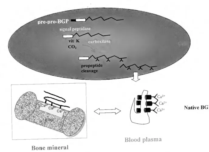
The current knowledge of the mammalian BGP biosynthetic pathway is partially summarized in Figure 1-2. A preproBGP, approximately twice the size of the mature peptide, is first synthesized, bearing a signal peptide (which direets the protein to the endoplasm.c reticulum) and a propeptide. both cleaved off in separate reactions prior to secretion (73, 320, 374 399 400). After removal of the 23-residue hydrophobic leader sequence by the signal peptidase'. concom.tant or immediately after translation (399), it appears that the intracellular proBGP (374, 375, 400) undergoes posttranslation modifications, leading to synthesis of the three Gla residues, by a processus that requires vitamin K as a cofactor and is C02- and 02- dependent (143, 286, 399, 400). This reaction is catalysed by the y-carboxylase enzyme, which also contains Gla residues (34), event that seems to be coincidem with membrane binding potentiai (182) and proceeds by a mechanism similar to that described for other vitamin K-dependent proteins (143, 542). The proregion contains a gamma-carboxylation recognition site homologous to corresponding regions in the vitamin K-dependent clottmg factors Arg at position -1 and Phe at -16 are strictly conserved and appear to be criticai for the binding of the carboxylase enzyme to its substratos (246, 399). Another homologous structure possibly involved in substrato recognition by the carboxylase is the invanant
INTRODUCTION
occurrence of the peptide sequence Glu-X-X-X-Glu-X-Cys (with X for variable amino acid) in ali Gla-containing clotting factors, BGPs and Matrix Gia proteins (MGPs) sequenced (442). The two glutamic acid residues in this consensus sequence are normally carboxylated to Gla, along with other specific glutamic acid residues, depending on the protein. This potential recognition site is part of the Gla-helix domain in BGP (214), and thus substrate conformation may play an important role in determining carboxylation specifrcity (218). After carboxylation, the propeptide is removed and the protein is secreted (198).
m signal peptidase ■■ V- VV, V t CO ,■ ■ ■ ■ propeptide cleavage Ca2 Ca2+ Ca > < (;íi2+ Ca m mH Vi b-A :V,' 2l-í Native BGP Blood plasma Boné mineral
Figure 1-2 General pathway of BGP biosynthesis and secretion by osteoblasts. Prepro BGP M « 10 000) consists of a 23-26 residue prepeptide, a 22-28-residue propeptide, and a 45 to 50-residue BGP sequence (this study, 117, 261, 371, 527), the exact size of each fragment bemg dependem on the species analysed. After cleavage by signal peptidase resulting P/e00? 13 presumably targeted for carboxylation by its propeptide (73, 246, 399, 442) Gla (symbolized by inverted and horizontal Ys) is normálly formed at residues 17, 21 and 24 (218). Adapted ftom
-lauschka et al. (218).
Two contiguous basic amino acids appear to tbe amino-terminal side of the propeptidase cieavage site in most of the vitamin K-dependent proteins. However, additional studies with vitamin K-dependent eoagulation factors and protein C mutants (30, 160) suggest that the propeptidase substrate specifícity is not restricted to the basic residues at -1 and -2, but also includes the carboxy-terminal four aminoacids of the propetide.
1.4. BIOSYNTHESIS. TISSUE DISTRIBUTION AND ABUNDANCE OF BGP
BGP in bone
BGP is synthesized in bone. where it represents 1-20% of ali non-collagenous proteins (90. 207, 209, 212, 216, 288, 436), being associated with the calcified extracellular matnx (214 236, 336, 337, 458). Several lines of evidence point to osteoblasts as the cells in bone which synthesize BGP (e.g., 73, 218, 295. 375, 503, 571) and the protein is also synthesized in vitro by bone-derived cells, such as primary cultures of osteoblastic cells (25, 33, 57, 273, 305. 368), and by clonal osteosarcoma cells which display features of the osteoblastic phenotype such as high PTH responsiveness and high alkaline phosphatase activity (5, 324, 374, 375).
BGP in teeth
Besides osteoblasts, BGP is also synthesized by odontoblasts in teeth (57, 123). In fact, rat incisor dentin has yielded a Gla protein identical to rat bone BGP (123, 303), in amounts comparable to those found in that tissue (302), an observation consistent with the fact that dentine has a calcified collagenous matrix generally similar to that of bone (304). Interestingly, BGP has never been detected in tooth enamel (420), a mineralized tissue that differs ffom dentine and bone by the average size of its hydroxyapatite crystals, which are far larger, and also by the absence of a collagenous matrix.
INTR0DUCT10N
BGP in athcrosclerotic lesions
BGP has been also identified in calcified athcrosclerotic plaques (158, 485). Already in 1863 the mineral component in calcified arteries was described as "an ossificalion, andnot a mere calcification; lhe plates which pervade ,he inner wall of the vessel are real plates of bone...We see ossification declare itself in precisely the same manner as when an osteophyte forms on the surface of hone...follomng the same course of development" (547). This bone associated with atherosclerosis inclusively possesses marrow (156). Not unexpectedly, we find in this athcrosclerotic lesions, besides BGP, the same proteins present in normal bony tissues, namely MGP (485). Bone Morphogenetic protein-2a (BMP-2a (a potent osteogemc differentiation factor (50, 259))) and osteopontin (445). The cells responsible for the presumable active calcification observed in atherosclerosis are not known. but it ts possible that vascular cells may be able to act in a manner similar to osteoblasts (445). In addition, at least some of these cells have morphological and immunocytochemical features of macrovascular pericytes (50; a type ol mural cell).
BGP in plasma
BGP has been detected in the plasma of ali vertebrates examined (e.g., 197, 425, 427, 433), suggesting that a significant amount of BGP synthesized by bone cells circulates in plasma. Studies have shown that the BGP found in the circulation derives from new protein synthesis and not from bone resorption or release of pre-existing BGP from the bone matnx (433). This fact, together with the evidences that serum BGP binds strongly to added hydroxyapatite [(433) which indicates that the plasma protein has a foll complement of y- carboxyglutamate residues] has raised the possibility that BGP found in bone represents serum BGP bound to bone hydroxyapatite (425). However, several evidences point against that conclusion (e.g., the fact that less than 7% of the 125I-labelled BGP injected into rats actually accumulates in bone (433)), and an altemative model appeared suggesting that the high BGP concentrations adjacent to hydroxyapatite were due to its secretion near mineralizing sites (425). The BGP found in serum would then represent the ífaction of newly synthesized BGP escaping from hydroxyapatite binding and diffirsing away from the mineralizing site. This model also accounts for the increase in serum BGP upon warfarin
administration, which creates an abnormal BGP that cannot bind to hydroxyapatite, or upon ethylhydroxydiphosphate injection, which competitively blocks the binding of BGP to hydroxyapatite (432).
The molecular weight of plasma BGP is identical to that for BGP extracted from bone (425-427). The turnover of BGP in plasma is quite fast (433), as indicated by the complete shift in the ability of serum BGP to bind to hydroxyapatite within 3 hours of warfann administration. Sinee warfarin inhibits the Y-carboxylation of BGP at the microsomal levei, this result indicates that the interval between new BGP synthesis within the cell and complete turnover of BGP in serum is only 3 hours (439).
BGP in blood
Sensitivo radioimmunoassays (RIAs) for BGP have shown that this protein circulates in blood at concentrations ranging from 2 to 15 ng/ml in normal adult humans (72, 103, 522, 524) lo as much as 900 ng/ml in young rats (2, 406, 430), Comparable leveis of BGP are found in both serum and plasma samples of humans and other species (197), at least in those checked until present by this method.
In contrast with the short half-life of the BGP protein in the circulation (around 5 minutes), being rapidly metabolized by the kidney (433), the lifetime of the BGP message in unstimulated or fully 1,25(OH)2D3-stimulated cells appears to be quite long, which is indicated by the fact that 15 hours of exposure to transcriptional inhibitors affects neither basal nor fully stimulated BGP synthesis (398).
Various fragments of BGP are known to circulate in blood. The majonty of circulating BGP is composed of the intact molecule and a large N-terminal mid-molecule fragment, which is thought to encompass residues 1-43 (199). Reeent studies indicate that there are other smaller (< 30 residues) N-terminal immunoreactive species of BGP in the serum (80, 178, 194). The origin and significance of the N-terminal mid-molecule fragment is uncertain. Some authors state, based on animal studies, that the BGP found in the circulation is derived from new protein synthesis rather than from bone resorption or release of existing BGP from the bone matrix. The dynamics of plasma BGP have been clarif.ed by studies on rats treated with various amounts of warfarin and vitamin K, (430, 433, 439). There are still those who
INTRODUCTION
believe that the breakdown of BGP may also occur during osteoclastic dissolution of bone (467). Proteins like cathepsins (24) and plasmin (379) are capable of degrading BGP, producing fragments of diverse lenghts. Osteoblastic degradation of BGP could also serve as a mechanism to regulate BGP concentration (199). Whether the generation of BGP fragments during bone resorption is related or not to BGP biological funetion in regulatmg bone turnover is not clear.
1.5. DEVELOPMENTAL APPEARANCE OF BGP
Investigations into the developmental appearance of BGP in calcifying t.ssues have relied either on direct bioehemical and immunochemical assays for BGP itself or on chemical analysis for y-carboxyglutamic acid. These two approaches, using human and rat models, yield different results for the developmental appearance of BGP. Direct assays for BGP in demineralized extracts of calcifying tissues indicate that the protein itself does not appear in parallel with the accumulation of mineral, but rather 1-2 weeks later, at the approximate Ume when the initial minera, phase matures to hydroxyapatite (439, 525). Accord.ngly, the levei of BGP in rat bone rises rapidly after birth, from 1.7% of the adult levei in newbom rats to 65% of the adult levei in 24-day-old rats (427). The same eonclusion was reached by studies analysing the developmental expression of the mouse and rat BGP gene by in situ hybridization and Northern techniques, with accumulation of mRNA for BGP occurring at a late stage of osteoblast differentiation, suggesting that the protein is not necessary for mtUal mineralization of the bone matrix (79, 331, 362, 497). In contrast, chemical analysis of y- carboxyglutamic acid in developing bone shows that the appearance of this ammo acid parallels the accumulation of mineral, rather than subsequent mineral maturation (427). These results are in agreement with those obtained in a previous study (210), where the appearance of y-carboxyglutamate in developing chicken bone was shown to correlate with the appearance of bone mineral, and with a later study (216) on the developmental appearance of BGP in bovine and chicken bone. However, this result is now attributable to the presence in bone matrix of at least one other Gla-containmg protein, MGP, whose accumulation is known
to occur much earlier than that of BGP during mouse development (19, 309, 316, 388. 497). Another interesting fact is that serum BGP in newbom rats is within the adult range, what makes clear that the 100-fold lower content of BGP in newbom rat bone is not due to an inhibition in expression of the BGP gene (427). The fact that a great number of cells stains strongly with anti-BGP antibodies in the bonés of 2- and 3-day-old rodents (32) reinforces the conclusion that BGP is synthesized at a high rate by osteoblasts in the newbom rat bone, but mostly fails to aceumulate in the mineral phase. The hypothesis that the initial bone mineral phase is deficient in a binding domain required for a strong interaction with BGP is strengthened by the finding that, in vitro, BGP does not bind to amorphous calcium phosphate (420), a less ordered mineral phase than hydroxyapatite.
From these results we can expect BGP not to be dispersed evenly throughout developing bonés. Accordingly, a study using tibial bonés of 2- and 4-week-old rats (431) has shown that BGP concentrates mostly in the midshafts, with the proximal and distai growth plates containing less that 5% of the midshaft leveis of BGP, a result consistent with the fact that most new mineral is deposited at the growth plate. Two weeks later, however, m this model, the segments previously located near the growth plate have become part of the bone diaphysis and the BGP leveis have risen to midshaft leveis. Other studies (451) have reached the same conclusion, i.e., in rat, mineral accumulation precedes BGP appearance by about 2 weeks. The explanation for this delay lies surely in the still not evident mode of action of BGP. which will be discussed in the nexl section.
1.6. BGP FUNCTION
The function(s) of BGP
Because BGP is one of the most abundant noncollagenous proteins in bone, an importam, but elusive, function has been inferred since the time of its diseovery. However, and despite the concerted efforts of an important number of scientists, the precise function of BGP in the formation and metabolism of bone has remained, until recently, unclear. Because of its specific interaction with hydroxyapatite, BGP was thought to affect the growth or
1NTR0DUCTI0N
maturation of Ca2+ -phosphate mineral phases. In agreement with this, BGP developmental appearance roughly parallels the onset of mineralization and the increase in synthesis of this protein is concomitant with hydroxyapatite deposition during skeletal growth (210, 2B8).
The adsorption affinity of BGP for hydroxyapatite may be an important factor in mineral dynamics of bone. The transition of brushite (CaHPO^HzO) to hydroxyapatite [Caio(P04)6(OH)2] is inhibited by very low concentrations of BGP (205). BGP also mhibits precipitation of hydroxyapatite from supersaturated solutions (414. 420, 537) and from seeded hydroxyapatite systems (416, 462), but has no effect on Ca2+ -phospholipid-PO,,- dependent crystallization (48). The degree to which BGP retards cystalhzation, at least m supersaturated solutions of calcium phosphate, depends critically on the concentration ot BGP. with a doubling of the time required for half-maximal crystal formation at a BGP concentration of 6 pM (414). Since the final amount of hydroxyapatite is not affected by BGP, the effect of BGP is exclusively on the kinetics of mineral formation, rather than on the thermodinamic end point (solubility product) of the mineral phase (439). Similar studies performed with supersaturated solutions of calcium phosphate seeded with a small amount of hydroxyapatite (416) led to the same conclusion; BGP is a potent inhibitor of mineralization, but only if it contains Gla residues and an intact disulfide bond. Its effect is kinetic, rather than thermodinamic. The protein binds poorly to amorphous calcium phosphate of unspecified surface area (420). BGP adsorption to fluorapatite [Ca^PCXOíFj] exhibits a fivefold greater affinity constant than hydroxyapatite (219, 560), which may account for some of the known disparate effects of fluoride in bone mineral metabolism,
Studies addressing the developmental appearance of BGP (see previous section) also give insight into the possible function(s) of BGP, The finding that fetal rat bone is nearly devoid of BGP (0.09 mg/g of bone; 427) indicates that this protein is not required to be present at this stage of rat development but also that any structure or property already present in fetal rat bone can be excluded as a possible biological function for BGP. For example, BGP cannot be required for the formation of the first mineral phase of rat bone, since a 20- day old fetal rat with 0.1% of the adult BGP levei has already 40% of the adult mineral leveis (427). BGP also cannot be required for osteoclastic bone resorption in response to hormones such as parathyroid hormone, since fetal rat bone is resorbed in response to such hormones
(449, 450). The same information is given by the differences in BGP content observed in different bone regions, according to its age (see previous section). The fact that, in rats? BGP accumulates in calcifying tissues only 1-2 weeks after the aecumulation of mineral may be related to the maturation of initially deposited mineral to hydroxyapatite.
When one tries to analyse changes in bone structure and physiology which occur between rat birth and the 24-day old-stage. when the BGP levei rises to 65% of the adult levei (427), we find that one change in bone structure which approximately parallels the appearance of BGP is the transition from the initially formed amorphous calcium phosphate mineral of fetal bone to the hydroxyapatite phase characteristic of adult bone (525). BGP may appear concurrently with this mineral transition because its function only requires it to be present at this stage. For example, its function could be to catalyze the transition to hydroxyapatite, to regulate the size or shape of the hydroxyapatite crystals formed, or to orient the crystals epitactically allong the collagen fiber (427). Altematively, BGP could appear in bone paralleling the appearance of hydroxyapatite because only this phase of the mineral binds BGP (414, 420). The first of these two hypothesis was strenghtened by the studies described in the next paragraph.
Depletion studies carried out in rodents have shed some light on the contribution of BGP in bone formation. In one of these studies (135), gene targeting has produced a mouse that has had the BGP gene "knocked out" (KO). These mice are characterized by a progressivo increase in bone mass, with an accelerated rate of bone formation without changes in osteoclast or osteoblast number. No changes in mineral content of the bonés of BGP-depleted mice were detectable by von Kossa staining or histomorphometry. However, a more sensitive assay of mineralization, Fourier transform infrared microspectroscopy, revealed differences in the size and perfection of the crystallites (49). In wild-type animais the crystals were larger and more "perfect" in the cortical bone than in trabecular bone. In contrast, in the BGP KO animais, the crystal size and perfection were the same in both the trabecular and cortical bone. These findings are consistent with impaired mineral maturation in the BGP-deficient bone and imply the presence of newer (less remodeled) mineral. Also interesting is the fact that the expression of other non-collagenous proteins, such as MGP, osteopontin and bone sialoprotein was not signif.cantly affected by the absence of BGP. Over
INTR0DUCT10N
time, the mutant developed abnormalities of bone remodeling which became noticeable in 6- month-old animais (135). These findings have led to the intriguing conclusion that, despite its abundance in skeletal tissues and its ability to bind calcium and apatite. BGP may actually serve as an inhibitor of bone formation.
Chronic treatment of rats for 2 months with the vitamin K antagonist warfarm reduced BGP in bone matrix to less than 2% of the control leveis, but did not affect bone formation or structure (430). A visible effect on bone structure was only detected when the same warfarm treatment was prolonged for 8 months (435), and resulted on excessive mineralization and closure of the growth plate, with cessation of ali longitudinal growth. This phenotype resembles the "fetal warlàrin syndrome", a defect characterized by radiological stippling of the growth plate in children bom to mothers who have received warfarin during gestation (204). The hypothesis that defective synthesis of vitamin K-dependent coagulation factors, which results in sporadic bleeding, is responsible for these phenotypes seems to have been ruled out, since fetal warfarin syndrome is absent when other anticoagulants. hke heparin. are used. One possible explanation for the absence of visible bone alterations may be the existence of backup systems that may compensate for a BGP deficiency in rodents. In any case, the elucidation of the BGP function in the rodent depletion model may require the imposition of an externai stress to bone metabolism other than those tested to date.
In fulfilling its yet conceptual role, it is possible that BGP may act in combination with other hydroxyapatite binding proteins, such as osteopontin, which potentiates osteoclast adhesion to mineral surfaces and forms a complex with BGP in vitro (452, 456). It is possible that this and other BGP-protein complexes may ftmction as a bone remodellmg signal. Further studies in animais depleted of combinations of matrix proteins should provide clues to the function of BGP and other bone speciftc proteins.
Several in vivo and in vitro studies reinforce the above stated conclusion. First, disrupted collagen fibrillogenesis in the cloned mouse calvarial cell line MCT3T3-E1 results in increased tumover of the collagenous matrix, a decrease in alkaline phosphatase, but a five-fold increase in BGP biosynthesis (567). Second, the pattem of BGP distnbution in human osteons changes with gender and age, and localized reductions of BGP in the extracellular matrix are associated with reduced cortical remodeling (237). Several early
sludies have suggested that BGP is involved in recruitment and aclivation of bone resorbing cells. The prolein is a chemoaltractant for peripheral mononuclear cells and gianl osteoclasl- like cells from tumours (81, 291, 325).
Immunolocalization sludies show lhal BGP is distributed Ihroughout lhe mineralized regions of bone matrix, dentin and calcified cartilage (330, 338). However, a growing accumulation of evidence indicates thal BGP is not related to events that allow mineral deposition to occur, but rather that it participates in regulation of mineralization or bone tumover. Alternatively, Thiede et ai (529) suggest that, since BGP can chelate calcium ions, it may act as a natural anticoagulant within bone.
It is interesting that the presently most credited hypolhetical BGP function, inhibition of hydroxyapatite crystal growth. seems to be also performed by a non-y carboxylated protein, osteonectin. This protein has a very acidic NfB lerminus containmg glutamic acid and aspartic acid residues which, if appropriately spaced, could interact with the hydroxyapatite crystal lattice (462). However, the inhibitory effect on crystal growth of a mixture oí BGP and osteonectin seems lo be additive (462), which indicates that not only osteonectin and BGP do not undergo any synergistic inleraction nor can compete tor binding to hydroxyapatite, but also that lhese proteins may play, even if slightly, different roles in prevenling hydroxyapatite crystal growth and maturation.
Finally, it is interesting to note that the odontoblasts already engaged in synlhesizing predentine but not yet in mineralizing it to form dentine can already be strongly stained with the anti-BGP antibody (57). This observation reveals a temporal dissociation between BGP synthesis and mineralization and suggests that the protein could act to delay the mineralization of predentine.
Bone pathologies and BGP
Although, as referred above, the first clear evidence conceming the exact biological function of BGP has been provided only recently (49, 135), the protein has been extensively studied since its first identification in bone, mostly as a climcally important diagnostic parameter of bone pathologies (e.g., as a marker of bone tumover; 58, 116, 218, 529, 557), role that is favoured by its short half-life in the circulation (around 5 minutes), being rapidly
1NTR0DUCT10N
metabolized by the kidney (433). Serum BGP concentrations are correlated with histomorphometric Índices of bone formation (58, 59, 91, 106, 139, 177), leading most investigators to agree that assay of serum BGP is a measure of bone formation in particular and bone tumover in general. The serum concentration of BGP reflects that portion of the newly synthesized protein that does not bind to the mineral phase of the bone but is released directly into the circulation, It is estimated that >90% of the newly synthesized protein is deposited in bone in one-month-old rats, but as the animal mature a greater proportion of protein is released directly into the serum (340). In normal human adults approximately one- third of BGP synthesis ends up in the circulation (340). Deviations from normal concentrations of circulating BGP are a consequencc of changes in lhe synthesis or degradation pathways of the protein. Such changes may result from physiological alterations in skeletal homeostasis that accompany normal development or may be associated with specific disease states. The rate of glomerular filtration or renal catabolism also influences circulating BGP leveis. Finally, serum concentrations may reflect drug- or disease-induced alterations in the normal hydroxyapatite-protein interaction, resulting in an altered proportion of existing or newly synthesised protein that binds to bone (218).
Plasma BGP content is elevated dramatically in patients with metabohc bone diseases characterized by increased bone turnover (426). Consequently, this protein can be of great utility as a diagnostic tool for such important diseases as (i) osteoporosis (a metabohc bone disease characterized by a defect in bone reraodelling and the loss of the normally mineralized bone), where it may serve as an early diagnostic critena (103, 104, 142, 372, 426), (ii) Pagefs disease, as a marker of response to treatment (107, 563), or (m) metastatic bone câncer (423). The levei of BGP carboxylation has also been proposed as an indicator of the nutritional state of bone with respect to vitamin K (279), which is supported by the results of studies showing that BGP can be up to 40% undercarboxylated in postraenopausal women when compared with premenopausal women (412).
Serum BGP is commonly measured by both in house methods and commercial kits with various assay formats (e.g., 419, 463). These include high perfonnance liquid chromatography (HPLC), radioiramunoassay (RIA), immunoradiometnc assay (IRMA), enzyme linked immunosorbent assay (ELISA), and luminescence immunoassay (LIA). Also,
BGP has been increasingly used as a highly specific osteoblastic marker produced during bone formalion, more explicilly as a marker of lale osteoblast ditterenliation (121, 333, 371, 524).
Is the role of BGP crucial to thc same extent in ali vcrtcbratcs?
There is a possibility lhal lhe role of BGP in bone developmenl/formation may not have the same degree of importanee in ali vertebrates. The faet lhal normal BGP leveis are dramatieally low in man. when eompared, for example, with ral and calf (see section 1.4) suggests lhal the prolein may play a less crucial role in the metabolism of the human skelcton when eompared lo other vertebrates, a feature lhal can provide an imporlant clue lo its function. However, additional studies using different species are required to address this question.
1.7. THE BONE GLA PROTEJN GENE
To date. only mammalian BGP genes have been characterized. Therefore it is not possible to compare stmctures and pattems of regulation across other than mammalian species.
Chromosome location and gene organization
The transcribed regions of the BGP genes expressed in bone of human, rat and mouse contain three introns and four exons. The promoters of these mammalian BGP genes have a similar overall organization and contain comparable promoter regulatory elements (II sapiens: 73; M. musculas (OG1): 117; R. norvegicus: 527). Thus these bone-specific BGP genes appear to be organized in a manner that supports analogous responsiveness to homeostatic physiological mediators and developmental expression in relation to bone cell differentiation.
The human BGP gene has been localized to the Iq distai region of chromosome 1
INTRODUCTION
(446). A mouse gene has been mapped to chromosome 3, which conlains genes homologous to those located in the distai region of the human chromosome Iq (244). From these and other studies, the BGP gene was initially described as a single copy gene. However, more recent analysis of several mouse and rat strains has indicated that, in these models, BGP is part of a gene cluster (116. 117. 448). In lhe mouse, three contiguous genes were identified in ali strains examined, while in the rat, either one or multiple copies were detected dependent on the slrain (448). Of the three genes in mouse, two have identical promoters, and one gene (ORG, for Osteocalcin-Relaled Gene) has a varianl promoler that is developmenlally expressed in vivo in several non-bony tissues, such as brain. lung and kidney (116, 117). The coding region of ORG has a similar intron/exon organization to the BGP gene expressed in bone but carries five amino acid substitutions, one at lhe propeptide cleavage site. ORG also contains an additional exon that is not translated, and a 3 Rb insertion separates the ORG coding sequence from its promoter. The inserted sequence has lhe structure of a typical retrovirus, an attribute that leads to the downregulation of transcription, possibly explaining lhe low leveis of expression of ORG in non-osseous tissues (117).
The function of the nonosseous expressed gene remains to be established. According to some authors (192), consideration should be given to the possibilities that it encodes (i) Gla-containing nephrocalcin, a calcium oxalate crystal growth inhibitor found in kidney, although never cloned (363-366), (ii) BGP associated with platelets (529) and the hematopoietic system (311), and (iii) BGP associated with cartilage and other tissues (281, 307). Relatively to the first hypothesis, the primary sequence of this protein remains unknown, and human specific BGP antibodies do not cross-react with partially purified human nephrocalcin (F.L. Coe, Y. Nakagawa and C. Gundberg, unpublished observations).
In contrast to other bone-related genes (e.g., type I collagen and alkalme phosphatase) (29, 577), only a single mRNA transcript has been observed from the BGP gene, in ali species analysed to date (mammals and chicken). It should be noted that although regulalion of expression does not appear to be modulated by changes in the organization of the mRNA transcripts, this does not preclude the presence of sequences in the transcnbed region of the BGP gene which contribute to regulation of transcription. A silencer element has been identified within the BGP gene (284) and is similar to silencer elements observed in several
prokaryotic (326) and eukaryotic genes (66, 175, 465, 560, 570). Interestingly, the BGP gene silencer was the first to be identified within the protein coding region of a gene in higher eukaryotes (504).
The bone GLA protein promoter
A model of the three dimensional organization of the BGP gene promoter is presented in Figure 1-3, showing postulated interactions between distinct promoter elements to support transcriptional control within a three dimensional context of cell structure and regulatory requirements at the cell and tissue levei.
llNÀCTIVE Nuclear Mairlx
Silo A Silo B ^lte C. prox prom TPs VCIÍt
Nudear Matr x BASAL
NMP-2= AML AML AML
NA POL II i NMP mRNA TFsjr—r Site Sue Site
VDRE prox. prom. DNascI HS QMase I HS VDR/RXR Murlnar M.-ifrl VITAMIN D INDUCED AML AML NMP-2= AML NA POL VDFV RNA Site RXR TF's
Site Site ,/M irr-; prox. prom VDME
DNasc HS DNasel HS
Figure 1-3. General model of BGP gene regulation. Schematic represen- tation of promoter organization and occupancy of regulatory elements by transcription factors to either i) suppress transcription in proliferating osteoblasts, ii) activate expression in differentiated osteoblasts, or iii) enhance transcription by vitamin D. Placement of nucleosomes is indi- cated, as well as remodelling of chromatin structure and nucleosome organization to support supression, basal expression and vitamin D- enhanced transcription of the BGP gene. Representation and magnitude of Dnase I hypersensitive sites (see 347) are shown by solid triangles. AML, AML-related nuclear matrix bound factor; NMP-1 and NMP-2, nuclear matrix protein-1 and 2; RNA POL II, RNA polymerase II; prox. prom., proximal promoter; TF's, transcription factors. Adapted from Stein et al. (504).
Regulatory sequenees in the proximal BGP promoter
Transgenic studies indicate that sequenees residing within the proximal 1,800 bp ot
1NTRODUCTION
the rat BGP gene promoter support tissue-specif.c transcription (12). In vitro deletion-mutant experiments have shown that a 200 bp fragment of the rat promoter (521) and a 160 bp fragment of the mouse OG2 promoter (134) are neeessary and suffieient to eonfer osteoblast- speeifie express.on to a repórter gene. Frendo et ai (167) have shown that 647 bp of the mouse OG2 promoter contain ali the regulatory elements neeessary and sufficrent to d.reet bone expression of a repórter gene, including the eis-acting elements required for t.me- speciftc expression of the BGP gene. However, th.s does not preelude the contnbuttons of additional upstream sequences to BGP gene promoter activity (504), When transgemc animais were constructed with 3,900 bp of the human BGP promoter fused wtth a CAT repórter (258) expression was observed predominantly in bone but addit.onally at reduced leveis in hypertrophic ehondrocytes and kidney. Subtleties in regulatory sequenees and/or nuclear proteins that account for these differences remain to be defined.
Typical sequences associated with most genes transcribed by RNA polymerase II are found in 5'-flanking regions of the rat BGP gene (e.g., TATA, CAAT, AP1 and AP2) ( 527 572) In addition, the BGP promoter also contains a sma.l stretch of altemating punnes and pyrimidines just 5' of the CCAAT sequence. This sequence has the potentral of form.ng Z-DNA a structure which may play a role in gene regulation (224). Table I.1I summarizes ali the factors known to regulate the expression of the BGP gene at the levei of transcnpt.on and corresponding DNA binding elements.
Developmental and tissue specific-control
Combined activities of overlapp.ng regulatory elements and associated transcnptton factors provide a mechanism for complex developmental control of BGP express.on durtng osteoblast growth and differentration (147, 367, 383, 392, 472, 504). One such examp.e ,s Osteoealcin box, an highly eonserved regulatory sequence required for basal expresston o t e rat (295) and mouse (134) BGP genes, which contains multiple regulatory elements, including AP-1 and homeodotnain binding sites, The OC box contributes to both bone ttssue- specific expression and speeies-specif.c regulation of the BGP gene (222, 223. 227, 228, 521, 530).
Table 1 11 Main regulatory factors of BGP gene expression and corresponding DNA binding sequences. Alse "nd.cated are the proposed mechan.sms of act.on of these regulatory factors and corresponding references.
REGULATION FACTOR
VIODE OF ACTION RESPONSIVE ELEMENT
REFERENCES
1,25 (OH)2D3 Direct and indirect transcriptional activation/ inhibition
VDRE 46, 393, 394
9-c/.s-retinoic acid Moslly indirect activation/inhibition.
Generally wilh no effect when alone (some exceptions; e.g.
384).
RARE/VDRE 47, 264,319,349, 376, 384. 408. 477.
480
Fos-jun proteins Transcriptional regulation
AP-l sites 296, 392, 503
Dexamelhasone Transcriptional inhibition/activation
GREs 9, 46, 222, 51 J
17p-Estradiol Induction of estrogen receptor gene transcription ERE 20 Id-HLH Transcriptional regulation, conferring tissue-specificity E box 357, 381, 487, 504. 521 AP1 Transcriptional inhibition; regulator of osteoblast-specific sene expression VDRE; OSCARE-2 111, 191, 296, 392, 395,396, 480 Cbfal Transcriptional activator OSE2 18, 134, 136, 254, 283 Osfl Transcriptional activator OSE1 474 i Parathyroid Transcriptional inhibitor PTHRE 33,276
Tumour Necrosis Transcriptional inhibitor TNFRE 283 TGF-P Transcriptional inhibitor TGRE 17, 299 Nuclear Matrix Protein-2 (NMP-2) Regulator of osteoblast-specific gene expression Nuclear matrix protein binding site (AML-1 recognitio motif) 39, 342, 346 s n 24