Printed version ISSN 0001-3765 / Online version ISSN 1678-2690 http://dx.doi.org/10.1590/0001-3765201620150454
www.scielo.br/aabc
Evaluation of antibiotics as a methodological procedure to inhibit
free-living and biofilm bacteria in marine zooplankton culture
VanEssa O. agOstini1,2*
, alExandrE J. MacEdO2
and Erik Muxagata1
1Laboratório de Zooplâncton, Universidade Federal do Rio Grande/FURG, Instituto de Oceanografia/
IO, Av. Itália, s/n, Km 8, Campus Carreiros, CP 474, 96203-900 Rio Grande, RS, Brasil
2Laboratório de Biofilmes e Diversidade Microbiana, Universidade Federal do Rio Grande do Sul/UFRGS, Faculdade
de Farmácia e Centro de Biotecnologia, Av. Ipiranga, 2752, Bairro Azenha, 90610-000 Porto Alegre, RS, Brasil
Manuscript received on June 24, 2015; accepted for publication on August 13, 2015
aBstract
There is a problem with keeping culture medium completely or partially free from bacteria. The use of prokaryotic metabolic inhibitors, such as antibiotics, is suggested as an alternative solution, although such substances should not harm non-target organisms. Thus, the aim of this study was to assess the
effectiveness of antibiotic treatments in inhibiting free-living and biofilm bacteria and their half-life in artificial marine environment using the copepod Acartia tonsa as bioindicador of non-harmful antibiotic combinations. Regarding to results, the application of 0.025 g L-1 penicillin G potassium + 0.08 g L-1 streptomycin sulphate + 0.04 g L-1
neomycin sulphate showed great potential for use in marine cultures
and scientific experiments without lethal effects to non-target organisms. The effect of this combination starts within the first six hours of exposure and reduces up to 93 % the bacterial density, but the half-life
is short, requiring replacement. No adverse changes in water quality were observed within 168 hours of exposure. As a conclusion, we can infer that this treatment was an effective procedure for zooplankton
cultures and scientific experiments with the aim of measuring the role of free-living and biofilm in the
marine community.
key words: Acartia tonsa, antimicrobials, experiment, marine culture, toxicity.
Correspondence to: Vanessa Ochi Agostini E-mail: nessaochi@gmail.com
intrOductiOn
Scientific laboratory experiments are often voided
by the presence of bacteria in the medium, which may be responsible for biased or dubious results (Spencer 1952). The real importance/contribution
of microorganisms in cultures is difficult to be
mea-sured because conditions totally (axenic) or partially (semi-axenic) free of bacteria are hardly attainable
for long periods. In this context, an efficient method
for the inhibition/destruction of bacteria without harming non-target organisms still needs to be developed to allow hypothesis testing and to assess their real contribution/role in the community.
Yetka and Wiebe (1974) proposed the use of prokaryotic inhibitors, such as antibiotics, as a tool for inhibiting bacteria in water and thus allowing
for scientific experiments. Subsequently, several *Programa de Pós-graduação em Oceanografia Biológica/
authors have reported success using these sub-stances to quantify bacterial respiration (Pringault et al. 2009), bacterial growth (Wheeler and Kirch-mann 1986), and nitrogen assimilation (Fouilland et al. 2007), as well as to understand aquatic trophic
interactions (DeLorenzo et al. 2001).
The use of inhibitors to prevent bacterial activity can improve our knowledge in experiments that assess their activities, such as those aimed to distinguish the absorption of nitrogen between phytoplankton and bacteria (Tungaraza et al. 2003). Inhibitors can also exclude competition among
groups benefiting from the growth of a particular group free from bacteria (Droop 1967), and improve
or accelerate the development of aquatic species such as invertebrates and fish larvae (Conover 1967, Tighe-Ford et al. 1970, Walne 1974).
Antibiotics have been used for a long time to control bacterial diseases in aquaculture, but massive use of these substances without care or control resulted in a limitation or even ban of these substances in some countries where production
was intended for human consumption (Defoirdt
et al. 2007). Nevertheless, with proper care, this
procedure could be used in scientific experiments
aimed at systems without bacterial contamination, such as semi-intensive cultivation of organisms for population studies (Tighe-Ford et al. 1970), to identify/measure the development stages/ rates of crustaceans (Rice and Ingle 1975, Harms 1986), and for hypothesis testing by checking the contribution of bacteria to the community (Yetka and Wiebe 1974, Veuger et al. 2004).
Combinations of antibiotics such as penicillin and streptomycin have been widely used in cultivation with positive survival and growth results in the species of interest (Spencer 1952),
with some authors adding neomycin (Droop 1967, agostini 2014) and chloramphenicol (Droop 1967), thus increasing the efficiency in inhibiting
bacterial growth. On the other hand, the individual application of oxytetracycline hydrochloride
has shown significant results in the reduction of bacterial diseases in fish (Pereira Jr. et al. 2006).
For the use of antibiotics on a large scale, such
as intensive fish ponds and carcinoculture, it is
nec-essary to estimate the real effect of such substances on the microbiological community, preferably after conducting an assessment of their effects on non-target organisms known as bioindicator with great sensitivity, such as copepods and mysids (Resgalla and Laitano 2002, Ihara et al. 2010).
The copepod Acartia tonsa Dana Calanoida
1849 certainly meets this premise: along with having a wide distribution, short life cycle and easy cultivation, it still has high sensitivity and is recommended for use in acute toxicity tests (ISO 1499 1999).
In addition, copepods are an important link in the aquatic food chain, playing a key role as primary consumers as well as secondary producers, generating the interest of the economic sector to
be used in aquaculture as live food for fish larvae (Drillet et al. 2006, Olivotto et al. 2008, 2009,
Buttino et al. 2012).
Thus, the aim of this study was to assess the effectiveness of different antibiotic treatments in
inhibiting free-living and biofilm bacteria
associ-ated with organisms and surfaces and their half-life in the marine zooplankton cultures.
MatErials and MEtHOds
This study was divided into two stages: the first
was to assess the survival of the of copepod Acartia
tonsa with different combinations of antibiotics, and the second was to estimate the half-life and efficacy for the combination of antibiotics with
the best survival results for the copepod A. tonsa
obtained in the first step of the experiment on the inhibition of free-living and biofilm bacteria.
To evaluate the survival of A. tonsa to
antibio-tics, we used 60 experimental units (EU)
represen-ted by five treatments with twelve replicates each.
Adults of the copepod Acartia tonsa that were used in the experiments were obtained from zooplankton samples collected from horizontal tows performed at the surf zone of Cassino Beach, Rio Grande, RS, Brazil. All samples were collected with a 30 cm cylinder-conical plankton
net fitted with a 200 micron mesh. In the laboratory,
Acartia tonsa specimens were identified according Bradford-Grieve (1999) and separated, with the aid of Pasteur pipettes and a dissecting microscope (Olympus SZ40), from the rest of the community. All Acartia specimens were checked according to Sabatini (1990) to verify the development
stage they were in (i.e., copepodites VI males and
females) and acclimated for 30-hours in similar conditions to which they were collected (salinity 30, temperature 25 °C and with a photoperiod 14:10 (light:dark)), so that any organism that had been injured or debilitated by the stress generated during collection could die or be removed. After
acclimation, 3 adults (i.e., Copepodite VI), without
distinction of sex, were placed in experimental
units (EU) with 60 mL (density of 1 org 20 mL-1)
(Gorbi et al. 2012) of the same conditions to which where they were acclimated. Copepods were fed
daily with the diatom Conticribra (=Thalassiosira)
weissflogii Stachura-Suchoples and Williams
(2009) at a concentration of 20,000 cell mL-1
(Teixeira et al. 2010). The culture medium of each EU was completely renewed every 24 hours to ensure the effect of the antimicrobial agent used. To evaluate the impact of treatments on copepod
survival, the number of copepods was recorded
every 24 hours for 3 days (Delupis et al. 1992).
The second stage was conducted in six EU represented by two treatments containing 120 mL of culture medium, with three replicas each: control
(without antibiotic) and T2) 0.025 g L-1 penicillin
G potassium + 0.08 g L-1 streptomycin sulphate
+ 0.04 g L-1 neomycin sulphate. Six substrates of
wood (20 × 20 mm) that had been cleaned with
70 % ethyl alcohol were placed in each eU. To prevent biofilm colonization on only one side of the wooden substrates, the eU were shaken five
times a day. All EU were kept at a salinity of 30, temperature of 25 °C and photoperiod of 14:10
inside a BOD incubator (Marconi 403), reflecting
the same conditions as the previous experiment. To assess the beginning effects of the antibiotics
as well as the half-life in artificial sea medium,
the experiment was static, with antibiotics being applied only once.
To estimate the density of free-living and
biofilm bacteria, three aliquots of the medium (1 mL) and of three substrates immersed in Mili-Q
water (after detachment) were taken after 6, 12, 24, 72, 120 and 168 hours of exposure to the treatments. Free-living bacteria were deposited directly inside Eppendorf tubes and preserved with
4 % buffered formaldehyde. Biofilm had to be
detached from the wooden substrates using three pulses of 20 kHz for 15 seconds on each side of the substrate (Oliveira et al. 2006) with the aid of a Cole-Parmer® ultrasound (series 4710 - ultrasonic
homogenizer). Detached biofilm was then placed taBlE i
summary of all combinations tested in each treatment (t).
Abbreviation Composition Control untreated
T1 0.75 g L-1
penicillin G potassium + 0.75 g L-1 streptomycin sulphate (modified of Spencer 1952)
T2 0.025 g L
-1
penicillin G potassium + 0.08 g L-1
streptomycin sulphate + 0.04 g L-1
neomycin sulphate (Agostini 2014)
T3 0.63 g L
-1
penicillin G potassium + 0.3 g L-1
streptomycin sulphate + 0.05 g L-1
neomycin sulphate + 0.01 g L-1 chloramphenicol (Droop 1967)
inside eppendorf tubes and fixed with 4 % buffered
formaldehyde. Both samples were then stored in the dark at 8 °C until analysis.
Free-living (org mL-1) and biofilm (org cm- 2)
bacterial density was estimated with a flow cyto-meter (BD FaCSVerseTM) at the Faculty of
Phar-macy of the Federal University of Rio Grande do Sul (UFRGS). To ensure the reliability of data, part of the samples were visually counted under epifluorescence microscope, applying the same methodology for the evaluation of the microbial community described below, and were subsequently compared with the counts obtained in cytometer, resulting in equivalent bacterial densities. The size (µm) and complexity of the cells were measured using the mode of Forward Light Scatter (FSC-A) and Light Side Scatter (SSC-A) parameters using 6 µm latex beads (Invitrogem®) as standard (Herzenberg et al. 2006, Bouvier et al. 2011, Picot
et al. 2012). The bacterial cell biovolume (μm³)
estimation and conversion to bacterial cell biomass (pg C-1
) were made using the allometric conversion factor of Sun and Liu (2003) and Norland et al. (1987), respectively.
For the evaluation of the microbial community, biological material (of water and substrate)
obtained from eU was filtered on polycarbonate
filters (darkened with Irgalan Black), stained
with acridine Orange (1 %) and viewed under
epifluorescence microscope (Zeiss Axioplan) at
100 x magnification. Bacterial morphotypes were classified according to Zaritski (1975). Fungi were observed and quantified by the same method.
Water samples of 100 mL were collected from each treatment at 6, 72 and 168 hours of exposure to
determine the amount of phosphate (PO4),
ammo-nium (NH4+), nitrite (NO2-) and nitrate (NO3-), and
filtered through 0.2 µm glass fibre filters (Whatman
GF/C) and frozen in polycarbonate bottles at -18° C to evaluate the water quality and estimate possible
changes caused by the antibiotics. Nutrients (PO4,
NO2- and NO3-) were analysed following the
methodology described by Strickland and Parsons
(1972), and ammonium (NH4+) analysis followed
the Blue Indophenol method (UNESCO 1983). After analysis, antibiotics were stored in sealed containers and collected by the Engineering and
Environmental Services, SANIPLAN©.
All data were analysed with parametric statistics. Two-way ANOVA was used to test the
survival results of Acartia tonsa over time, and
the comparative density results of microorganisms between treatments at different exposure times. A post-hoc Tukey test followed the analyses.
rEsults
Significant Acartia tonsa survival rates between
treatments (F(12,220)=12.618; p<0.001) were found
(Fig. 1). T1 and T3 treatments caused the death of
all organisms within the first 24 hours of exposure.
In contrast, T2 and T4 treatments showed no
significant differences from the control (100 %
survival) in the same period of observation. After
48-hours, 8, 17 and 25 % mortality was observed
in the control, T2, and T4, respectively, but without
significant differences between them. at the end of
the experiment (72-hours of exposure), no statistical difference was observed between the control and the two treatments. Survival at the end of the
experiment was 83, 83 and 75 %, respectively, for
the control, T2 and T4.
From the Acartia tonsa survival results
presented, the treatment for T2 (0.025 g L-1
peni-cillin G potassium + 0.08 g L-1 streptomycin
sul-phate + 0.04 g L-1 neomycin sulphate) was selected
to check its potential for inhibiting free-living and
biofilm bacteria.
In figure 2, it can be observed that the T2 treatment showed a lower density of free-living
bacteria (org mL-1) compared with the control
only in the first few hours of exposure, where a significant difference was detected at 6 and 12 hours (p<0.001) (Fig. 2a). as for the biofilm bacteria (org
cm-2), it was observed that the selected treatment
12, 24 and 120 hours (p<0.001) with an average
bacterial reduction of 78 % throughout the duration
of the experiment (Fig. 2b).
For free-living bacteria, a reduction of 88 %
was observed in the density with 6 hours of exposure
(org mL-1) while in the biofilm, the reduction was
75 % (org cm-2
). The highest inhibitory effect in
relation to control of biofilm and free-living bacte-ria occurred at 12 (93 %) and 6 hours (88 %) of
ex posure, respectively.
Differences in morphotypes and bacteria size
were found in the bacterial community composition when the control was compared with those found
under the influence of treatment T2, especially in the first 24 hours of the experiment. In the control,
free-living bacteria were represented by coccobacilli forms (Fig. 3a), while in the T2 treatment, rods were the dominant form with different sizes (Fig.
3b). The bacterial biofilm found in the control and
T2 were described to be of the coccus form with similar sizes (Fig. 4).
In the biofilm of the control, it was possible to
observe the recalcitrance of the attached bacteria even after the detachment process with ultrasound. It was also possible to observe the extracellular polymeric matrix and transport channels, whereas
F(12, 220)=12.618, p<0.001
Control T1 T2 T3 T4
24 h 48 h 72 h
Exposure time 0% 20% 40% 60% 80% 100% 120% 140% Survival of (%) Acartia tonsa a a a c c
c e e e
b b d d f f
Figure 1 - Two-way ANOVA indicating the survival of Acartia tonsa exposed to different treatments (Control, T1, T2, T3 and T4) after 24, 48 and 72 hours, where control = treatment without antibiotics; T1 = 0.75 g L-1
penicillin G potassium + 0.75 g L-1
streptomycin sulphate; T2 = 0.025 g L-1
penicillin G potassium + 0.08 g L-1
streptomycin sulphate + 0.04 g L-1 neomycin sulphate; T3 = 0.63 g L-1 penicillin G potassium + 0.3 g L-1
streptomycin sulphate + 0.05 g L-1
neomycin sulphate + 0.01 g L-1
chloramphenicol; T4 = 0.02 g L-1
oxytetracycline hydrochloride. Bars represent confidence intervals (95 %) and lowercase letters indicate similarities or statistical differences between treatments for each time evaluated.
F(5, 24)=12.115; p<0.001
Control T2 6 h 12 h 24 h 72 h 120 h 168 h
Exposure time 0 0.5x10⁶ 1x10⁶ 2x10⁶ 2.5x10⁶ 3x10⁶ a Bacterial densit y
F(5, 16)=13.966; p<0.001
6 h 12 h 24 h 72 h 120 h 168 h 0 10x106 20x106 30x106 40x106 50x106 60x106
Free-living (org mL )-1
Biofilm (org cm )-2
b c e
e f f g g h h a a b c d e f f g h i d i
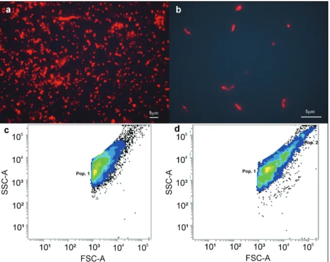
Figure 3 - Comparison of free-living bacterial communities between control (a) and T2 (b) with the evaluation of the population of bacteria by flow cytometer after 10,000 acquired events on control (c) and T2 (d). Lighter colours are related to higher density cells. For interpretation of the references to color in this figure legend, the reader is referred to the web version of this article.
in the T2 treatment, the bacteria were well dispersed and individualized.
After 6 hours of exposure, the time of greater free-living bacterial inhibition by the T2 treatment, a higher biomass was noted for this treatment
(0.127 pg C-1) compared to control (0.120 pg C-1).
The free-living bacteria showed an average
biovolume (µm³) and biomass (pg C-1) of 26.157
e 0.120 for the control and 35.108 and 0.123 at the T2, respectively.
The relative size (FSC-A) and complexity (SSC-A) of the cells of the free-living bacteria with 6 hours of exposure (Figs. 3c and 3d) were also evaluated. It was possible to detect differences
between the control and T2, such as the presence of a second population forming in the antibiotic treatment (Fig. 3d).
The free-living bacterial community also showed variations in cell size throughout the expe-riment. The control was represented by bacteria relatively smaller than average (~ 1.39 µm) compared with those observed at the T2 (~ 1.86 µm), and with lower average cell complexity (Figs. 3c and 3d).
complexity. In the FSC-A, similar sizes were found in the control and T2 (~ 1.11 µm) (Figs. 4c, d).
After 12 hour exposure, the time for higher
biofilm bacterial inhibition by the T2 treatment, it was observed that the bacterial biofilm had an
average biovolume and biomass of 20.926 and 0.118, respectively, for both treatments.
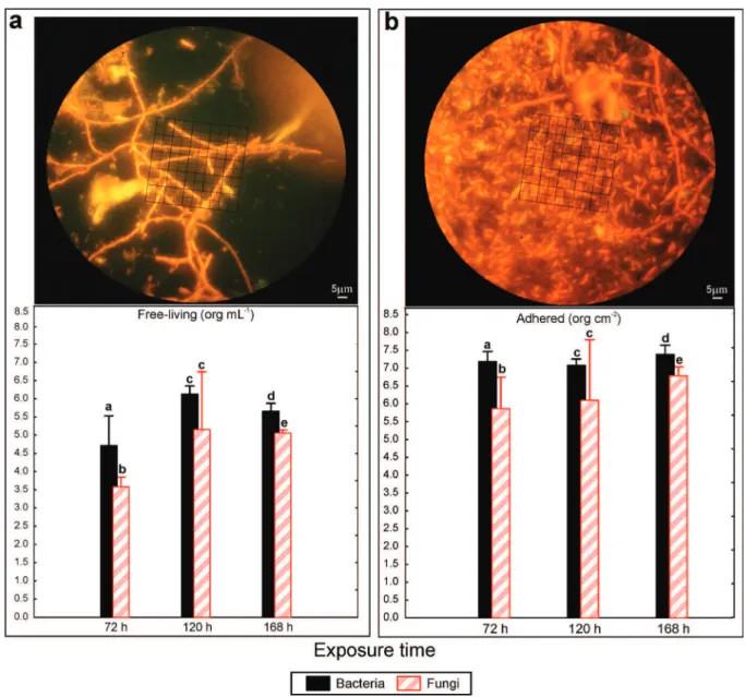
at 72 hours, fungi of the Deuteromycota
group was observed in the substrate (spores), while in the water column its presence was less
significant and was mostly in the form of hyphae.
Nevertheless, due to the absence of reproductive structures (that are essential for the taxonomic
identification genera/species), it was not possible
to reach a lower taxonomic level, with only conidia of Fusarium sp. being observed in both biofilm and the water column. However, it was a single
individual filament (hyphae), and each displayed a
reproductive structure (spore).
Figure 5 shows a comparison of relative fungal and bacterial density. Free-living and adherent fungi were not observed in the control at any exposure time or in the T2 at 6, 12 or 24 hours. An
increase of bacteria in the biofilm between 24 and
72 hours of exposure was observed in T2 in both conditions. The free-living community showed
significantly more bacteria than fungi at 72 hours
and 168 hours of exposure (F(1,4)=23.755; p=0.016;
Figure 5 - Two-way ANOVA indicating density of free-living (org mL-1
) (a) and adhered (org cm-2) microorganisms (b). Top figures are pictures, while bar graphs compare the density of bacteria and fungi after 72, 120 and 168 hours of exposure to T2. The bars represent the confidence intervals (95 %) and lowercase letters indicate similarities or statistical differences between treatments for each time evaluated.
F(1,4)=50.539; p=0.002, respectively), while at 120
hours, no significant difference was observed in
either population (F(1,4)=6.082; p=0.069). However,
the average bacterial density was higher than that of fungi for all times evaluated.
Regarding the biofilm community, a similar trend was observed with significantly higher
bacte-rial density at 72 hours and 168 hours (F(1,4)=152.640;
p=0.001; F(1,4)=121.150; p<0.001, respec tively) and no difference was observed in either popu lation at
120 hours of exposure (F(1,4)=6.883; p=0.058).
In relation to nutrients, there was only a slight va riation of nitrite between the control (0.001±0.00
mg L-1) and the T2 (0.000±0.00 mg L-1) with 168
hours of exposure, but without any significant
discussiOn
The T2 and T4 treatments, based on the proportions proposed respectively by Agostini (2014) and
Pereira Jr. et al. (2006), each represent a treatment
with a combination of antibiotics (penicillin G potassium, streptomycin sulphate and neomycin sulphate) or a single substance (oxytetracycline hydrochloride). The two treatments did not show significant differences in the survival of Acartia tonsa when compared with the control. Nevertheless, T2 showed a slightly higher average survival, similar to that found in the control, than T4. Since Kinne (1977) reported that a combination of antibiotics should be more effective than a single substance to prevent the excessive growth of micro-organisms, and due to time constraints, the T2 was selected for the next experiment instead of T4.
Apart from T4, the T1, T2 and T3 treatments are mainly based on different concentrations of the same antibiotics, and surprisingly T1 and T3 treatments caused the death of all organisms. If we take a closer look at the concentrations used, it is clear that T1 and T3 are based on a much higher concentration of antibiotics used than T2 and T4, and this could be making the culture medium toxic to the copepods.
Despite the fact that the combination tested
on T1 had been previously used (Spencer 1952, Borojevic 1966, Harms 1986, Roberts et al. 2007, Pringault et al. 2009, Trotted et al. 2011), the concentration used had never been tested on invertebrate cultures. Spencer (1952) used 0.32 g L-1
penicillin G potassium + 0.83 g L-1 streptomycin
sulphate in microalgae culture, however, the concentration of penicillin used in the current study for T1 was higher, which might have caused toxicity for the copepods. The T3 treatment, apart from the higher concentration used and the addition of chloramphenicol, also had not been tested on
invertebrate cultures, but only with diatoms (Droop
1967). Those results only reassure the classic
toxicology maxim “Sola dosis facit venenum” (From
Latin: The dose makes the poison”) attributable to Paracelsus (1493–1541), who said “all things are poison and nothing is without poison; only the dose makes a thing not a poison,” indicating that a substance can be harmless or poisonous depending on the dose.
According to Sherr et al. (1986), antibiotics used in the aquatic environments should have broad-spectrum inhibition without affecting non-target organisms. Based on this requirement, T1
and T3 are not recommended for use in scientific
experiments where harmful effects to non-target organisms are not acceptable.
The use of the combination of penicillin + streptomycin sulphate + neomycin sulphate in different concentrations has been widely used to evaluate the assimilation of nitrogen in
natural aquatic communities (Middelburg and
Nieuwenhuize 2000a, b, Veuger et al. 2004, Cozzi and Cantoni 2006) and trophic interactions
(DeLorenzo et al. 2001). However, none of these
studies tested the effects of these antibiotics on sensitive non-target organisms, such as the copepod Acartia tonsa.
The use of A. tonsa for ecotoxicological tests
and marine pollution studies have been suggested since 1977 (Lee 1977) because they are sensitive organisms and, therefore, useful as biological
indicators (Carotenuto et al. 2002, Medina and
Barata 2003, Ihara et al. 2010, Gorbi et al. 2012).
The sensitivity of the copepod A. tonsa to different
toxic substances such as metals, antibiotics, insecticides, antifouling products, and surfactants has been demonstrated by acute and chronic bioassays (Sosnowski and Gentile 1978, Lanzky and Halling-Sørensen 1997, Barata et al. 2002,
Medina et al. 2002). For this reason, the International
Organization for Standardization (ISO 14699
1999) proposed the use of the copepod A. tonsa
to a substance, other organisms in the marine community should also be resistant. Treatment with antibiotics may increase the survival, growth and lifetime of crustaceans (Fisher and Nelson 1978, Pelletier and Chapman 1996); for this reason, we believe that the survival results observed in T2, similar to those found in the control, could be an indication that the use of antibiotics in long term cultures could reduce the lethal damage caused by microorganisms.
When the T2 combination was tested to evaluate its bacterial inhibition potential, it was observed that
it significantly inhibited bacterial biofilm compared
with the control in almost all exposure times. Regarding free-living bacteria, this treatment only
significantly inhibited bacterial growth at 6 and
12 hours. In general, all antimicrobial agents are most effective to kill rapidly growing cells (Paraje 2011). Our results indicate that the inhibition effects of the antibiotics used in this experiment in the free-living bacteria started before 6 hours, reaching its inhibitory peak within 12 hours and lasting for approximately 72 hours. However, when
comparing only the efficiency of T2 without the
control, we observed that for free-living bacteria,
greater inhibition efficiency occurs at 12 hours of exposure, as observed for the bacteria biofilm. The
contrast observed between the treatment and control of the free-living bacteria may be due to depletion of nutrients, such as phosphorus and nitrogen (Smith and Praire 2004), by the high-density observed in the control with 6 hours of exposure. This high density considerably diminishes in the control with 12 hours of exposure, masking the effect of antibiotics when compared with the control.
If we consider the concentration observed at the control at 6 hours as the initial bacterial concentration, it is observed that there is a decrease in the amount of bacteria within 24 hours, and at 72 hours there is a trend of bacterial increase. For the
biofilm community, the greatest density reduction
occurred at 12 hours and then the effect decreased.
After 72 hours, it was significantly reduced, suggesting that either the antibiotic effects ceased or they developed resistance to the combination used. Antibiotics act as inducers for the expression of bacterial genes encoding drug resistance mechanisms (Butaye et al. 2003). However, there is the possibility that antibiotic resistance varies between regions and environments, mainly depending on the selection pressure imposed by
the ecosystem (Pereira Jr. et al. 2006) or time of
exposure of the bacteria to the antibiotic (Ali Abadi and Lees 2000). It is likely that the single dose administered could explain the increase in bacterial density observed in this study.
Analysing the results, it is clear that there is a delay of several hours to obtain the highest
inhibition efficiency of bacteria (between 7 and
12 hours). We believe that the antibiotics loses its full effect between 25 and 71 hours after the initial ministration because it is clear that there is an increase in bacterial density after 24 hours of exposure when compared with the control. However, we also need to note that a community response delay to this loss effect should also be taken into account.
The species of bacteria found in the water
column are the same ones found in the biofilm. The transition from the water column to biofilm occurs
in response to environmental changes and involves multiple regulatory networks that translate signals to the concerned expression change genes (Abel-Aziz and Aeron 2014). Bacteria are capable of
multicellular behaviours (associations) that benefit
the bacterial community as a whole. This association can be viewed as a survival mechanism, in which
bacteria are benefited by the facilitated acquisition
of nutrients and biocidal protection (Paraje 2011). Therefore, it is likely that the inhibition effect of
antibiotics should affect biofilm bacteria later when
compared with the free-living bacteria.
the constituents of microbes to antibiotics. The
structural nature of the biofilm and the characteristics
of sessile cells produce antimicrobial resistance, which leads to a protected environment against adverse conditions and host defences (Costerton et al. 2003). On the other hand, free-living bacteria are usually susceptible to antibiotics; however, the minimum inhibitory concentration of antibiotics in
bacterial biofilm can be up to 1,000 times higher
than that for the free-living bacteria (Patel 2005). For biomass, it was observed that the free-living bacteria in the treatment with antibiotics showed a higher amount of carbon. Thus, we believe that T2 selected a population that was allowed to grow either because they were more resistant to antibiotics or due a lack of competition because most of the bacterial community was inhibited by the treatment.
We speculate that the fungi observed in this work was favoured by the reduction of bacteria under the effect of the combination of the different prokaryote inhibitors used. Fisher and Nelson (1978) found that the elimination of bacteria adhered to a substrate with antibiotics restricts the connection of other microorganisms, such as fungi. However, in the current research, fungi occurred with 72 hours of exposure in both communities, contradicting the earlier study. The presence of fungi could be masking the real end of the effect
of the antibiotics in this artificial marine system
because it is known that competition between those microorganisms occurs and the use of a fungicide should be taken into account in future studies.
DeLorenzo et al. (2001) successfully used the
combination of 0.025 g L-1 penicillin G potassium
+ 0.08 g L-1 streptomycin sulphate + 0.04 g L-1
neomycin sulphate + 0.5 g L-1 cycloheximide as
a specific eukaryote inhibitor of fungi. However,
the use of this combination should be tested before application to marine cultures to check its effects on non-target organisms and their efficiency in inhibiting fungi because it was reported that this
fungicide can cause mortality on microalgae and protozoa (Lampen and Arnow 1961).
Within 168 hours, a lower amount of nitrite was observed in T2 when compared to the control, which can be related to the absence of nitrifying
bacteria (Nitrosomonas and Nitrobacter) (Chen et
al. 2006) caused by the action of antibiotics. This
result contradicts the findings of Silva et al. (2012), who found that nitrite was significantly higher in
the treatment with antibiotic, but did not affect
the development of Penaeus (=Farfantepenaeus)
brasiliensis Latreille 1817.
klavei and Matthews (1994) noted that the
use of antibiotics in farming can completely inhibit
the nitrification process within seven days, which
could lead to an accumulation of toxic ammonium and nitrite. Nitrite, for example, can cause a lag in the growth and survival of cultured organisms (Lin and Chen 2003) in high concentrations, and for this reason must be controlled.
Based on the results of this experiment, we suggest some possible modifications on future considerations depending on the conditions
available: (i) addition of a fungi inhibitor; (ii)
diminish the sampling time to 3 for 24 hours, to determine with greater accuracy the start and end of the effect a new dosage (probably between 6 and
24 hours); (iii) the chemical parameters must be
observed because nitrite and ammonium can reach toxic levels in aquatic systems.
Thus, the treatment used in this experiment can be considered an effective methodological tool
for scientific experiments about the role of bacteria
in marine communities.
cOnclusiOn
With the data obtained at the end of the experiment, we determined the absence of negative effects on non-target organism and efficiency in the
inhibition of free-living and biofilm bacteria using
a combination of 0.025 g L-1 penicillin G potassium
neomycin sulphate diluted in sea water. This treatment seems to be a solution for cultures where there is a need to decrease the free and adhered bacterial density without the absolute necessity of inhibiting growth completely. It was found that
the onset of action occurs within the first six hours
of exposure and that the time where the greatest reduction in bacterial density occurred was 12 hours.
acknOWlEdgMEnts
The authors acknowledge the support of the
Marine Phytoplankton and Microorganisms
Lab of the Federal University of Rio Grande, to
Conselho Nacional de Desenvolvimento Científico
e Tecnológico (CNPq). We also want to thank the
Veterinarian alice Meirelles Leite for the help
in supplying the prescriptions for the antibiotics and Waldemar Appolinário Amaral and Priscila Teixeira-Amaral for help in the development of the experiment.
rEsuMO
Existe um problema em se manter meios de cultivos completa ou parcialmente livres de bactérias. O uso de inibidores metabólicos de procariontes, tais como antibióticos, é sugerido como uma solução alternativa, no entanto tais substâncias não podem prejudicar os organismos não alvo. Assim, o objetivo deste estudo
foi avaliar a eficácia de tratamentos com antibióticos na inibição de bactérias livres e associadas ao biofilme e a sua meia-vida em meio marinho artificial, usando o
copépode Acartia tonsa como organismo bioindicador de combinações de antibióticos não prejudiciais. Em relação aos resultados, a aplicação de 0,025 g.L-1
de penicilina G potássica + 0,08 g.L-1 de sulfato de estreptomicina + 0,04 g.L-1
de sulfato de neomicina apresentou grande potencial de aplicação em cultivos e experimentos
científicos marinhos sem causar efeitos letais a
organismos não alvo. O efeito desta combinação inicia dentro das primeiras seis horas de exposição e reduz em
até 93% a densidade bacteriana, mas a sua meia-vida é
curta, necessitando reposição. Não ocorreram alterações
adversas na qualidade da água de cultivo no prazo de 168 horas de exposição. Como conclusão, este tratamento foi
um procedimento eficaz para cultivos de zooplâncton e em experimentos científicos com o objetivo de mensurar o papel de bactérias livres e associadas ao biofilme na
comunidade marinha.
Palavras-chave: Acartia tonsa, antimicrobianos, experi-mento, cultivos marinhos, toxicidade.
rEFErEncEs
Abel-Aziz SM And Aeron, A. 2014. Bacterial Biofilm:
Dis-persal and Inhibition Strategies. SaJ Biotechnol 1(1): 105.
AgoStini VO. 2014. Avaliação dos efeitos do uso de anti-microbianos em cultivos de plâncton marinho. Dissertação de Mestrado. Programa de Pós-Graduação em Oceanografia Biológica). Universidade Federal do Rio Grande, Brasil. 124 p. (Unpublished).
Ali AbAdi FS And leeS, P. 2000 Antibiotic treatment for
animals: effect on bacterial population and dosage regimen optimization. Int J antimicrob agents 14: 307-313.
bArAtA C, MedinA M, telFer t And bAird dJ. 2002.
Determining demographic effects of cypermethrin in the marine copepod Acartia tonsa: Stage-specific short tests versus life-table tests. Arch Environ Contam Toxicol 43: 373-378.
boroJeviC R. 1966. Éstude espérimentale de la differenciation des cellules de l’êponge au cours de son développement. Devl Biol 14: 130-153.
bouvier t, trouSSellier M, Anzil A, CourtieS C And
ServAiS P. 2011. Using Light Scatter Signal to Estimate Bacterial Biovolume by Flow Cytometry. Cytometry 44: 188-194.
brAdFord-grieve, JM. 1999. To replace Fiches d’identifica-tion du Zooplancton No. 12. In: Lindley Ja (ed), ICeS Identification Leaflets for Plankton, 181, Copenhagen, 19 p.
butAye P, CloeCkAert A And SChwArz S. 2003. Mobile
genes coding for efflux-mediated antimicrobial resistance in Gram-positive and Gram-negative bacteria. Int J Antimicrob Agents 22: 205-210.
buttino i, iAnorA A, buono S, vitiello v, MAlzone
Mg, riCo C, lAngellotti A, SAnSone g, gennAri
l And MirAlto A. 2012. Experimental cultivation of the mediterranean calanoid copepod Temora stylifera
and Centropages typicus in a pilot re-circulation system. Aquaculture 43: 247–259.
CArotenuto y, iAnorA A, buttino i, roMAno g And
MirAlto A. 2002. Is postembryonic development in the copepod Temora stylifera negatively affect by diatom diets? J exp Mar Biol ecol 276: 49-66.
Chen S, ling J And blAnCheton JP. 2006. Nitrification
Conover RJ. 1967. Reproductive cycle, early development,
and fecundity in laboratory populations of the copepod
Calanus hyperboreus. Crustaceana 13: 61-72.
CoSterton w, veeh r, ShirtliFF M, PASMore M, PoSt C
And ehrliCh g. 2003. The application of biofilm science to the study and control of chronic bacterial infections. J Clin Invest 112: 1466-1477.
Cozzi S And CAntoni C. 2006. Partition nitrogen uptake in phytoplankton and bacteria using bactericidal agents and light dependent incubations. Period Biol 108: 145-150.
deFoirdt t, boon n, SorgelooS P, verStrAete w And
boSSier P. 2007. Alternatives to antibiotics to control bacterial infections: luminescent vibriosis in aquaculture as an example. Trends Biotechnol 25: 472-479.
delorenzo Me, lewituS AJ, SCott gi And roSS
PE. 2001. Use of metabolic inhibitors to characterize ecological interactions in an estuarine microbial food web. Microb ecol 42: 317-327.
deluPiSGDDI, MACri A, CivitAreAle C And Migliore
L. 1992. Antibiotics of zootechnical use: Effects of acute high and low dose contamination on Daphnia magna
Straus Aquatic Toxicol. 22: 53-60.
drillet g, JorgenSen NOG, SorenSen tF, rAMlov
h And hAnSen bw. 2006. Biochemical and technical observations supporting the use of copepods as live feed organisms in marine larviculture. Aquac Research 37: 756-772.
drooP MR. 1967. a procedure for routine purification of
algal cultures with antibiotics. Br Phycol Bull 3: 295-297.
FiSher wS And nelSon RT. 1978. Application of antibiotics in the cultivation of Dungeness Crab, Cancer magister. J
Fish Res Board Can 35: 1343-1349.
FouillAnd e, goSSelin M, rivkin rb, vASSeur C And
MoStAJir B. 2007. Nitrogen uptake by heterotrophic bacteria and phytoplankton in arctic surface waters. J Plankton Res 29(4): 369-376.
gorbi g etAl. 2012. Standardized methods for acute and semichronic toxicity tests with the copepod Acartia tonsa. Environ Toxicol Chem 31: 2023-2028.
hArMS J. 1986. effects of temperature and salinity on
larval development of Elminius modestus (Crustacea, Cirripedia) from Helgoland (North Sea) and New Zealand. Helgoländer Meeresun 40: 355-376.
herzenberg lA, tung J, Moore wA, herzenberg lA
And PArkS DR. 2006. Interpreting flow cytometry data: a
guide for the perplexed. Nat Immunol 7(7): 681-685.
ihArA PM, PinhoGLLAnd FillMAnn G. 2010. Avaliação
do Copépodo Acartia tonsa (Dana, 1849) como
organismo-teste para ensaios de toxicidade crônica. J Braz Soc Ecotoxicol 5(1): 27-32.
ISO - internAtionAl orgAnizAtionFor StAndArdi
-zAtion. 1999. ISO 14669:1999, Water Quality -
Determi-nation of acute lethal toxicity to marine copepods (Copepoda, Crustacea). Geneva.
kinne O. 1977. Research cultivation. In: Kinne O (Ed), Marine ecology: a comprehensive, integrated treatise on life in oceans and coastal Waters. J Wiley & Sons, Ltd., Germany, p. 579-1293.
klAvei Al And MAtthewS RA. 1994. Effects of oxytetra-cycline on nitrification in a model aquatic system. Aqua-culture 123: 237-247.
lAMPen, Jo And Arnow, P. 1961. Inhibition of algae by nystatin. J Bacteriol 82: 247-251.
lAnzky PF And hAlling-SørenSen B. 1997. The toxic effect of the antibiotic metronidazole on aquatic organisms. Chemosphere 35: 2553-2561.
lee W. 1977. Some laboratory cultured crustaceans for marine pollution studies. Mar Poll Bull 8: 258-259.
lin yC And Chen JC. 2003. acute toxicity of nitrite on
Litopenaeus vannamei (Boone) juveniles at different salinity levels. Aquaculture 224: 193-201.
MedinA M And bArAtA C. 2003. Static-renewal culture of
Acartia tonsa (Copepoda:Calanoida) for ecotoxicological testing. Aquaculture 229: 203-213.
MedinA M, bArAtA C, telFer t And bAird DJ. 2002.
Age- and sex-related variation in sensitivity to the pyrenoid cypermetrin in the marine copepod Acartia tonsa Dana.
Arch Environ Contam Toxicol 42: 17-22.
Middelburg JJ And nieuwenhuize J. 2000a. Uptake of
dissolved nitrogen in turbid, tidal estuaries. Mar ecol-Prog Ser 192: 79-88.
Middelburg JJ And nieuwenhuize J. 2000b. Nitrogen
up-take by heterotrophic bacteria and phytoplankton in the ni-trate-rich Thames estuary. Mar ecol-Prog Ser 203: 13-21.
norlAnd S, heldAl M And tuMyr O. 1987. On the relation between dry matter and volume of bacteria. Microbial ecol 13(2): 95-101.
olivotto i, AvellA AM, buttino i, borroni M, CutignAno A And CArnevAli O. 2009. Calanoid copepod administration improve yellow tail clownfish (Amphiprion clarkii) larviculture: biochemical and molecular implications. AACL Bioflux 2: 355-367.
oliveirA SS, wASieleSky JRW, bAlleSter ELC And
Abreu PC. 2006. Caracterização da assembléia de bactérias nitrificantes pelo método “Fluorescent in situ Hybridization” (FISH) no biofilme e água de larvicultura do Camarão-rosa Farfantepenaeus paulensis. Atlântica 28(1): 33-45.
olivotto i, buttino i, borroni M, MAlzone Mg
And CArnevAli O. 2008. The use of the mediterranean calanoid copepod Centropages typicus in yellowtail clownfish (Amphiprion clarkii) larviculture. Aquaculture 284: 211-216.
PArAJe M. 2011. antimicrobial resistance in biofilms. In:
PAtel R. 2005. Biofilms and antimicrobial resistance. Clin Orthop Relat Res 437: 41-47.
Pelletier Jk And ChAPMAn JW. 1996. Use of antibiotics
to reduce variability in amphipod mortality and growth. J Crust Biol 16: 291-294.
PereirA JRDJ, Figueiredo HCP, CArneiro do And
leAl CAG. 2006. Concentração inibitória mínima de oxitetraciclina para isolados de Aeromonas hydrophila
obtidos de diferentes fontes. Ciênc Agrotec Lavras 30(6): 1190-1195.
PiCot J, guerin Cl, le vAn kiM C And boulAnger CM.
2012. Flow cytometry: retrospective, fundamentals and recent instrumentation. Cytotechnology 64: 109-130.
PringAult o, teSSon S And roChelle-newAll E. 2009. Respiration in the light and bacterio-phytoplankton coupling in a coastal environment. Microbial ecol 57(2): 321-334.
reSgAllA Jr C And lAitAno KS. 2002 Sensibilidade dos organismos marinhos utilizados em testes de toxicidade no Brasil. Notas Téc Facimar 6: 153-163.
riCe Al And ingle RW. 1975. The larval development of
Carcinus maenas (L.) and C. medi terraneus Czerniavsky (Crustacea, Brachyura, Portunidae) reared in the laboratory. Bulletin of the British museum 28(3): 103-107.
robertS rd, kAwAMurA t And hAndley CM. 2007.
Factors affecting settlement of abalone (Haliotis iris) larvae on benthic diatom films. J Shellfish Res 26: 323-334.
SAbAtini Me. 1990. The developmental stages (Copepodids I
to VI) of Acartia tonsa Dana, 1849 (Copepoda Calanoida).
Crustaceana 59: 53-61.
Sherr b, Sherr e, Andrew t, FAllon r And newell S.
1986. Trophic interactions between heterotrophic Protozoa and bacterioplankton in estuarine water analysed with selective metabolic inhibitors. Mar ecol-Prog Ser32: 169-179.
SilvA EFB, FróeS Cn, SouzA dM, SoAreS r, Peixoto
S, wASieleSky w And bAlleSter ELC. 2012. Uso de probióticos na produção de pós-larvas de camarão-rosa. Pesq Agropec Bras47(6): 869-874.
SMith eM And PrAire YT. 2004. Bacterial metabolism and growth efficiency in lakes: the importance of phosphorus availability. Limnol Oceanogr 49: 137-147.
SoSnowSki Sl And gentile JH. 1978. Toxicological
comparison of natural and cultured population of Acartia
tonsa to cadmium, copper and mercury. J Fish Res Board
Can 35: 1366-1369.
SPenCer CP. 1952. On the use of antibiotics for isolating bacteria-free cultures of marine phytoplankton organisms. J Mar Biol assoc Uk 31: 97-106.
StriCklAnd JDHAnd PArSonS TR. 1972. A practical handbook of seawater analysis. 2nd
ed., Fisheries Research Board of Canada, Ottawa. Bulletin, p. 167-311.
Sun J And liu D. 2003. Geometric models for calculating cell
biovolume and surface area for phytoplankton. J Plankton Res 25(11): 1331-1346.
teixeirA PF, SoniA Mk, AvilA tr, CArdozo AP, berSAnoJGFAnd biAnChini a. 2010. Diet influence
on egg production of the copepod Acartia tonsa (Dana,
1896). An Acad Bras Cienc 82(2): 333-339.
tighe-Ford dJ, Power MJd And vAile DC. 1970.
Laboratory rearing of barnacle larvae for antifouling research. Helgoland Wiss Meer 20: 393-405.
trotted A, FouillAnd e, leboulAnger C, lAnouguère
e And bouvy M. 2011. Use of inhibitors for coastal
bacteria and phytoplankton: Application to nitrogen uptake measurement estuar Coast Mar Sci 93: 151-159.
tungArAzA C, brion n, rouSSeAu v, bAeyenS w And
goeyenS L. 2003. Influence of bacterial activities on nitrogen uptake rates determined by the application of antibiotics. Oceanologia 45(3): 473-489.
UNESCO. 1983. Chemical methods for use in marine environ-mental monitoring. Intergovernenviron-mental Oceanographic Comission. Manual and Guides 12, 337 p.
veuger b, Middelburg J, boSChker h, nieuwenhuize
J, riJSwiJk P, roChelle-newAll e And nAvArro N. 2004. Microbial uptake of dissolved organic and inorganic ni trogen in Randers Fjord. Estuar Coast Shelf S 61: 507-515.
wAlne PR. 1974. Culture of Bivalve Molluscs. 50 Years:
Experience at Experience at Conwy-llod. England, Fishing News Books Limited, 189 p.
wheeler PA And kirChMAn DL. 1986. Utilization of
inorganic and organic nitrogen by bacteria in marine systems. Limnol Oceanogr 31: 998-1009.
yetkA Je And wiebe WJ. 1974. ecological application of
antibiotics as respiratory inhibitors of bacterial populations. appl Microbiol 28: 1033-1039.