ORI GI NAL ART I CLE
Abstract:
Background: Uropathogens have an ability to form biofilm in urinary tract. Microorganisms growing in biofilm are associated with chronic and recurrent UTI. They are highly resistant to a variety of antimicrobial agents. There are different phenotypic methods to detect biofilm production like Tube Adherence Method (TAM), Congo Red Agar Method (CRAM), Tissue Culture Plate Method (TCPM), etc. Aim and Objectives: The purpose of the study was to observe biofilm formation by uropathogens, their antibiotic resistance pattern and to correlate biofilm formation with drug resistance. Material and Methods: Total 168 isolates were collected from urine over six months. They were subjected to AST by Kirby Bauer disc diffusion method. Detection of biofilm production was done by TAM, CRAM, and TCPM. Results: Escherichia coli was the commonest isolate. Of the 68 clinical isolates, 54% were positive for biofilm production by TAM, 58% by CRAM, and 66% by TCPM. Compared to non-biofilm producers higher antibiotic resistance was observed among biofilm producers. TCPM was found to be more accurate. Conclusion: E. coli was the most frequent isolate. Biofilm producers were found to be resistant for multiple drugs. TCPM was found to be more quantitative and reliable.
Keywords: Recurrent UTI, biofilm, TCPM
Introduction:
Biofilm, also known as plaque, are complex community of millions of adherent bacterial cells embedded within a self produced matrix of extracellular polymeric substances [1]. The cells
In Vitro
Biofilm Formation by Uropathogenic Bacteria and their Antibiotic
Susceptibility Pattern
1 1*
Somya Verma , Santosh S. Patil
1
Department of Microbiology, Bharati Vidyapeeth Deemed University, Medical College and Hospital, Sangli-416416 (Maharashtra) India
within biofilm are exposed to different environmental conditions including decreased oxygen tension, availability of key nutrients, and therefore differ from bacteria living on the surface. This results in different phenotypes, gene expression, etc that makes bacteria survive in unfavourable circumstances [1]. Availability of nutrients, chemotactic behaviour towards surface, motility, presence of surfactants, all this may contribute to biofilm formation. Microorganisms growing in such environment are intrinsically more resistant to antibiotics. Organisms producing such type of infections are therefore difficult to treat. Such infections may require higher concentration of antibiotics, as concentration of bacteria can increase upto thousand folds [2]. Previous studies stated that 80% of infections are merely because of biofilm formation [3, 4]. Biofilm formation is associated with variety of medical conditions like upper respiratory tract infections, endocarditis, thrombophlebitis, Urinary Tract Infections (UTI), etc and is most commonly associated with indwelling medical devices [3].
As Multi Drug Resistance (MDR) is the major community health problem these days, it is important for us to determine the causes for MDR. Therefore our study focuses on biofilm production and antibiotic resistance pattern of uropathogens in hospitalised patients. In our study, we screened UTI cases for etiological agents and strains isolated were tested for biofilm production by Tube Adherence Method (TAM), Congo Red Agar Method (CRAM), Tissue Culture Plate Method (TCPM). We correlated our findings of biofilm production with that of drug resistance.
Material and Methods:
This prospective analytical study was carried out for six months in the department of microbiology after obtaining institutional ethical committee clearance and informed written consent from patients. Total 168 isolates were collected from patients admitted in our hospital with symptoms of UTI for at least two days. Patients of all age groups and of both sexes were included in the present study. Midstream urine samples were obtained after proper anogenital toilet. Samples were inoculated in blood agar and Mac Conkey's agar with calibrated loop to determine Colony Forming Units (CFU). Patients with significant bacteriuria were included in the present study. Organisms were identified on the basis of their growth characters, gram staining and biochemical tests as per the standard recommended procedures [5]. AST was carried out by Kirby Bauer disc diffusion method on Muller Hinton Agar as per Clinical and Laboratory Standard Institute guidelines [6]. Detection of biofilm production was done by using three methods, namely Tube Adherence Method (TAM), Congo Red Agar Method (CRAM) and by Tissue Culture Platte Method (TCPM).
1) Tube Adherence Method:
The test was performed in Brain Heart Infusion (BHI) broth, in which the test organism was inoculated and incubated for 48
0
hours at 37 C. Tube was then decanted and stained by 1% crystal violet solution. Tubes were washed with distilled water three times and dried. Presence of layer of stained material adhered to inner wall of the tube was considered as a positive result. Presence of stained ring only at the liquid air interface was considered negative [7, 8] (Fig. 1).
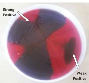
2) Congo Red Agar Method:
BHI broth supplemented with 5% sucrose and congo red was used for CRAM. The medium was composed of BHI (37gm/litre), sucrose (50gm/litre), agar agar (10mg/litre) and congo red stain (0.8gm/litre). Aqueous solution of congo red was autoclaved separately and was used for media preparation. Plates were inoculated and incubated aerobically for 24 to 48 hours at
0
37 C. Positive results were noted by observing formation of black colonies with dry crystalline consistency, whereas weak biofilm producers usually remained pink. Darkening of colonies with absence of dry crystalline consistency indicated intermediate result [9] (Fig. 2).
3) Tissue Culture Plate Method:
Tissue culture method was performed as described originally by Christensen et al [10] and modified by others [8, 11]. Briefly, for
0
0
37 C of static incubation wells were washed with phosphate buffer saline, dried in inverted position and stained by 1% crystal violet for 15 min. The wells were rinsed once more and crystal violet was solubilised in 200µl of ethanol and acetone mix (80:20 v/v). The A595 filter of Biorad ELISA reader was used to determine optical density (OD) using microtitre plate reader. Biofilm formation was scored as non biofilm forming (negative), weak (+), moderate (++), strong (+++), corresponding to optical density values <1, 1 to <2, 2 to £3, and 3 respectively obtained by using A filter (Fig. 3).595
Statistical Analysis:
All the tests mentioned above were performed in triplicates. E. faecalis ATCC 29212 was taken as positive control. Statistical analysis was carried out using paired and unpaired't' test. p values <0.05 was considered significant.
Fig. 1: Tube Adherence Method for biofilm detection: Left side of the image shows positive and negative controls. The right side
shows negative test and positive tests
Fig. 2: Congo Red Agar Method for Biofilm Detection, Positive: Black Colonies
Fig.3: Tissue Culture Plate Method for Biofilm Detection
Results:
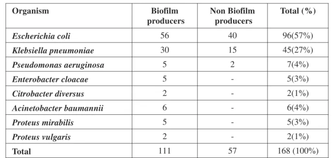
Total 168 isolates were identified by conventional biochemical methods. Biochemical identification of uropathogens up to species level revealed, E. Coli to be the predominantly isolated pathogen (57%), followed by Klebsiella pneumoniae (27%). (Table 1)
Strong Positive
Total 111 (66%) of the strains were biofilm producers and 57 (34%) were non biofilm producers by TCPM. However, by TAM and CRAM 91(54%) and 98(58%) isolates were found to be biofilm producers, whereas 46% and 42%were non biofilm producers respectively. (Table 2)
Sensitivity and specificity of TAM was found to be 75% and 86% and that of CRAM was found to be 81% and 86% when compared to TCPM. Biofilm producing strains showed relatively high drug resistance against all the antibiotics tested as compared to non biofilm producing strains. The correlation between biofilm production and antibiotic resistance was found statistically significant (p<0.05). Maximum resistance was observed with Amoxicillin (153/168), Ampicillin (153/168), Cephalexin (141/168), Least resistance was noted with Imipenem (22/168), Amikacin (54/168) (Graph 1).
In the present study, 100% biofilm producing strains were resistant to two or more number of antibiotics, and were considered MDR phenotypes [12]. Amongst these, maximum numbers of isolates were resistant to five or more number of antibiotics used. In contrast, resistance was much less in non biofilm producing strains. Of the total 57 non biofilm producer strains 42 (74%) strains showed resistance to two or more number of antibiotics whereas 15 (26%) non biofilm producing strains were sensitive to all antibiotics used.
Organism Biofilm
producers
Non Biofilm producers
Total (%)
Escherichia coli 56 40 96(57%)
Klebsiella pneumoniae 30 15 45(27%)
Pseudomonas aeruginosa 5 2 7(4%)
Enterobacter cloacae 5 - 5(3%)
Citrobacter diversus 2 - 2(1%)
Acinetobacter baumannii 6 - 6(4%)
Proteus mirabilis 5 - 5(3%)
Proteus vulgaris 2 - 2(1%)
Total 111 57 168 (100%)
Table 1: Distribution of 168 Urinary Isolates Positive for Biofilm Formation by TCPM
Number of Isolates
Method
TAM CRAM TCPM
83 + + +
7 - + +
6 + +
-21 - - +
2 + -
-2 - +
-47 - -
-168 91(54%) 98(58%) 111(66%)
Discussion:
UTI is the major public health problem in the developing countries and it is one of the most commonly encountered clinical conditions. Present study showed E. coli was the most frequently isolated pathogen followed by Klebsiella pneumoniae. Both are known to be responsible for high percentage of UTI and causes symptomatic UTI [1, 4, 10]. These bacteria have multiple virulence factors including biofilm formation to establish them in the urinary tract. Biofilm formation helps the organisms to survive in adverse conditions even in the presence of antibacterial agents.
followed by E. coli (58%) (Table 1). Our results are in agreement with previous studies, where E. coli and Klebsiella were found to be predominant biofilm producers in the catheterised as well as non catheterised UTI patients [11, 13].
Biofilm producing strains showed relatively high drug resistance against all antibiotics tested as compared to non biofilm producing strains. Maximum number of strains showed resistance to 5-11 numbers of antibiotics tested. This is a worrisome trend and UTI caused by resistant strains poses challenge for clinicians to treat the patients. It has been observed that biofilm production is often associated with long term persistence of organisms in urinary tract. Dramatically increased resistance to antibiotics makes the situation more complicated [14, 15]. In the present study, strong correlation was noted between biofilm production and resistance to multiple antibiotics, where 100% biofilm producing strains were MDR phenotypes (resistant to two or more number of antibiotics) [12]. This was in accordance with other studies [2, 10, 16,].
Proximity of cells within a biofilm can facilitate exchange of plasmid as well as poor response to conventional antibiotic therapy may contribute in the spread of antibiotic resistant traits by biofilm producing uropathogens [3]. An elevated expression of efflux pump is another mechanism described earlier for the development of antibiotic
resistance among biofilm producing bacteria [2, 17]. Furthermore, trapping of antibiotics in the hexo-polysaccharides matrix, ability of bacteria to escape from the host immune system when coated with biofilm, quorum sensing, altered metabolism and decreased growth rate all may responsible for development of antibiotic resistance among them. [2]. These organisms may not be resistant to antibiotics per se but becomes resistant when associated with biofilm.
Detection of biofilm producing bacteria in UTI influences the treatment plan. Blocking biofilm production by uropathogens in vivo provides alternative methods of therapy, which in turn will reduce the use of antibiotics. This will ultimately result in the prevention of development of multi drug resistance among the uropathogens.
Conclusion:
References
1. Oli AK, Raju S, Rajeshwari, Nagaveni S and Kelmani CR. Biofilm formation by Multidrug resistant Enterococcus faecalis (MDEF) originated from clinical samples. J Microbiol Biotech Res 2012; 2(2): 284-88.
2. Stewart PS. Mechanisms of antibiotic resistance in biofilms. Int J Med Microbiol 2002; 292(2):107-13. 3. Soto SM. Importance of biofilms in urinary tract
infections: new therapeutic approaches. Adv Biol 2014; 2014:543974.
4. Hwang-Soo Joo, Michael Otto. Molecular basis of in-vivo biofilm formation by bacterial pathogens. Chem Biol 2012;19(12): 1503-13
5. Koneman E, Allen SD, Janda WM, Schreckenberger PC. Color atlas and textbook of diagnostic microbiology, 5th edn, San Fransisco, Lippincott 2006: 624-71.
6. CLSI. (2012). Performance Standards for Antimicrobial Susceptibility Testing; Twenty-Second Informational Supplement. CLSI document M100-S22.Wayne, PA: Clinical and Laboratory Standards Institute.
7. Christensen GD, Simpson WA, Bismo AL, Beachery EH. Adherence of slime–producing strains of staphylococcus epidermidis to smooth surfaces. Infect Immun 1982; 37(1): 318-26.
8. Afreenish Hassan, Javaid Usman, Fatima Kaleem, Maria Omair, Ali Khalid, Muhammad Iqbal. Evaluation of different detection methods of biofilm formation in the clinical isolates. Braz J Infect Dis
2011; 15(4):305-11
9. Freeman DJ, Falkiner FR, Keane CT. New method for detecting slime production by coagulase negative staphylococci. J Clin Pathol 1989; 42(8): 872-74. 10. Christensen GD, Simpson WA, Younger JA et al.
Adherence of coagulase negative Staphylococci to plastic tissue cultures: a quantitative model for the adherence of Staphylococci to medical devices. J Clin Microbiol 1995; 22(6): 996-1006.
*
Sangli-416416 Maharashtra Email: [email protected] Cell: 9422691523
Author for Correspondence: Dr. Santosh S. Patil, Department of Microbiology, BVDU Medical College and Hospital,
11. Anbarasu Priyadharshini, Mangaiyarkarasi T, Balasubramaniam.R, Dhandapany Senthil Pragash, Gopal. R. Biofilm production and antibiotic resistance among uropathogens causing bacteriuria in diabetic individuals. Sch J App Med Sci 2014; 2(2A):568-71. 12. Levy SB. Factors impacting on the problem of
antibiotic resistance. J Antimicrob Chemother 2002; 49(1): 25-30.
13. Subramanian P1, Shanmugam N, Sivaraman U, Kumar S, Selvaraj S. Antibiotic resistance pattern of biofilm forming uropathogens isolated from catheterised patients in Pondicherry, India. Australas Med J 2012; 5(7):344-48.
14. Atray D, Atray M. Correlation between biofilm production and antibiotic resistance pattern in uropathogenic Escherichia coli in tertiary care hospital in Southern Rajasthan, India. Int J Curr Microbiol App Sci 2015; 4(7): 640-46.
15. Chen L, Wen YM. The role of bacterial biofilm in persistent infections and control strategies. Int J Oral Sci 2011; 3(2): 66-73.
16. Awasthi TR, Pant ND, Dahal PR. Prevalence of multidrug resistant bacteria in causing community acquired urinary tract infection among the patients attending outpatient department of Seti Zonal Hospital, Dhangadi, Nepal. Nepal J Biotechnol 2015; 3(1): 55-9. 17. Neupane S, Pant ND, Khatiwada S, Chaudhary R,
Banjara MR. Correlation between biofilm formation and resistance toward different commonly used antibiotics along with extended spectrum beta lactamase production in uropathogenic Escherichia coli isolated from the patients suspected of urinary tract infections visiting Shree Birendra Hospital, Chhauni, Kathmandu, Nepal. Antimicrob Resist Infect Control