S CIE
NT OR
U
M
ACTA
Acta Sci. Pol., Technol. Aliment. 10(3) 2011, 303-312
ISSN 1644-0730 (print) ISSN 1889-9594 (online)© Copyright by Wydawnictwo Uniwersytetu Przyrodniczego w Poznaniu
Corresponding author – Adres do korespondencji: Dr in . Kamila Myszka, Department of
Bio-EFFECT OF STARVATION STRESS
ON MORPHOLOGICAL CHANGES AND PRODUCTION
OF ADHESIVE EXOPOLYSACCHARIDE (EPS)
BY PROTEUS VULGARIS
Kamila Myszka, Katarzyna Czaczyk
Poznań University of Life Sciences
Background. Proteus vulgaris attach to available surfaces in industrial environments, can
develop into extensive biofilm. Such bacterial layer is a potential source of contamination of foods that may lead to spoilage or transmission foodborne pathogens. The purpose of these investigations was to evaluate the influence of limited nutrients availability in the medium on the morphological changes and biosynthesis of bacterial surface-associated EPS by P. vulgaris. The relationship between the dimension of cells, EPS production and
P. vulgaris biofilm development process on stainless steel surfaces (type 316L) was also
examined.
Material and methods. P. vulgaris ATCC 6380 was used in this study. The cultures were
incubated at 37°C on the Enterobacteriaceae enrichment broth according to Mossel [1962]. During the investigations the medium with optimal and 10 times diluted optimal of nutrient availability were used. For cells dimension analysis a Carl-Zeiss Axiovert 200 inverted microscope and a scanning electron microscope (LEO 435VP) was applied. Iso-lation of exopolysaccharides was based on the procedure employed by Forde and Fitzger-ald [1999]. To determine the level of P. vulgaris adhesion to the surface of stainless steel, the method described by Le Thi et al. [2001] was used.
Results. In all experimental variants the area of P. vulgaris cells was changed upon
long-term starvation. Altering of physical dimension of bacteria was effected by the decreasing value of the cell length. The change of P. vulgaris morphology promoted the beginning stages of biofilm formation process on the surface of stainless steel. Under starvation con-ditions P. vulgaris produced more EPS. It was observed with an increase of incubation pe-riod. These extracellular molecules initiated more advanced stages of P. vulgaris biofilm formation on examined surfaces.
Conclusion. The data support the notion thatcellular factors influencing P. vulgaris
se-cretion by marine bacteria under starvation conditions will help to eradicate the attached bacteria.
Key words: Proteus vulgaris, starvation, biofilm, image analysis, exopolysaccharides
INTRODUCTION
Biofilm formation process on food contact surfaces can have detrimental effect on the microbial status of the food. The presence of biofilm on abiotic materials can contaminate the product through direct contact. As a consequence, there is an increased chance of food spoilage that may lead to reduced shelf life and an increase in the risk of food poisoning from pathogens [Gram et al. 2002, Fuster-Valls et. al. 2008]. Bacteria colonizing the processing equipments are extremely difficult to overcome. Biofilms can tolerate antimicrobial agents at concentrations of 10-1000 times that needed to inacti-vate genetically equivalent planktonic bacteria [Jefferson 2004]. A better understanding of bacterial adhesion process is needed for production of microbiologically safe and good quality products in the food industry.
In food processing plants abiotic materials are often colonized by Gram-negative bacteria that originally exist in aquatic environment (with limited nutrients availability) [Gram et al. 2002]. Among marine bacteria species, Proteus vulgaris is one of the most important food spoilage and human opportunistic microorganism [Ró alski et al. 1997].
P. vulgaris may cause textural changes resulting in sensory rejection of fresh meat,
poultry and seafood [Ró alski et al. 1997, Kumar and Anand 1998]. These bacteria have also been described as etiological agents in urinary tract infections, as well as in gastro-enteritis resulting from the consumption of contaminated food [Ró alski et al. 1997].
The bacteria biofilm expansion process on food contact surface is due to morpholog-ical changes of the cells and to extracellular polysaccharide (EPS) production [Wai et al. 1999, Dunne 2002]. Changes in the physical dimensions of cells improved initial adhe-sion process to solid surfaces [Hood and Zottola 1997]. Pores and crevices at the abiotic materials increased the surface area available for cell contact. Moreover, bacteria locat-ed inside pores are shelterlocat-ed from shear forces [Kumar and Anand 1998]. The produc-tion of exopolysaccharide is responsible for both adhesion and cohesion interacproduc-tions and play a crucial role in maintaining structural integrity of mature biofilms [Sutherland 2001, Chen and Stewart 2002]. In some cases EPS can promote a preconditioning of surface, making the adhesion process more favourable [Dunne 2002].
The presence of P. vulgaris biofilm on food contact surfaces has not been eliminated yet. Most investigations have focused so far on the mechanisms determining the bacte-ria attachment process under optimal nutrients availability in the medium. These culti-vation conditions do not correspond to natural environment, where Proteus spp. is wide-ly distributed [Ró alski et al. 1997]. Anawide-lysis of both physical dimension of cells and EPS secretion by marine bacteria under starvation conditions will help in the prevention of biofilm development process on solid materials [Bower et al. 1996].
MATERIAL AND METHODS
Bacterial strains and growth conditions
P. vulgaris ATCC 6380 (American Type Culture Collection, Rockville, MD, USA)
was used in this study. P. vulgaris is a straight gram negative rod, 0.5 µm in width and 1.5-5.0 µm in length. During the investigations the microorganisms were passaged three times after every 48 h on Enterobacteriaceae enrichment medium according to Mossel [1962]. The medium used in the work is included in official (ISO) standard for detecting
Enterobacteriaceae in food products. In the study from each passage 10ml of inoculum
of P. vulgaris was added to the fresh medium. The cultures were incubated at 35°C
under shaking conditions (100 rpm/min) on the media with optimal and 10 times diluted of nutrients availability. The pH value of the culture medium at the beginning of incu-bation was 7. The incuincu-bation lasted in total 144 h.
Microscopic preparation for cells dimensions analysis
Microscopic preparations were carried out after 24, 72, 120 and 144 h of each exper-iment. The simple stain method with crystal violet was used. To avoid distortion of
P. vulgaris cells, heat fixing process was not conducted. Images were captured using
a Carl-Zeiss Axiovert 200 inverted microscope with a digital camera Carl-Zeiss Axio-Cam attached to a computer.
Cell dimensions analysis
Photographs were prepared from 30 randomly selected microscopic fields from each sample and examined using KS-300, Carl-Zeiss Soft. The image analysis steps includ-ed: image acquisition, image segmentation and measurement of the detected objects. Image acquisition enabled to create a 100 × 100 pixel numerical image, each pixel being coded into 256 gray levels. Image segmentation improved selection the objects of inter-est from the background. Measurements of the detected cells included cells area, cells length, cells width and width to length ratio.
Scanning electron microscopy
P. vulgaris morphology was also examined by a scanning electron microscopy.
The bacteria were harvested by centrifugation at 3000 g for 20 min. The pelleted cells were mounted on the steel plates and fixed for 2 h in 2% glutaraldehyde (v/v) and 2% paraformaldehyde (v/v). After rinsing, the samples were dehydrated in 99.8% (v/v) ethanol. The plates were mounted on aluminum stubs and coated with gold-palladium. The samples were then examined in the scanning electron microscope (LEO 435VP) at an accelerating voltage of 5 kV [Arnold and Bailey 2000].
Isolation and quantification of bacterial surface-associated EPS
at the room temperature after 24, 48, 72, 96, 120 and 144 h of each experiment. The cells were resuspended in 1.5 ml of 30% (w/v) NaOH. Samples were boiled for 15 min, centrifuged at 15 000 g for 15 min and the supernatants fluid were added dropwise to 60% (v/v) ethanol. The total EPS (expressed as μg/CFU) was determined using acid hydrolysis method of Parkar et al. [2001]. The precipitated EPS was collected by cen-trifugation (15 000 g, 20 min) and resuspended in 1 ml of sterile water. The samples were mixed with 7 ml of 77% (v/v) H2SO4 and cooled for 10 min in an ice-bath. 1 ml of
1% (w/v) of cold tryptophan was added and the samples were heated in a boiling bath for 20 min to effect hydrolysis. The acid hydrolysis of EPS produced a furan which condenses with the tryptophan and forms a coloured product. This was evaluated after cooling the samples by measuring O.D.500. Calibration curves were prepared against
standard dextran (Mp. 40 000) solutions (Sigma, USA).
Stainless steel surface preparation
Stainless steel plates (type 316L) sized 1 cm × 6.5 cm × 1 mm was treated with 50% solution of HNO3 for 10 min at 70°C. After soaking under distilled water the plates were
put into glass containers and sterilized at 121°C for 15 min [Parkar et al. 2001].
Bacterial adhesion analysis
P. vulgaris adhesion analysis was started after 144 h in each experiments. The
stain-less steel plates were put into P. vulgaris cultures. At 145 h the plates were removed from the glass containers and washed with PBS solutions (pH 7.2) in order to remove unattached cells from the surfaces. The plates were stained with 0.01% solution of acri-dine orange (2 min at room temperature). For observation of bacteria adhering to the stainless steel surface a fluorescence microscope was used (Carl-Zeiss, Axiovert 200). To determine the level of P. vulgaris adhesion to the surface of stainless steel the meth-od proposed by Le Thi et al. [2001] was used. This technique is based on the estimation of randomly selected 50 visual fields according to a 9-degrees scale:
1st degree: from 0 to 5 bacteria in visual field. 2nd degree: from 5 to 50 bacteria in visual field.
3rd degree: only single bacteria (above 50 bacteria cells in visual field); no microcol-onies.
4th degree: single bacteria cells + small microcolonies.
5th degree: large but not confluent microcolonies + single bacteria cells. 6th degree: confluent microcolonies + single bacteria cells.
7th degree: 1/4 visual field covered by the biofilm. 8th degree: 1/2 visual field covered by the biofilm. 9th degree: visual field totally covered by the biofilm.
Statistical analysis
Presented results are the average of three independent experiments. Effect of differ-ent nutridiffer-ents availability in the medium on P. vulgaris morphology was analysed using one-way ANOVA with post-hoc comparison (Tukey’s test; program Statistica).
RESULTS
P. vulgaris morphology
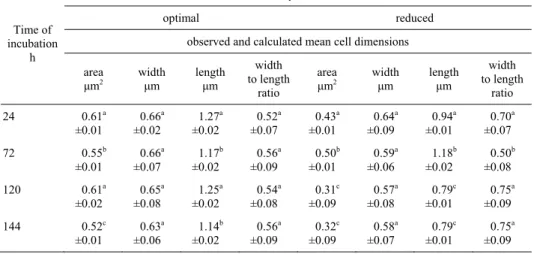
To characterise the ability of P. vulgaris to accumulate on microroughness of the abiotic surface under starvation conditions, the cells dimension analysis were carried out. P. vulgaris dimension at different nutrients availability in the medium is shown in Table 1. Scanning electron micrographs of P. vulgaris cells on stainless steel surface upon nutrionally favourable and starvation conditions are presented in Figure 1. P.
vul-garis cells alter their morphology in response to nutrients deprivation. In the first 72 h
of the process, the area of examined cells equaled between 0.43 µm2 and 0.50 µm2. From 120 h of cultivation the cell area was significantly decrease to the level of 0.31 µm2. The effect of change in P. vulgaris area upon starvation depends on shortening of the cell length from 0.94 µm to 0.79 µm. The width to length ratio indicates that
P. vulgaris starved of nutrients form swollen cells. In contrast, favourable conditions
induced a non-detectable or a small change in the examined cells morphology. During the cultivation process, mean values of P. vulgaris area, cells width and cells length equalled: 0.57 µm2, 0.65 µm and 1.20 µm respectively.
Table 1. P. vulgaris dimension under different nutrients availability in the medium
Time of incubation
h
Nutrients availability in the culture medium
optimal reduced
observed and calculated mean cell dimensions
area μm2
width
μm length μm
width to length ratio area μm2 width
μm length μm
width to length
ratio
24 0.61a
±0.01 0.66a ±0.02 1.27a ±0.02 0.52a ±0.07 0.43a ±0.01 0.64a ±0.09 0.94a ±0.01 0.70a ±0.07
72 0.55b
±0.01 0.66a ±0.07 1.17b ±0.02 0.56a ±0.09 0.50b ±0.01 0.59a ±0.06 1.18b ±0.02 0.50b ±0.08
120 0.61a
±0.02 0.65a ±0.08 1.25a ±0.02 0.54a ±0.08 0.31c ±0.09 0.57a ±0.08 0.79c ±0.01 0.75a ±0.09
144 0.52c
±0.01 0.63a ±0.06 1.14b ±0.02 0.56a ±0.09 0.32c ±0.09 0.58a ±0.07 0.79c ±0.01 0.75a ±0.09
Average values ±standard deviations.
A B
Fig. 1. Scanning electron micrographs of P. vulgaris cells on stainless steel surface (316L) upon different nutrients availability in the medium, ×10 000: A – optimal nutrients availability, B – reduced nutrients availability
Quantitative determination of bacterial surface-associated EPS
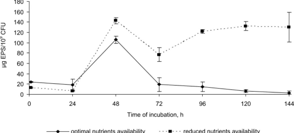
To define the potential of P. vulgaris cells to form mature biofilm structure on abiot-ic surfaces, the quantitative determination of bacterial surface-associated EPS was con-ducted. Figure 2 presents the EPS production capacity of P. vulgaris at different nutri-ents availability in the medium. P. vulgaris synthesized more EPS with an increased incubation period upon starvation. In the first 24 h of the experiment, the EPS produc-tion by examined bacteria was not higher than 10 µg/109 CFU. At 48 h of incubation the EPS synthesis was significantly increased to the value of 140 µg/109 CFU. From 120 h of the cultivation the EPS secretion by P. vulgaris remain relatively constant at the level of 125 µg/109 CFU. Upon nutrient-rich conditions, the maximum EPS production by P.
vulgaris (105 µg/109 CFU) was observed in 48 h of the process. From 72 h the EPS
synthesis decreased to the level of 18 µg/109 CFU.
Fig. 2. EPS synthesis by P. vulgaris under different nutrients availability in the medium (error bars represent standard deviation)
0 20 40 60 80 100 120 140 160 180
0 24 48 72 96 120 144
Time of incubation, h
μ
g EPS
/10
9 CF
U
Adhesion
In the study to define the rate of P. vulgaris biofilm development process, the 9- -degree scale according to Le Thi et al. [2001] was used. The results of the influence of nutrients availability on the attachment of P. vulgaris cells to stainless steel (type 316L) are presented in Table 2. In the work, the adhesion analysis started when P. vulgaris
alter their morphology and produced the high amount of EPS upon starvation condi-tions. Approximately 105-106 CFU/ml P. vulgaris cells were present in the culture me-dium during the experiments.
Table 2. The influence of nutrients availability on P. vulgaris adhesion to stainless steel surfaces (type 316L)
Time of incubation
h
Nutrients availability in the culture medium
optimal reduced
cell number in the medium CFU/ml
dominant adhesion degrees
appearance of higher adhe-sion degrees
(6, 7, 8, 9)
cell number in the medium CFU/ml
dominant adhesion degrees
appearance of higher adhe-sion degrees
(6, 7, 8, 9)
145 1.5 × 10⁶ 1, 2 – 8.2 × 10⁵ 3, 4 6
In this study when a particular degree of adhesion occurred with a minimum amount of 20% that degree became the dominant one. Stainless steel was efficiently colonized
by P. vulgaris upon nutrients limited conditions. Starvation conditions induced more
advanced stages of examined bacteria biofilm formation process on the surface of stain-less steel (6th adhesion degree). P. vulgaris grown under nutrient-rich conditions colo-nized the stainless steel surface at the 1st and 2nd adhesions degree and no developed stages of adhesion (6th-9th degrees) was noticed in this work.
DISCUSSION
The microbiological attachment to solid surfaces is a multi-step process. To predict the rate of biofilm formation process on abiotic materials under starvation conditions, both morphology and bacterial surface-associated EPS production by marine bacteria must be accounted for. In this work we aimed to evaluate the influence of limited nutri-ents availability in the medium on the morphological changes of P. vulgaris cells. Ac-cording to Wai et al. [1999] and Shau-Yan et al. [2009], altering of physical dimension of cells is favoured by nutrients deprivation. However, in the literature very little infor-mation is available on this starvation-induced mechanism. Nutrient limited condition represents the natural aquatic habitat of P. vulgaris cells [Ró alski et al. 1997].
Under starvation conditions rod-shaped bacteria, may change their size and become coccoid [Shau-Yan et al. 2009]. In our study, long-term starvation decreased the P.
vul-garis area. Altering of physical dimension of P. vulgaris depended on shortening of the
cell length. Haznedaroglu et al. [2008] noticed similar effects when monitoring
Esche-richia coli morphology upon starvation. According to this study, the change of
et al. 2008]. Siegele and Kolter [1992] reported that changes in cell size and shape are accompanied by changes in the subcellular compartments; cytoplasm is condensed and the volume of the periplasm increases. The change of marine bacteria morphology is believed to be a means of minimizing the requirements for cells maintenance and pro-tects non-spore-forming bacteria against environmental stresses [Chaiyanan et al. 2007]. In this work, the EPS synthesis by P. vulgaris cells during growth on low nutrients availability in the medium was also investigated. In the food industry and clinical condi-tions, EPS-rich strains are difficult to overcome [Bower et al. 1996]. Fuster-Valls et al. [2008] reported that EPS surrounding the bacteria protect the cells from the effects of antimicrobial agents. This feature of microorganisms may seriously affect the quality and safety of the processed food and suppose a potential risk to patients [Dunne 2002]. In our work, EPS synthesis was affected by the increasing incubation period. The high-est yield of EPS production by examined bacteria was observed after 120 h of cultiva-tion process. Also Kiliç and Dönmez [2008] noticed that long-term starvacultiva-tion influenced higher productivity of EPS by marine bacteria. The highest production of extracellular matrix by the examined cells was observed after incubation of 96 h [Kiliç and Dönmez 2008]. Siegele and Kolter [1992] and Dunne [2002] concluded that extensive production of exopolysaccharides by marine bacteria is a starvation-induced mechanism. It helps in trapping and retaining the nutrients by the cells from surrounding environments [Siegele and Kolter 1992, Dunne 2002].
Researchers have concluded that low-nutrient environments may enhance adherence [Hood and Zottola 1997]. The main biofilm expansion is due to bacterial morphology and to extracellular polysaccharide production [Dunne 2002]. These features help the microorganisms become more closely associated with a surface where nutrient accumu-lation can take place [Bower et al. 1996]. In the work the adhesion analysis started when
P. vulgaris alter their morphology and produced the high amount of EPS upon
starva-tion condistarva-tions. This knowledge may improve the eliminastarva-tion the particular pathogenic and spoilage promoting mechanisms [Gram et al. 2002]. In this study, starvation condi-tions increased the adhesion of P. vulgaris to abiotic surfaces. Our study performed that changes in marine bacteria morphology are required for the first step in biofilm for-mation process (3rd adhesion degree). According to Haznedaroglu et al. [2008] the mor-phological changes of cells affect microbial penetration in porous surface. Altering of physical dimension of cells also helps optimize interactions between cells and the sur-faces to which they attach [Young 2006]. In our study we observed that high quantities of EPS are needed to develop a true biofilm matrix (6th adhesion degree). Similar effects were noticed for a wild type of Pseudomonas fluorescens and a nonpolysaccharide-producing mutant [Allison and Sutherland 1987]. Both bacteria adhered to a glass sur-face, but over time, the wild strains formed three-dimensional structure while the mutant remained as single adherent cells on the surface.
CONCLUSION
Our data support the notion that cellular factors influencing P. vulgaris adhesion process to abiotic materials should be examined under conditions in which marine bac-teria are widely distributed. In response to nutrient limitation in the medium the size of
P. vulgaris cells was changed. Altering of physical dimension of bacteria was effected
EPS molecules by P. vulgaris cells under long-term starvation has greater importance in advanced stages of cells attachment process on the examined materials.
REFERENCES
Allison D.A., Sutherland I.W., 1987. The role of exopolysaccharide in adhesion of freshwater bacteria. J. Gen. Microbiol. 133, 1319-1327.
Arnold J.W., Bailey G., 2000. Surface finishes on stainless steel reduce bacterial attachment and early biofilm formation: scanning electron and atomic force microscopy study. Poultry Sci. 79, 1839-1845.
Bower C.K., McGuire J., Deaschel M.A., 1996. The adhesion and detachment of bacteria and spores on food-contact surfaces. Trends Food Sci. Technol. 7, 152-157.
Chaiyanan S., Grim C., Maugel T., Huq A., Colwell R.R., 2007. Ultrastructure of coccoid viable but non-culturable Vibrio cholerae. Environ. Microbiol. 9, 393-402.
Chen X., Stewart P.S., 2002. Role of electrostatic interactions in cohesion of bacterial biofilms. Appl. Microbiol. Biotechnol. 59, 718-722.
Dunne W.M., 2002. Bacterial adhesion: seen any good biofilms lately? Clin. Microbiol. Rev. 15, 155-166.
Forde A., Fitzgerald G.F., 1999. Annalysis of exopolysaccharide (EPS) production mediated by the bacteriphage adsorption blocking plasmid, pCI658, isolated from Lactococcus lactis ssp.
Cremoris HO2. Int. Dairy J. 9, 465-472.
Fuster-Valls N., Hernández-Herrero M., Marin-De-Mateo M., Rodríguez-Jerez J.J., 2008. Effect of different environmental conditions on the bacterial survival on stainless steel surfaces. Food Control. 19, 308-314.
Gram L., Ravn L., Rasch M., Bruhn J.B., Christensen A.B., Givskov M., 2002. Food spoilage – interactions between food spoilage bacteria. Int. J. Food Microbiol. 78, 79-97.
Haznedaroglu B.Z., Bolster C.H., Walker S.L., 2008. The role of starvation on Escherichia coli
adhesion and transport in saturated porous. Water Res. 42, 1547-1554.
Hood S.K., Zottola E.A., 1997. Adherence to stainless steel by foodborne microorganisms during growth in model food systems. Int. J. Food Microbiol. 37, 145-153.
Jefferson K.K., 2004. What drives bacteria to produce a biofilm? FEMS Microbiol. Lett. 236, 163-173.
Kiliç N.K., Dönmez G., 2008. Environmental conditions affecting exopolysaccharide production
by Pseudomonas aeruginosa, Micrococcus sp., and Ochrobacterium sp. J. Hazard Mater. 154,
1019-1024.
Kumar G.C., Anand S.K., 1998. Significance of microbial biofilms in food industry: a review. Int. J. Food Microbiol. 42, 9-27.
Le Thi T.T., Prigent-Combaret C., Dorel C., Lejeune P., 2001. First stages of biofilm formation: characterization and quantification of bacterial functions involved in colonization process. Met. Enzymol. 336, 152-159.
Mossel D.A.A., 1962. Use of a modified MacConkey agar medium for the selective growth and enumeration of all Enterobacteriaceae. J. Bacteriol. 84, 381-386.
Parkar S.G., Flint S.H., Palmer J.S., Brooks J.D., 2001. Factors influencing attachment of ther-mophilic bacilli to stainless steel. J. Appl. Microbiol. 11, 675-685.
Ró alski A., Sidorczyk Z., Kotełko K., 1997. Potential virulence factors of Proteus bacilli.
Mi-crobiol. Mol. Biol. Rev. 61, 65-89.
Shau-Yan C., Wann-Neng J., Yi-Shin C., Hin-Chung W., 2009. Morphological changes of Vibrio
parahaemolyticus under cold and starvation stresses. Int. J. Food Microbiol. 129, 157-165.
Siegele D.A., Kolter R., 1992. Life after log. J. Bacteriol. 174, 345-348.
Sutherland I.W., 2001. The biofilm matrix – an immobilized but dynamic microbial environment. Trends Microbiol. 9, 222-227.
Young K.D., 2006. The selective value of bacterial shape. Microbiol.Mol. Biol. Rev. 70, 660-703. Wai S.N., Mizunoe Y., Yoshida S., 1999. How Vibrio cholerae survive during starvation. FEMS
WPŁYW STRESU GŁODOWEGO NA ZMIANY MORFOLOGICZNE I BIOSYNTEZ ADHEZYJNYCH EGZOPOLISACHARYDÓW (EPS) PRZEZ PROTEUS VULGARIS
Wst p. W warunkach przemysłowych adhezja Proteus vulgaris do powierzchni stałych
rozpoczyna proces tworzenia się biofilmów. Powstawanie błon bakteryjnych stwarza ry-zyko zanieczyszczenia ywności drobnoustrojami, powodującymi zepsucie organolep-tyczne produktu, oraz mikroorganizmami chorobotwórczymi. Celem badań była ocena wpływu ograniczonej dostępności składników od ywczych w środowisku wzrostu na zmiany morfologiczne i biosyntezę EPS, związanych z powierzchnią komórki P.
vul-garis. Oceniono równie zale ność pomiędzy morfologią komórek, biosyntezą EPS i
two-rzeniem się biofilmów P. vulgaris na powierzchni stali nierdzewnej (typ 316L).
Materiał i metody. W pracy wykorzystano gatunek P. vulgaris ATCC 6380. Hodowlę
drobnoustrojów prowadzono w temperaturze 37°C na podło u namna ająco-wybiórczym, zaproponowanym przez Mossel [1962]. W doświadczeniach zastosowano podło a o op-tymalnej i 10-krotnie zredukowanej dostępności składników od ywczych. W badaniach nad właściwościami morfologicznymi komórek wykorzystano mikroskop odwrócony (Carl-Zeiss, Axiovert 200) oraz skaningowy mikroskop elektronowy (LEO 435VP). Do izolacji egzopolisacharydów bakteryjnych zastosowano metodę, którą opracowali Forde i Fitzgerald [1999]. Dynamikę procesu adhezji P. vulgaris do powierzchni stali nie-rdzewnej oceniano na podstawie metody, którą zaproponowali Le Thi i in. [2001].
Wyniki. We wszystkich wariantach doświadczenia zaobserwowano zmianę powierzchni
komórek P. vulgaris pod wpływem stresu głodowego. Zmiana wymiarów komórek wyni-kała ze zmniejszania się długości drobnoustrojów. Zmiany morfologiczne P. vulgaris
promowały początkowe etapy tworzenia biofilmów na powierzchni stali nierdzewnej. W warunkach głodowych zaobserwowano intensyfikację biosyntezy EPS przez P. vulga-ris. Było to szczególnie widoczne w końcowych etapach procesu hodowlanego. Biosynte-za zewnątrzkomórkowych polisacharydów inicjowała tworzenie się dojrzałych matryc biofilmu na testowanych powierzchniach stałych.
Podsumowanie. Wyniki przeprowadzonych doświadczeń wskazują, e czynniki wpł
ywa-jące ze strony komórek P. vulgaris na proces adhezji drobnoustrojów do powierzchni abiotycznych powinny być badane w warunkach, w których bakterie wodne występują na-turalnie. Badania nad zmianami morfologicznymi oraz biosyntezą EPS przez bakterie wodne w warunkach głodowych mogą usprawnić procedury eliminacji błon biologicz-nych z powierzchni stałych.
Słowa kluczowe: Proteus vulgaris, warunki głodowe, biofilm, cyfrowa analiza obrazu,
egzopolisacharydy
Received – Przyjęto: 24.01.2011
Accepted for print – Zaakceptowano do druku: 28.03.2011