ABSTRACT
Purpose: To evaluate the efficacy and safety of trabeculectomy and non-penetrating deep sclerectomy (NPDS) in eyes with advanced glaucoma.
Material and Methods: Retrospective review of patients with advanced glaucoma submitted to surgery between January 2015 and May 2017. Absolute success was defined as postoperative intraocular pressure (IOP) <18 mmHg without application of hypotensive medications and qualified success as postoperative IOP <18 mmHg with or without hypotensive medications.
Results: 52 eyes were submitted to glaucoma surgery (34 NPDS and 18 trabeculectomy). The mean follow-up time was 16.2 months. 76.9% of the procedures were combined with cataract surgery. The absolute success was achieved in 82.7% of the cases and the qualified success in 96.2%. The mean IOP pre and postoperative was 22.24 and 11.93 mmHg, respectively. The mean number of preoperative hypotensive medications was 2.13 and the postoperative 0.25. The mean preoperative visual acuity was 0.42logMAR and the postoperative 0.22 logMAR. In the postoperative period, 7 cases of early hypotony, one of choroidal detachment and two of bleb cysts were recorded. Five patients required goniopuncture and two selective laser trabeculoplasty for IOP control.
Conclusions: NPDS and trabeculectomy were effective in the treatment of patients with advanced glaucoma. There was a low rate of complications, and no cases of visual acuity decrease during the follow-up period. Control of IOP without hypotensive medications is associated with an improvement in quality of life and lower IOP fluctuations, which are crucial aspects for these patients.
Keywords: trabeculectomy, non-penetrating deep sclerectomy, advanced glaucoma, efficacy, quality of life.
RESUMO
Objetivo: Avaliar a eficácia e segurança da trabeculectomia e esclerectomia profunda não penetrante (EPNP) em olhos com glaucoma avançado.
Material e Métodos: Foram revistos os casos de glaucoma avançado submetidos a cirurgia entre janeiro de 2015 e maio de 2017. O sucesso absoluto foi definido como pressão intraocular (PIO) no pós-operatório <18 mmHg sem aplicação de fármacos hipotensores oculares (FHO) e o sucesso qualificado como PIO no pós operatório <18 mmHg com ou sem colocação FHO. Resultados: 52 olhos foram submetidos a cirurgia de glaucoma (34 realizaram EPNP e 18 trabeculectomia). O tempo médio de follow-up foi de 16,2 meses. 76,9% dos procedimentos foram combinados com cirurgia de catarata. Atingiu-se o sucesso absoluto em 82.7% dos casos e o sucesso qualificado em 96,2%. A PIO média pré e pós cirurgia foi 22,24 e 11,93 mmHg, respetivamente. O número médio de FHO pré cirúrgico foi 2,13 e o pós cirúrgico 0,25. A acuidade visual média pré cirúrgica foi 0.42logMAR e a pós cirúrgica 0.22 logMAR. No pós operatório registaram-se 7 casos de hipotonia precoce, um de descolamento coroideu e dois de quistos da ampola. 5 pacientes realizaram goniopunção e dois trabeculoplastia laser seletiva para controlo da PIO.
Conclusões: A EPNP e a trabeculectomia foram eficazes no tratamento de pacientes com glaucoma avançado. Registou-se uma baixa taxa de complicações, não se verificando diminuição da acuidade visual em nenhum dos casos. O controlo da PIO sem FHO está associado a uma melhoria da qualidade de vida e menores flutuações da PIO, aspetos fundamentais para estes pacientes.
Palavras-chave: Trabeculectomia, esclerectomia profunda não penetrante, glaucoma avançado, eficácia, qualidade de vida.
INTRODUCTION
Glaucoma is the leading cause of irreversible blindness in the world.23, 26
There is no universally accepted definition of advanced glaucoma. Regardless of the definition used, it is recognized that this group of patients presents a particular challenge for the ophthalmologist, due to their greater susceptibility for visual field and visual acuity loss.21 Furthermore, the diagnostic tests are frequently more challenging and less reliable in advanced glaucoma, leading to a greater number of complementary tests to confirm disease progression.21 In patients with advanced glaucoma, to avoid disease progression and further visual fields loss, the target intraocular pressure (IOP) is typically
lower. To accomplish that, the ophthalmologist has three alternatives: medical therapy, laser and surgical procedures, which can be combined.
All of these particularities dictate a closer monitoring for advanced glaucoma patients. The greater number of visits, complementary examinations, hypotensive agents and surgical procedures is associated with a higher personal and national health system financial cost, and more importantly, to a deterioration of patient’s quality of life.21
Intraocular pressure fluctuation is a risk factor for visual fields progression in patients with advanced glaucoma, even with lower IOP.8 In this particular group of subjects, less IOP fluctuation is expected in patients
submitted to trabeculectomy compared to patients undergoing ocular hypotensive drugs.17
The risk of visual impairment after glaucoma surgery has been described in some studies.1,13,18 Visual compromise may be related to cataract, cystoid macular edema, suprachoroidal or vitreous hemorrhage, retinal detachment and uncontrolled intraocular pressure (IOP).10 However, even after an otherwise uncomplicated surgery, loss of central vision has been described.1,10,13 In the scientific community, this “wipe-out” phenomenon is debatable.
To date there are few publications with surgical results in patients with advanced glaucoma. The aim of the "Treatment of Advanced Glaucoma Study” a multicenter, randomized study, is to compare medical treatment with primary trabeculectomy for patients with newly diagnosed advanced glaucoma. This trial is expected to be concluded in the year 2020.
The present study was undertaken to retrospectively review the results of glaucoma surgery (trabeculectomy and NPDS) in eyes with advanced glaucoma.
METHODS
This is a retrospective study of patients with advanced glaucoma submitted to surgery (trabeculectomy or NPDS) at the Department of Ophthalmology of Hospital Pedro Hispano (Matosinhos, Portugal) between january 2015 and may 2017.
The methodology of the study was designed in accordance with the tenets of the Helsinki Declaration.
Indications for surgery were insufficient IOP control, non-compliance to topical treatment or progression of the disease. Data recorded from each patient included age, sex, type and stage of glaucoma, number of IOP-lowering medications. Glaucoma type was defined by gonioscopy and slit-lamp examination, and advanced glaucoma was defined according to the Hodapp-Parrish-Anderson’s criteria.14
All IOP measurements were performed with Goldmann applanation tonometry. Absolute success was defined as IOP < 18 mmHg 28 without glaucoma medical treatment,
and qualified success as IOP < 18 mmHg with or without ocular hypotensive agents.
The surgeries were performed by 2 surgeons.
Pre-operative evaluation
The preoperative evaluation included: best corrected visual acuity (BCVA), anterior segment slit-lamp
examination, Goldmann applanation tonometry,
gonioscopy, evaluation of the ocular fundus after pharmacological mydriasis, visual fields (Humphrey visual field analyzer) and optical coherence tomography of optic disc (Optovue RTVue OCT®).
The preoperative BCVA, IOP, mean deviation (MD) of visual fields (VF) and retinal nerve fiber layer (RNFL) thickness were those evaluated at the last visit before surgery. BAVC was measured with a Snellen chart and was converted to the logMAR scale for comparison.
Surgical technique
NPDS
1- Either local anesthesia with monitored anesthesia care or general anesthesia was performed.
2- A 8-0 nylon traction suture was placed in the cornea adjacent to the superior limbus.
3- A fornix-based conjunctival flap centered at 12 o’clock was prepared, and the conjunctiva and Tenon’s capsule were dissected.
4- Light cautery was applied as needed.
5- Sponges soaked in 0.2 mg/mL Mitomycin-C (MMC) were placed under the conjunctiva for 2,5 minutes, and then the subconjunctival space was carefully washed with balanced salt solution.
6- A 5 x 5 mm superficial (one-third-thickness) scleral flap, with a limbus-based approach, was created and dissected anteriorly until clear cornea was reached. 7- A smaller (4 x 4mm) deep scleral flap, with a
limbus-based approach, was created, and dissection was continued anteriorly to the Schlemm’s canal.
8- The inner wall of Schlemm’s canal was carefully peeled off with forceps and the deep scleral flap excised.
9- An implant (AquaFlow® or Esnoper®) was placed on the exposed scleral stroma bed and secured with one 10-0 nylon suture.
10- The superficial scleral flap was sutured with interrupted 10-0 nylon suture.
11- The conjunctival flap was closed with 10-0 nylon suture.
12- At the end of the procedure, we checked to see if the conjunctival closure was watertight.
13- Cefuroxime and metilpednisolone subconjuntival injections were administered in the inferior quadrants.
Trabeculectomy
1- Either local anesthesia with monitored anesthesia care or general anesthesia was performed.
2- A 8-0 nylon traction suture was placed in the cornea adjacent to the superior limbus.
3- A fornix-based conjunctival flap centered at 12 o’clock was prepared, and the conjunctiva and Tenon’s capsule were dissected.
4- Light cautery was applied as needed.
5- Sponges soaked in 0.2 mg/mL Mitomycin-C (MMC) were placed under the conjunctiva for 2, to 35 minutes, and then the subconjunctival space was carefully washed with balanced salt solution.
6- A 5 x 5 mm half- thickness scleral flap, with a limbus-based approach, was outlined with a surgical blade, and then dissected.
7- A paracentesis in the peripheral cornea was performed. 8- Sclero-corneal tissue, including the trabecular
meshwork, was then removed with microsurgical knife and Vannas scissors;
9- A peripheral iridectomy was performed;
10- The scleral flap was sutured with 10-0 nylon suture. The 11th, 12th and 13th steps of NPDS technique were performed.
In the cases of combined surgery, phacoemulsification with intraocular lens (IOL) implantation was performed before the glaucoma surgery.
Postoperatively, all patients received a standard regimen of:
- topical antibiotic (quinolone), 5 times daily, discontinued after 2 weeks;
- opical corticosteroids (Dexamethasone phosphate 1mg/ml), 5 times daily during the first 4 to 6 weeks, and then tapered as needed.
Topical nonsteroidal anti-inflammatory was
administrated during the first month after combined surgery.
Follow-up visits were scheduled at: 1 day, 1 week; 2 weeks, 1 and 3 months; after that, patients were followed within a 3-6 month-interval. Additional visits outside this regimen were scheduled according to the IOP or the presence of complications.
Postoperative laser suture lysis and associated ocular massage were conducted in a step-by-step manner until the expected target pressure was achieved. Laser suture lysis was conducted postoperatively when the IOP increased and/or the filtering bleb became more localized and flattened. Ocular massage was added routinely after laser suture lysis.
Main outcome measures of this study are BCVA, IOP, number of hypotensive medications, MD and RNFL thickness evaluated at the latest examination after surgery (range: 4 to 26 months).
The incidence of intraoperative and also of any postoperative complications such as flat anterior chamber, hypotony (defined as IOP < 5 mmHg), macular edema, choroidal detachment and status of bleb was recorded at all visits.
Statistical analysis was performed using SPSS (Version 21.0, SPSS Inc., Chicago, IL, USA). Shapiro-Wilk test was used to test for normality. Paired samples t-test was used to compare measurements before and after surgery, including mean IOP values and mean number of IOP-lowering medications. A value of p<0.05 was considered to be statistically significant.
RESULTS
Records of 52 eyes were reviewed in this study. Demographic and baseline clinical characteristics of patients are summarized in Table 1 and 2. None of the eyes had previous glaucoma surgery.
Mean preoperative: BCVA was 0.42 ± 0,2 logMAR; IOP was 22.24 ± 6.2 mmHg; hypotensive medication was 2.13 ± 0.9. Average MD of the preoperative VF was
-22.79 ± 4.26 dB. Mean preoperative RNFL thickness was 66,4 ± 12,13 µm.
Table 1 - Baseline clinical characteristics of patients undergoing glaucoma surgery for advanced glaucoma
Total (eyes) 52 Age (mean) 76,2 ± 7,2 Gender Male Female 28 (53,8%) 24 (46,2%) Type of glaucoma Pseudoexfoliation Primary open-angle 20 32
BCVA (mean, LogMAR) 0.42 ± 0,2
IOP (mean, mmHg) 22.24 ± 6.2
Number of hypotensive
medications (mean) 2.13 ± 0.9
VF MD (mean, dB) -22.79 ±4.26
RNFL thickness (mean, µm) 66,4 ± 12,13
Table 2 - Baseline clinical characteristics of patients undergoing non-penetrating deep sclerectomy (NPDS) and trabeculectomy for advanced glaucoma.
NPDS Trabeculectomy t-test Total (eyes) 34 18 Age (mean) 75,4 ± 6,9 77,8 ± 7,5 p = 0,094 Gender Male Female 20 (58,8%) 14 (41,2%) 8 (44,4%) 10 (55,6%) Type of Glaucoma Pseudoexfoliation Primary open-angle 13 (38,2%) 21 (61,8%) 7 (38,9%) 11 (61,1%) BCVA (mean, LogMAR) 0,43 ± 0,21 0,39 ± 0,23 p = 0,096 IOP (mean, mmHg) 21,58 ± 5,21 23,47 ± 6,73 p = 0,041 Number of hypotensive medications (mean) 1,94 ± 0,81 2,5 ± 1,21 p = 0,001 VF MD (mean, dB) -21,9 ± 4,91 -24,85 ± 6,2 p = 0,036 RNFL thickness (mean, µm) 70,83 ± 12,62 63,33 ± 13,86 p =0,028
MD and RNFL thickness after filtration surgery in our patients. Mitomycin C was used in all surgeries. Thirty four eyes had NPDS and eighteen had trabeculectomy. Most of the surgeries (76,9%) were combined with phacoemulsification and IOL implantation. There were no intraoperative complications.
n 52 Follow-up (months) Range Mean 4 - 26 16,2 ± 4,8
BCVA (mean, logMAR) 0,22 ± 0,14
IOP (mean, mmHg) 11,93 ± 4,45
Number of hypotensive
medications (mean) 0,25 ± 0,17
Absolute sucess (%) 82,7
Qualified sucess (%) 96,2
IOP reduction (mean, %) 46,4
VF MD (mean, dB) -23,62 ± 5,51
RNFL thickness (mean, µm) 65.9 ± 11,24
Data from last visit after glaucoma surgery
Table 4 - NPDS group - Preoperative and postoperative data.
NPDS(n=34)
Preoperative Postoperative p
BCVA (mean, logMAR) 0,43 ± 0,21 0,21 ± 0,13 0,001
IOP (mean, mmHg) 21,58 ± 5,21 11,07 ± 4,1 0,001 Number of hypotensive medications (mean) 1,94 ± 0,81 0,15 ± 0,12 0,001 Absolute sucess (%) 88,24 Qualified sucess (%) 100
IOP reduction (mean,
%) 48,7
VF MD (mean, dB) -21,9 ± 4,91 -22,84 ± 4,63 0,61
RNFL thickness (mean,
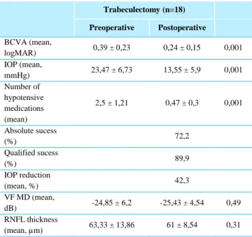
Table 5 - Trabeculectomy Group - Preoperative and postoperative data. Trabeculectomy (n=18) Preoperative Postoperative BCVA (mean, logMAR) 0,39 ± 0,23 0,24 ± 0,15 0,001 IOP (mean, mmHg) 23,47 ± 6,73 13,55 ± 5,9 0,001 Number of hypotensive medications (mean) 2,5 ± 1,21 0,47 ± 0,3 0,001 Absolute sucess (%) 72,2 Qualified sucess (%) 89,9 IOP reduction (mean, %) 42,3 VF MD (mean, dB) -24,85 ± 6,2 -25,43 ± 4,54 0,49 RNFL thickness (mean, µm) 63,33 ± 13,86 61 ± 8,54 0,31
Seidel occurred in 8 (15,4%) eyes, 4 (11,8%) after NPDS and 4 (22,2%) after trabeculectomy. Early hypotony occurred in 7 (13,5%) eyes, 3 (8,8%) after NPDS and 4 (22,2%) after trabeculectomy. These
complications were successfully managed with
conservative medical treatment, except one eye that needed a revision of conjunctival closure with 8-0 vicryl suture.
One (5,5%) choroidal detachment and two (11%) cases of bleb cysts occurred after trabeculectomy. The choroidal detachment was conservatively managed, and bleb cysts were successfully managed with bleb needling, with no further complications.
In the NPDS group, 5 (15,6%) eyes required Nd:YAG Laser goniopuncture for IOP control. One of this patients developed transient hypotony after this procedure. No other complications occurred.
Two (3,8%) patients (one in each group) required selective laser trabeculoplasty for IOP control.
There were no cases of postoperative flat anterior chamber, macular edema, blebitis or endophthalmitis. No patient experienced wipe-out phenomenon.
At the last follow-up visit, the mean BCVA was 0,22 ± 0,14 logMAR (p = 0.001, paired t test). After a mean follow-up of 16,2 months, a significant reduction of IOP (46,4%) was achieved (from 22.24 ± 6.2 to 11,93 ± 4,45mmHg postoperatively, p = 0.001, paired t test). Mean
postoperative hypotensive medications was significantly reduced 0,25 ± 0,17 (p = 0.001, paired t test).
Absolute and qualified successes were achieved in 82,7% and 96,2% of the eyes, respectively.
At the last visit, the mean postoperative VF MD and RNFL thickness were -23,62 ± 5,51 dB and 65.9 ± 11,24 µm , respectively. No statistically significant differences were found between VF MD and RNFL thickness values before and after glaucoma surgery (respectively, p = 0.56, p= 0.35, paired t test).
DISCUSSION
The goal of glaucoma treatment is to control IOP and maintain a stable visual function, with minimal impairment of our patient’s quality of life. 15
Inherent to this strategy is the concept of the "target IOP" level, corresponding to an IOP interval which minimizes further damage to the visual field and a rate of progression unlikely to affect the quality of life of the patient. As such, it should be individualized and adjusted during the course of the disease.15, 25 The greater the pre-existing glaucoma damage, the lower the target IOP should be. For advanced glaucoma damage was suggested an intraocular pressure ≤ 12mmHg. 31
It is important to diagnose glaucoma at an early stage to preserve the maximum visual function. The majority of patients included in this study was first diagnosed with advanced glaucoma. So our goal was no longer to protect a large field of vision, but rather a critical one, crucial for our patients quality of life and self-care. Patients in this stage of glaucoma have a high risk of further disease progression, so a 40% decrease of IOP from baseline is proposed by some authors.22 To achieve this target IOP, surgical procedures are often required.24 Even if advanced glaucoma could be controlled medically, the lack of compliance with medication regimens may induce significant short and long term IOP fluctuations, with a deleterious effect on the visual outcomes. Glaucoma procedures are often associated with a more effective IOP control and minimal IOP fluctuations, protecting what's left of the visual field.17
Due to a lower neuronal reserve in advanced glaucoma, the risk of visual impairment after glaucoma surgery remains a concern and has been described in some
studies. There is no consensus in the ophthalmological community regarding the risk of immediate unexplained visual field loss (wipe-out phenomenon) after filtering procedures in patients with glaucoma.1,2,10,16,19,20,22,29 It is thought that sudden, intraoperative ocular hypotony is the underlying mechanism of this phenomenon. 29 On the other hand, some authors2,19,20,29, with a large number of surgeries performed do not describe any case of wipe-out phenomenon.
A recent study30 found that the absence of postoperative laser suture lysis is a significant risk factor for ocular hypotony after trabeculectomy. Awai M. et al 6 defends that trabeculectomy with MMC, tight sutures for the closure of scleral flaps, followed by postoperative laser suture lysis and ocular massage, reduces the risk of fixation loss in eyes with advanced glaucoma, avoiding the occurrence of hypotony and excess IOP fluctuations after surgery. We performed glaucoma surgery following this principles. In our study no patient experienced wipe-out phenomenon during the follow-up period.
Some ophthalmologists do not perform cataract surgery in patients with advanced glaucoma with low preoperative visual acuity regarding that surgery as an high risk procedure, unlikely to bring great benefit to the patient. Other groups, however, demonstrated that cataract surgery improves visual acuity and quality of life in patients with advanced glaucoma.32 Different surgical options are available for patients with cataract and concomitant glaucoma. If the IOP is controlled, cataract surgery can be performed as a stand alone procedure, and glaucoma surgery postponed. After phacoemulsification pressure spikes may occur, which should be taken in account by the ophthalmologist. 28 Other option consists in performing glaucoma surgery first. Most patients, even those without cataract at the time of the glaucoma surgery, will need phacoemulsification in the future, especially when MMC is used3,28. But the cataract surgery performed after a filtration procedure has a risk of reducing the efficiency of first operation.9 Thus, a combination of glaucoma and cataract surgeries seems a reasonable approach in these patients, many of them elderly, with comorbidities that preclude multiple surgical procedures. Other advantages of performing combined surgeries include the possible immediate increase of visual acuity and economical aspects.7 In this study, combined surgery
was performed in more than three quarters of the patients, with good functional and safety profiles.
In this study we reviewed the outcome of advanced glaucoma patients submitted to trabeculectomy or NPDS. Both procedures achieved good IOP control (mean IOP reduction >40%), with overall absolute and qualified successes achieved in 82,7% and 96,2% of the eyes, respectively. Setting as the threshold for surgical success values of IOP ≤ 12 mmHg postoperatively, would correspond an absolute success of 51,9% and qualified success of 53,8%.We found statistically significant differences among the baseline characteristics of the patients in the different groups. That means that we can not compare the postoperative data of NPDS with trabeculectomy. However, in both groups a reduction of ocular hypotensive medications and no statistically significant disease progression during the follow-up time was noted. The safety profile was also good with both techniques, with low rate of complications and with no cases of visual acuity decrease during the follow-up period. The NPDS group presented a lower relative number of postoperative complications compared to the trabeculectomy group.
To our knowledge, in the literature there are limited published data of subjects with advanced glaucoma submitted to NPDS.4,5,12 The results of our study suggest that NPDS is effective in this particular group of patients. Since this is classically a safer procedure when compared to trabeculectomy (the results of our study also indicate that), NPDS may be a valuable surgical alternative in the treatment of advanced glaucoma, providing a significant reduction in IOP and hypotensive medication with a good profile risk.
The IOP control without ocular hypotensive agents is associated with an improvement in the quality of life and lower IOP fluctuations, which may be crucial for patients with advanced glaucoma.
There are some limitations in our study such as: retrospective methodology, small sample size, variable follow-up length and differences between baseline characteristics of patients submitted to NPDS and trabeculeculectomy. More trials are required, with larger number of patients, longer time of follow up to confirm the conclusions suggested by this study.
BIBLIOGRAPHY
1. Aggarwal S. P., Hendeles S. Risk of sudden visual loss following trabeculectomy in advanced primary open-angle glaucoma. Br J Ophthalmol 1986,70:97–99
2. Agrawal S, Agrawal J, Agrawal et al. Risk of Sudden Visual Loss Following Filtration Surgery in End-stage Glaucoma. Am J Ophthalmol,142(1):199 – 200 author reply 200
3. Asamoto A, Yablonski M. E., Posttrabeculectomy anterior subcapsular cataract formation induced by anterior chamber air. Ophthalmic Surg 1993;24:314–319.
4. Ates H., Andac K., Uretmen, O. Non-penetrating deep sclerectomy and collagen implant surgery in glaucoma patients with advanced field loss. Int Ophthalmol 1999 May;23(3):123–128
5. Ates H., Andac K., Uretmen O. Non-penetrating deep sclerectomy in the treatment of advanced cases of open
angle glaucoma. Int Ophthalmol 1999;23:123.
https://doi.org/10.1023/A:1010745204480
6. Awai M, Koga T. Inatani et al. Stability of the Central Visual Field After Modern Trabeculectomy Techniques in Eyes with Advanced Glaucoma. Jpn J Ophthalmol, 2007 March;51(2):116–120
7. Bilgin, G., Karakurt A., Sinan S. Combined Non-Penetrating Deep Sclerectomy with Phacoemulsification Versus Non-Penetrating Deep Sclerectomy Alone.
Seminars in ophthalmology. 2014, 29.
10.3109/08820538.2013.874466.
8. Caprioli J., Coleman A. L. Intraocular pressure fluctuation a risk factor for visual field progression at low intraocular pressures in the advanced glaucoma intervention study. Ophthalmology. 2008 July;115(7):1123-1129.
9. Casson R, Rahman R, Salmon JF. Phacoemulsification with intraocular lens implantation after trabeculectomy. J Glaucoma 2002;11:429–433
10. Costa V. P., Smith M., Spaeth G. L. et al. Loss of visual
acuity after trabeculectomy. Ophthalmology
1993;100:599–612
11. Galin M. A., Lin L. L., Obstbaum S. A. Cataract extraction and intraocular pressure. Trans Ophthalmol Soc U K 1978;98:124–127.
12. Guedes P., Ricardo & Maria Paletta Guedes, Vanessa & Chaoubah, Alfredo. Resources use, costs and effectiveness of non-penetrating deep sclerectomy according to glaucoma stage. ArqBrasOftalmol. 2011 74:400-4.
13. Hesse R. J. Risk of sudden visual loss after filtration
surgery in end-stage glaucoma. Am J Ophthalmol 2006;141:983
14. Hodapp E, Parrish R. K. II, Anderson D. R. Clinical Decisions in Glaucoma. St. Louis: CV Mosby Co; 1993:52–61.
15. Jampel H. Target IOP in clinical practice. In: Weinreb RN, Brandt JD, Garqay-Heath D, Medeiros FA. Intraocular Pressure. Kugler Publications, Amesterdam 2007:121-125
16. Kolker A.E. Visual prognosis in advanced glaucoma: a comparison of medical and surgical therapy for retention of vision in 101 eyes with advanced glaucoma. Trans Am Ophthalmol Soc 1977;75:539 –555.
17. Konstas A. G., Topouzis F., Leliopoulou O. et al. 24-hour intraocular pressure control with maximum medical therapy compared with surgery in patients with advanced
open-angle glaucoma. Ophtalmology. 2006
May;113(5):761-5.e1
18. Law S. K., Nguyen A. M., Coleman A. L. et al Severe loss of central vision in patients with advanced glaucoma undergoing trabeculectomy. Arch Ophthalmol. 2007, 125:1044–1050
19. 19 - Lichter P. R., Ravin J. G; Risks of sudden visual loss after glaucoma surgery. Am J Ophthalmol 1974;78:1009– 1013.
20. Martinez J. A., Brown R. H., Lynch M. G. et al. Risk of postoperative visual loss in advanced glaucoma. Am J Ophthalmol 1993;115:332–337.
21. Moraes C. G., Liebmann J. M., Medeiros F. A. et al. Management of advanced glaucoma: Characterization and monitoring. Surv of Ophthalmol 2016 September-October;61(5):597–615
22. Otto J. Loss of point of fixation after glaucoma surgery [in German]. Klin. Monatsbl. Augenheilkd. 1957;131:178– 195.
23. Quigley H. A., Broman A. T. The number of people with glaucoma worldwide in 2010 and 2020. Br J Ophthalmol 2006;90(3):262– 267.
24. Simon K. L., Law S. K., Nguyen A. M., Coleman A.L. et al Severe Loss of Central Vision in Patients With Advanced Glaucoma Undergoing Trabeculectomy. Arch Ophthalmol. 2007;125(8):1044-1050.
25. Singh K, Shrivastava A. Early aggressive intraocular pressure lowering, target intraocular pressure, and a novel concept for glaucoma care. Surv Ophthalmol 2008;53, Sup 1:S33-38
26. Tham Y. C., Li X., Wong T. Y. et al Global prevalence of glaucoma and projections of glaucoma burden through
2040: a systematic review and meta-analysis.
Ophthalmology. 2014 121(11):2081–2090.
27. Tharwat H. Mokbel. End Stage Glaucoma, Glaucoma - Basic and Clinical Concepts, 2011 ISBN:
978-953-307-591-4, InTech, Available from:
http://www.intechopen.com/books/glaucoma-basicand-clinical-concepts/end-stage-glaucoma
28. The AGIS Investigators. The advanced glaucoma intervention study (AGIS): 7. The relationship between control of intraocular pressure and visual field deterioration. Am J Ophthalmol 2000;130:429-40.
29. Topouzis F, Tranos P., Koskosas A. et al Risk of Sudden Visual Loss Following Filtration Surgery in End-Stage Glaucoma, Am J Ophthalmol 2005 October;140(4): 661.e1–661.e7
30. Tseng, V.L., Kim, C.H., Romero, et al. Risk Factors and Long-Term Outcomes in Patients with Low Intraocular Pressure after Trabeculectomy. Ophthalmology 2017 October;124(10):1457-1465
31. World Glaucoma Association Consensus Statement: Intraocular Pressure. The Netherlands: Kluger; 2007.
32. Xu X., Sun Q., Ma Y. Y. et al. Vision-related Quality of Life Outcomes of Cataract Surgery in Advanced Glaucoma Patients. Health and Quality of Life Outcomes. 2017 15:175
CONTACT
Tiago Maio
Rua de Santa Iria nº64 3º esquerdo 5000 – 446 Vila Real
E-mail: [email protected]
Sem conflitos de interesse. Trabalho não publicado previamente Cedemos os direitos de autor à SPO